Client Article | Cell Reports | The team of Mian Peng and Xiang Li from Zhongnan Hospital, Wuhan University, reveals that dexmedetomidine inhibits fear memory consolidation via the astrocyte-specific Srebf1-Phgdh pathway.
Release time:2026-04-30 10:36:56
Post-traumatic stress disorder (PTSD) is a severe mental disorder that occurs after exposure to traumatic events. However, effective prevention and treatment options are still lacking. Dexmedetomidine is a widely used sedative in clinical practice, with sedative, analgesic, anxiolytic, and sympatholytic effects. Studies have shown that low-dose dexmedetomidine sedation during or after trauma exposure can significantly reduce the incidence of PTSD. However, the effects of dexmedetomidine on fear memory and its underlying mechanisms remain unclear.
On March 17, 2025, the team of Mian Peng and Xiang Li from Zhongnan Hospital, Wuhan University, published a research paper titled “Dexmedetomidine inhibits fear memory consolidation via the astrocyte-specific Srebf1-Phgdh pathway in the prelimbic prefrontal cortex” in Cell Reports. The paper reveals that dexmedetomidine specifically inhibits the nuclear translocation of sterol regulatory element-binding protein 1 (Srebf1) in astrocytes of the prelimbic prefrontal cortex (PLPFC), thereby reducing its active form, which has transcriptional activity. This change further leads to the downregulation of its target gene, phosphoglycerate dehydrogenase (Phgdh). Phgdh, the major synthesizing enzyme of D-serine, a co-agonist of the N-methyl-D-aspartate (NMDA) receptor, plays a key role in regulating synaptic stability and ultimately participates in the suppression of fear memory consolidation. This study elucidates the molecular mechanism by which dexmedetomidine inhibits fear memory consolidation, providing a theoretical basis for its preventive application and revealing a new target for PTSD intervention.
Dexmedetomidine alleviates fear in mice by inhibiting fear memory consolidation without affecting fear memory extinction
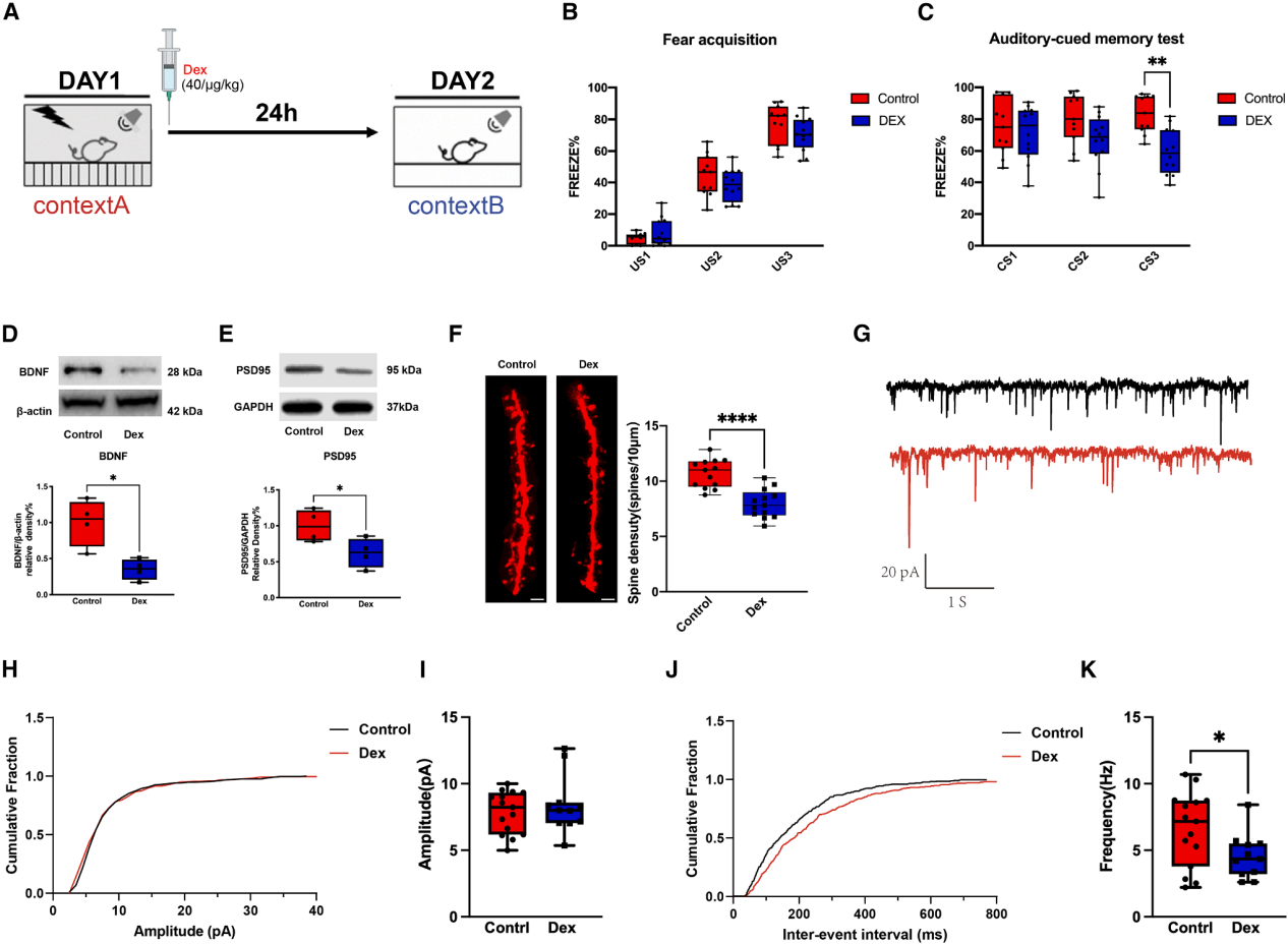
To investigate whether subanesthetic doses of dexmedetomidine can reduce fear and PTSD-like behaviors in mice, 40 μg/kg dexmedetomidine was intraperitoneally injected immediately after fear acquisition on day 1. Memory retention testing was performed on the second day at the same time (Figure 1A). All mice showed more than 50% freezing behavior during the final conditioned stimulus (CS) exposure (Figure 1B). Compared to the control group, dexmedetomidine significantly reduced the freezing time during the last 120 seconds of CS exposure 24 hours later (Figure 1C) and one week later. Further investigation of the effects of dexmedetomidine on synaptic stability showed that, under fear conditions, dexmedetomidine treatment decreased the levels of brain-derived neurotrophic factor (BDNF) and postsynaptic density protein 95 (PSD95), as well as dendritic spine density in the prelimbic prefrontal cortex (PLPFC) (Figure 1D-1F). Whole-cell patch-clamp recordings from PLPFC brain slices (Figure 1G) showed no difference in the amplitude of spontaneous excitatory postsynaptic currents (sEPSCs) between the dexmedetomidine and control groups, but the frequency was significantly reduced (Figure 1H-1K). These results suggest that subanesthetic doses of dexmedetomidine inhibit fear memory consolidation, impair synaptic stability, and do not affect fear extinction memory.
Figure 1 | Dexmedetomidine alleviates fear responses in mice by inhibiting fear memory consolidation, accompanied by impaired synaptic stability
Dexmedetomidine upregulates Srebf1 mRNA expression and reduces the nuclear levels of transcriptionally active nSrebf1 protein in the PLPFC of mice
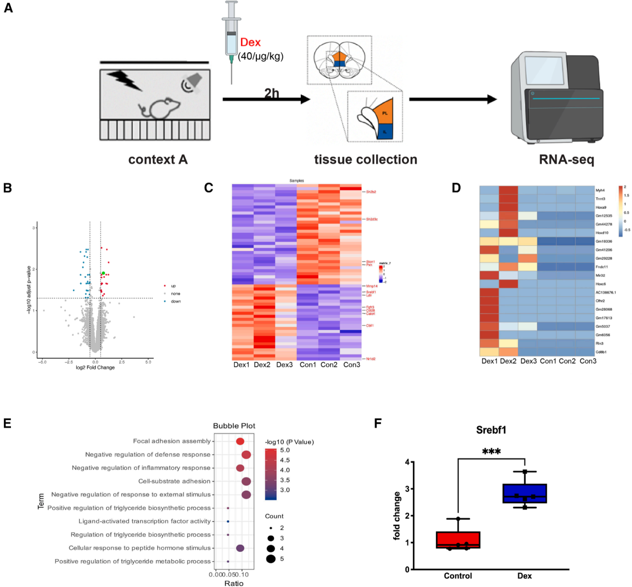
To explore the mechanism by which dexmedetomidine inhibits fear memory consolidation, dexmedetomidine was administered 2 hours after fear acquisition, and transcriptomic sequencing was performed on the PLPFC and infralimbic prefrontal cortex (ILPFC) tissue (Figure 2A). Differential expression analysis revealed that 55 genes were significantly altered in the PLPFC, while no differentially expressed genes were detected in the ILPFC (Figure 2B-2D). Gene ontology (GO) analysis of the protein-coding genes with differential expression during the memory consolidation phase showed significant enrichment in terms related to "ligand-activated transcription factor activity," "negative regulation of defense response," "vesicle membrane," and "regulation of triglyceride biosynthetic process" (Figure 2E). Among the differentially expressed genes, Srebf1 showed the most significant change, with a 2.8-fold upregulation in mRNA levels (Figure 2F).
Figure 2 | Transcriptomic features of PLPFC and ILPFC in dexmedetomidine-treated fear-conditioned mice
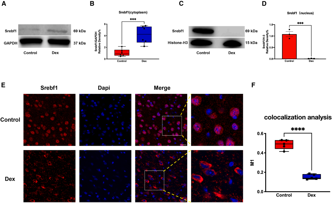
Further analysis of whether this transcriptional upregulation translates into changes in protein expression and subcellular localization was performed. Western blotting results showed that dexmedetomidine treatment increased the cytoplasmic Srebf1 protein levels in the PLPFC but reduced its nuclear translocation (Figure 3A-3D). Consistent with this, immunofluorescence staining revealed that, under fear conditions, dexmedetomidine treatment significantly reduced the nuclear localization of active Srebf1 (Figure 3E, 3F), suggesting that dexmedetomidine at least partially inhibits the nuclear translocation of Srebf1 in the PLPFC, thereby weakening fear memory consolidation.
Figure 3 | Dexmedetomidine inhibits the nuclear translocation of Srebf1
Knockdown of Srebf1 in Astrocytes Inhibits Fear Memory Consolidation in Mice
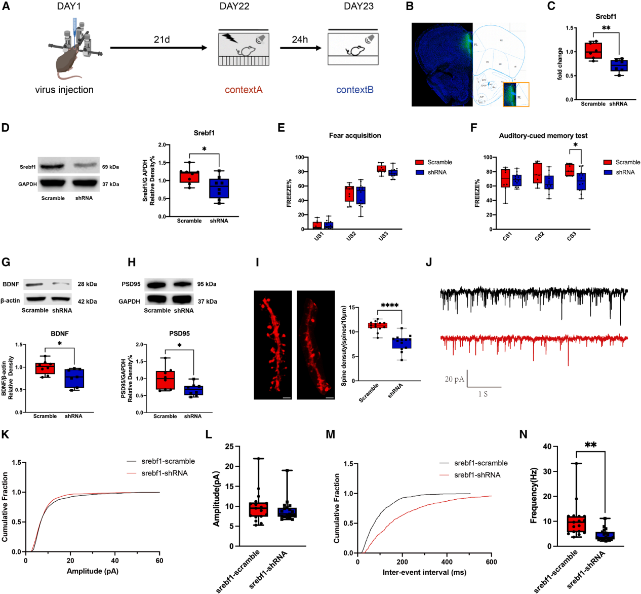
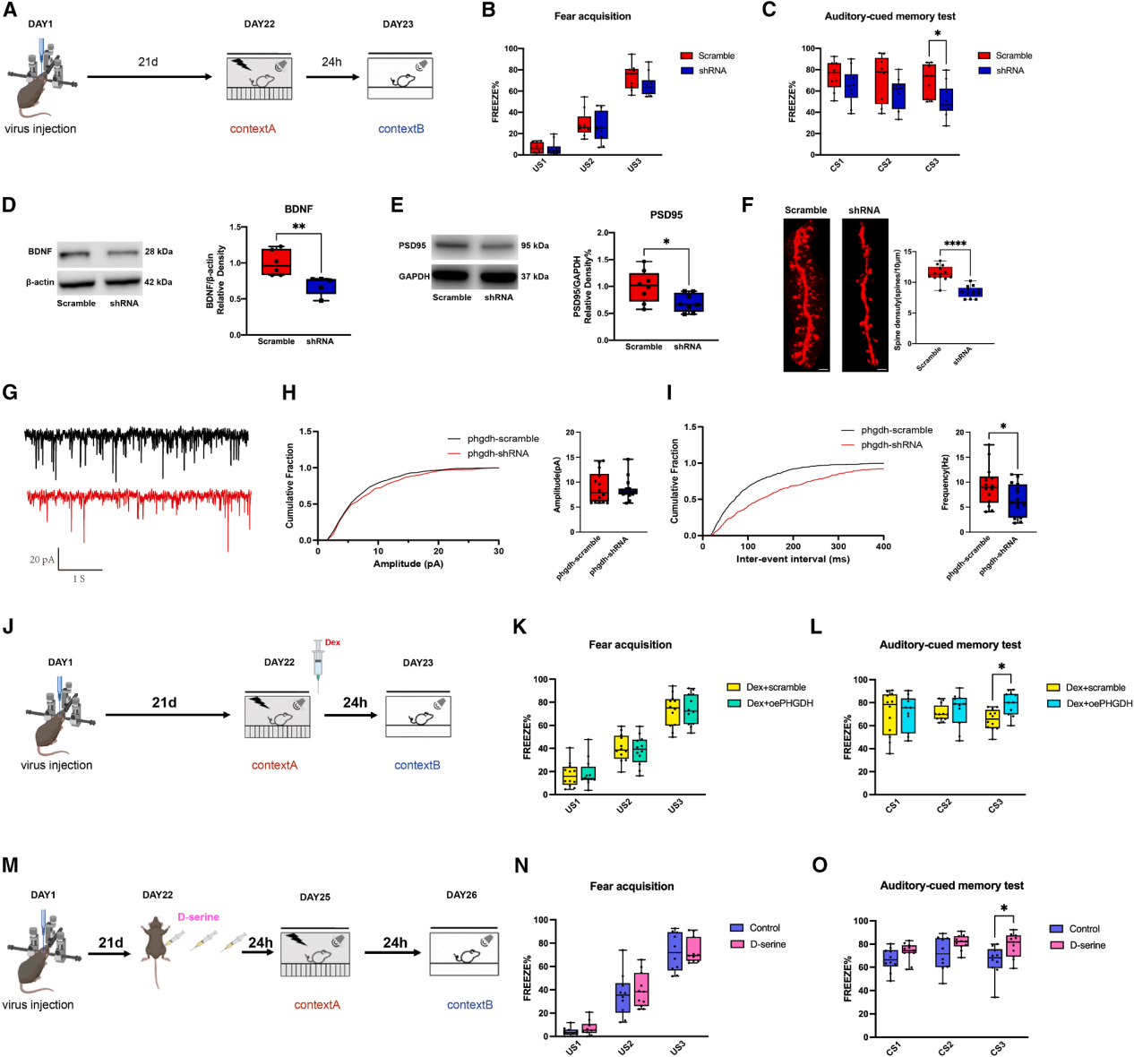
To clarify the cell-specific behavioral phenotype, Srebf1 immunofluorescence colocalization analysis was first performed. The results showed that dexmedetomidine specifically reduced nuclear Srebf1 (nSrebf1) expression in astrocytes of the PLPFC in fear-conditioned mice. Bilateral injection of rAAV-GfaABC1D-EGFP-5'miR-30a-shRNA (mSrebf1)-3'miR-30a-WPREs into the PLPFC of mice was used to specifically knock down Srebf1 in astrocytes. The results showed significant decreases in both mRNA and protein levels (Figure 4A-4D). This knockdown did not affect fear acquisition but significantly reduced the freezing time during the memory retention phase (Figure 4E, 4F). At the molecular and structural levels, astrocyte-specific knockdown of Srebf1 reduced BDNF and PSD95 expression in the PLPFC and decreased dendritic spine density. Electrophysiological recordings from PLPFC brain slices showed no change in the amplitude of sEPSCs, but the frequency of sEPSCs was significantly lower in the knockdown group compared to the random sequence control group (Figure 4G-4N). Taken together, Srebf1 in astrocytes of the PLPFC plays a key role in the dexmedetomidine-mediated inhibition of fear memory consolidation in mice.
Figure 4 | Knockdown of Srebf1 in astrocytes alleviates fear in mice by inhibiting fear memory consolidation, accompanied by impaired synaptic stability
Dexmedetomidine Reduces the Binding of Srebf1 to the Phgdh Promoter
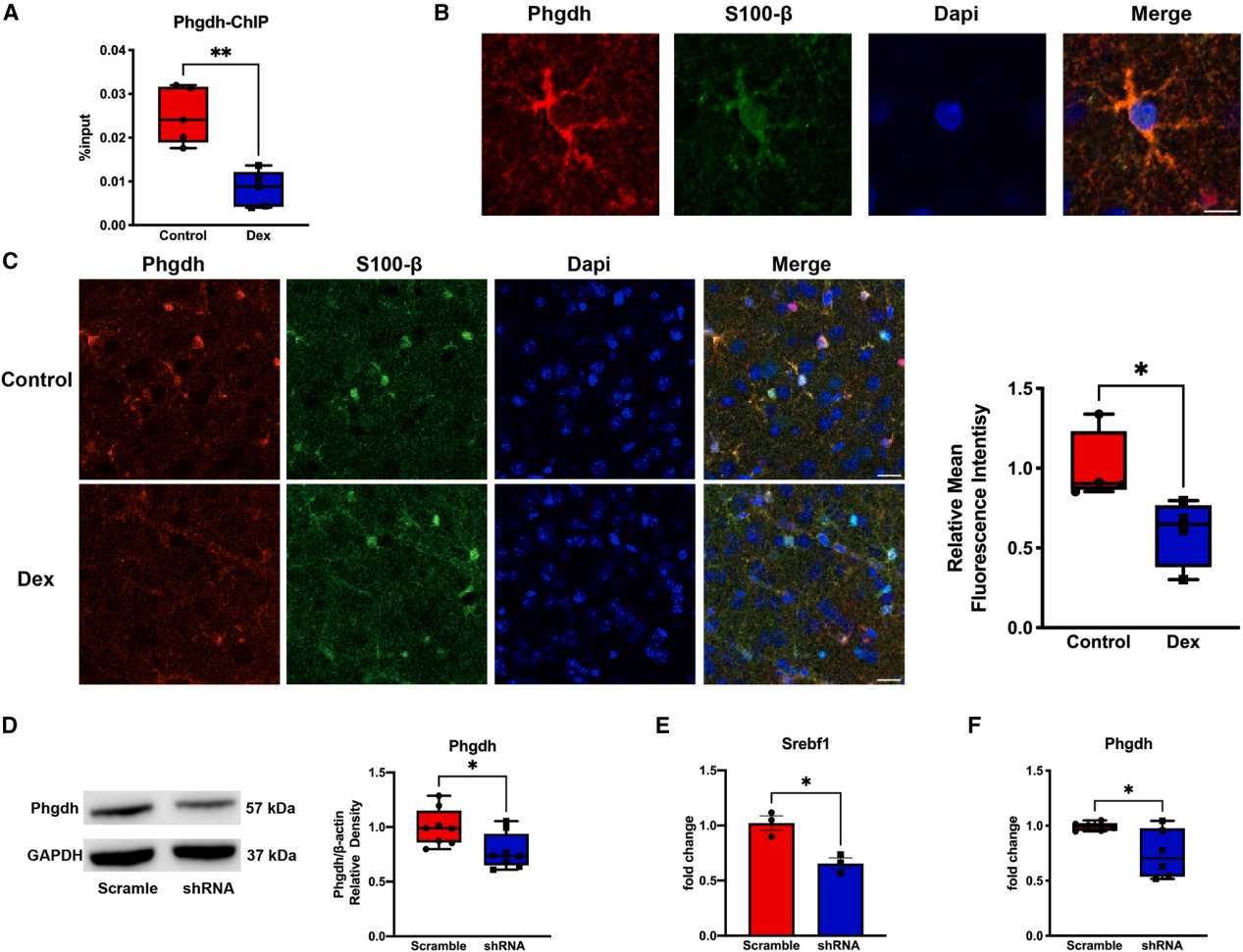
To explore the role of Srebf1 in fear memory consolidation, its downstream target genes were examined. Based on previous Srebf1 chromatin immunoprecipitation sequencing (ChIP-seq) data, the focus was placed on Phgdh (a gene whose promoter region Srebf1 can bind to). Compared to the control group, dexmedetomidine treatment reduced the binding of Srebf1 to the Phgdh promoter (Figure 5A). Immunofluorescence staining showed that Phgdh was mainly expressed in astrocytes, and dexmedetomidine could inhibit the upregulation of Phgdh expression in the PLPFC of fear-conditioned mice (Figure 5B, 5C). To clarify the relationship between Srebf1 and Phgdh in fear memory consolidation, astrocyte-specific knockdown of Srebf1 in the PLPFC significantly reduced Phgdh protein levels (Figure 5D). In primary cultured astrocytes, transfection with Srebf1 shRNA for 4 days significantly decreased Phgdh mRNA levels compared to the random sequence control group (Figure 5E, 5F). Srebf1 plays a key role in regulating Phgdh expression in astrocytes, and Phgdh is crucial for fear memory consolidation.
Figure 5 | Dexmedetomidine enhances the binding of Srebf1 to the Phgdh promoter and affects its expression
Phgdh Plays a Key Role in Dexmedetomidine-Mediated Inhibition of Fear Memory Consolidation, with D-Serine Likely Acting as Its Downstream Effector Molecule
After confirming that dexmedetomidine inhibits the active nSrebf1-mediated regulation of Phgdh expression, further evaluation was conducted on the role of Phgdh in fear memory consolidation in the PLPFC astrocytes. Prior to behavioral training, pAAV-GfaABC1D-EGFP-3xFLAG-miR30-shRNA (Phgdh)-WPRE was bilaterally injected into the PLPFC of mice to effectively reduce Phgdh mRNA and protein levels (Figure 6A). Phgdh knockdown did not affect fear acquisition but significantly reduced the freezing time during the memory retention phase. Additionally, BDNF and PSD95 expression were reduced, and dendritic spine density decreased (Figure 6B-6F). Whole-cell patch-clamp recordings showed that Phgdh knockdown reduced the frequency of sEPSCs without affecting their amplitude (Figure 6G-6I). In a rescue experiment, Phgdh was overexpressed in astrocytes of the PLPFC, and dexmedetomidine was administered (Figure 6J). The results showed that overexpression of Phgdh did not affect fear acquisition (Figure 6K) but reversed the dexmedetomidine-induced reduction in fear, with the freezing time significantly higher than that in the dexmedetomidine control group (Figure 6L). In summary, Phgdh in astrocytes plays a key downstream role in Srebf1-mediated inhibition of fear memory consolidation by dexmedetomidine. Given that Phgdh is the key rate-limiting enzyme for D-serine synthesis, further investigation was conducted to determine if its downstream product, D-serine, mediates dexmedetomidine’s effect on fear memory consolidation. In Phgdh knockdown mice in the PLPFC, 500 mg/kg D-serine was intraperitoneally injected for 3 consecutive days prior to fear conditioning (this concentration effectively regulates NMDA receptor function), with the control group receiving the same volume of saline. The results showed that exogenous supplementation of D-serine did not affect fear acquisition but significantly increased freezing time in the fear test, similar to the results of Phgdh overexpression (Figure 6M-6O), proving that Phgdh participates in fear memory consolidation by regulating D-serine availability.
Figure 6 | Phgdh plays a key role in dexmedetomidine-mediated inhibition of fear memory consolidation, with D-serine likely acting as its downstream effector molecule
Conclusion
The study reveals that dexmedetomidine alleviates PTSD-like behaviors by inhibiting the Srebf1-Phgdh pathway in astrocytes of the PLPFC, reducing D-serine-related synaptic stability, and subsequently weakening fear memory consolidation.
The viral tools used in this study are available from BrainCase.