E-mail:BD@ebraincase.com
Tel:+8618971215294
English 中文版
Pioneering Innovation in Gene Vector Research - BrainCasePioneering Innovation in Gene Vector Research - BrainCase
  • Home
  • Pre-made AAV Library
    CRISPRRNAiHSV-helperRV-helperNeurotropic virusCalcium SensorsOptogenetics activationOptogenetics inhibitionChemical geneticsSparse labelingFluorescent proteinBiosensorsRecombinaseApoptosis & AutophagyDisease ModelNeurotoxicityOther
  • Products & Service

    Product Center

    Virus

    Vesicular Stomatitis Virus-VSV
    RCAS-TVA
    Lentivirus
    Rabies Virus-RABV
    Herpes simplex virus-HSV
    Pseudorabies virus-PRV
    Adeno-Associated Virus-AAV
    Neurotropic virus list
    Baculovirus

    Kits

    EndoFree Plasmid Maxi Kit
    Lentivirus Concentration Kit

    Plasmid Construction & Vector cloning

    Lentiviral Vector
    AAV Vector
    Plasmid

    Popular Applications

    Gene Regulation

    Gene Overexpression-Brain Case
    RNA interference(RNAi)-siRNA-Brain Case
    Gene Editing - CRSIPR cloning - BrainCase

    Neural Circuit Function Research

    Optogenetics - BrainCase
    Chemical genetics-DREADDs-Brain Case
    Calcium signal recording-Gels- Brain Case
    GRAB Neurotransmitter Fluorescent Sensor- Brain Case
    Functional Magnetic Resonance Imaging Technology- Brain Case

    Research on the structure of neural circuits

    Direct Input and Output-viral vectors- Brain Case
    Anterograde Mono-synaptic Tracing -HSV- Brain Case
    Antrograde Muti-synaptic Tracing-HSV & VSV-Brain Case
    Retrograde Mono-synaptic Tracing-Rabies Virus-Brain Case
    Retrograde Muti-synaptic Tracing-PRV-Brain Case

    Featured Services

    AAV Serotype Screening

    AAV Serotypes screening-gene therapy

    Collaboration Products

    Kiryl Piatkevich lab
    Yulong Li lab
    Zhifei Fu lab
    Woo-ping Ge lab

    Oncolytic Virus Modification

    Oncolytic virus-for cancer therapy-Brain Case
    Herpes Virus Vector-anti-tumor- BrainCase
    Vesicular stomatitis virus-killing tumor cells-Brain Case
  • News
    Corporate News New Product Launch Investor News
  • Support
    Literature interpretation Customer article FAQs Blog Legal
  • About Us
    Pre-made AAV Library Products & Service News Support About Us Contact
  • Contact
    Contact Us Join us
  • 中文
    English 中文版
  • Home
  • Support
  • Customer article
  • Support
  • Literature interpretation
  • Customer article
  • FAQs
  • Blog
  • Legal

Customer Article|Diet-induced inflammation in the anterior paraventricular thalamus induces compulsive sucrose-seeking

Release time:2025-09-24 11:46:08

When animals encounter life-threatening situations, they often resort to compulsive behaviors to sustain their survival. For instance, research has reported that high-fat diet (HFD) consumption impedes threat-cue-induced suppression of sucrose-seeking in mice. The study found that HFD activates microglia, triggering neuronal activity in the anterior paraventricular thalamus (aPVT), which leads to a series of metabolic inflammatory responses and results in compulsive food-seeking behavior.

C-Fos validation of neurons responding to aPVT

Using c-fos immunohistochemistry, researchers compared neuronal activation patterns in various brain areas/nuclei of HFD mice and chow-fed mice (control) after the compulsive sucrose-seeking test. One of the brain areas that showed large differences in the number of c-fos-positive (c-fos+) cells between HFD mice and chow mice was the aPVT, which is known to play a role in the regulation of food-seeking. More than 70% of the c-fos+ cells in the aPVT were also positive for CaMKIIα.

Activation of aPVT nucleus neurons during forced feeding

To further examine how aPVT neurons respond in different aspects of compulsive eating behavior, researchers used fiber photometry to record Ca2+ transients in CaMKIIα+ neurons of the aPVT in mice during the compulsive sucrose-seeking paradigm. When lever presses were followed by a cue light, the light in HFD mice triggered a larger Ca2+ transient in the aPVT than in chow-fed mice (Fig. 1f–h, right). The increase in Ca2+ transients in the aPVT was consistent with the increased number of c-fos+ cells in this region in HFD mice. This result suggests that the HFD-induced increase in c-fos+ neuronal activation in the aPVT may underlie compulsive eating behavior.

Optogenetic activation of responsive neurons in aPVT

Next, researchers used optogenetic activation of glutamatergic neurons in the aPVT and found that photo stimulation significantly increased the number of lever presses in HFD mice, but not in control chow mice (Fig. 1k). This result indicates that activating aPVT CaMKIIα+ neurons is sufficient to drive compulsive sucrose-seeking behavior in mice.

Chemogenetic inhibition of responsive neurons in aPVT

Conversely, researchers used clozapine-N-oxide (CNO) to inhibit aPVT excitatory neurons expressing hM4Di-eGFP in fear-conditioned HFD mice performing the operant task. The results showed that CNO administration reduced the firing frequency of neurons expressing hM4Di and decreased the number of lever presses in HFD mice expressing hM4Di-eGFP, but not in control HFD mice expressing eGFP, when these mice were presented with the light cue (Fig. 1m). This led to a lower ratio of lever presses in the test session to lever presses in the baseline session (Fig. 1n). These results indicate that chemogenetic inhibition of aPVT CaMKIIα+ neurons reduces compulsive sucrose-seeking in fear-conditioned HFD mice.
Overall, the activity level of excitatory neurons in the aPVT determines the motivation to obtain a food reward in the presence of a cue predicting an aversive stimulus.

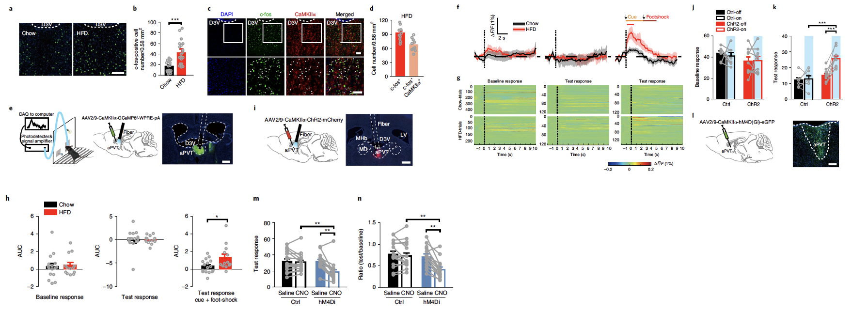
Fig1. Investigating the responses of neurons in the aPVT nucleus to different aspects of compulsive eating behavior in chow mice and HFD mice.(more details https://www.nature.com/articles/s41593-022-01129-y)

HFD induces microglial proliferation, leading to compulsive eating behavior in mice

The aPVT, as a nucleus of the midline thalamus, may contain sentinel cells that detect metabolic factors in the cerebrospinal fluid (CSF). Researchers indeed found that HFD significantly increased the number of Iba-1+ microglia in the aPVT (Fig. 2a, b), and c-fos+ CaMKIIα neurons were close to and had a large contact area with Iba-1+ microglia in the aPVT. Using the colony-stimulating factor 1 receptor antagonist PLX3397 (which depletes microglia) inhibited neuronal activation in the aPVT of HFD mice (Extended Data) and eliminated compulsive sucrose-seeking behavior in these mice (Fig. 2c, d). Additionally, local injection of a CSF-1 antibody (which specifically reduces microglia in the aPVT, Fig. 2f, g) significantly reduced the number of c-fos+ neurons in the aPVT of HFD mice and their contact with Iba-1+ microglia. Depleting a portion of the microglia in the aPVT also eliminated compulsive sucrose-seeking behavior in HFD mice (Fig. 2h, i) without altering their freezing behavior and pain sensitivity (Extended Data). These results indicate that HFD-induced microglial proliferation in the aPVT causes compulsive eating behavior in HFD mice.

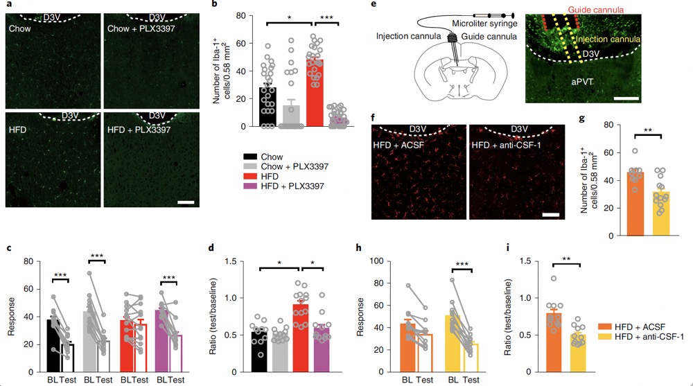
Fig2. HFD induces microglial proliferation in the aPVT and increases their contact area with c-fos+ CaMKIIα neurons, triggering compulsive eating behavior in mice

For more details, please refer to the original article at https://www.nature.com/articles/s41593-022-01129-y

Products Used in the Study

Catalog Number. BC-SL014
Product Name. AAV-CaMKIIα-FCSSP-EYFP-5E4 (Sparse labeling virus for CaMKIIα+ cells)

Fig3. The product used in the study to assess whether the local injection of CSF-1 antibody causes neuronal damage. This product can sparsely and brightly label the morphology and structure of neurons, and in some scenarios, it can replace the Golgi staining method.

Brain Case Biotech provided various serotypes of AAV viruses used in this study. If interested, please contact us at BD@ebraincase.com

Service Type :

Select the service you'd like to purchase.

Order Information(Premade-AAVs)

Please provide us some information about the service you'd like to order.

How did you hear about BrainCase Biotech or our products?(Fill in this field to get a discount coupon)

Detailed requirements:

scroll

Order Information(Custom AAV/Lentivirus)

Please provide us some information about the service you'd like to order.

Gene ID or gene information:

Selection of the reporting gene:

Special Instructions:

scroll

Order Information(Others)

Please provide us some information about the service you'd like to order.

Virus name/Detailed requirements:

scroll

Related products

400+ Customer Publications Collection

400+ Customer Publications Collection

Client Article | Neuron | Xinan Liu/ Zuxin Chen Team from Shenzhen Institute of Advanced Technology Reveals "Gut-Brain" Pathway Mediating the Antiepileptic Effects of Probiotics

Client Article | Neuron | Xinan Liu/ Zuxin Chen Team from Shenzhen Institute of Advanced Technology Reveals "Gut-Brain" Pathway Mediating the Antiepileptic Effects of Probiotics

Customer Article | Cell | Fudan University’s YiZhou Jiang, ZhiMin Shao, JinFei Ni, and Yi Xiao Team Reveal the Role of Sensory Neurons in Regulating the Tumor Microenvironment in Triple-Negative Breast Cancer (TNBC),Providing New Targets for Immunotherap

Customer Article | Cell | Fudan University’s YiZhou Jiang, ZhiMin Shao, JinFei Ni, and Yi Xiao Team Reveal the Role of Sensory Neurons in Regulating the Tumor Microenvironment in Triple-Negative Breast Cancer (TNBC),Providing New Targets for Immunotherap

New Approaches in Epilepsy Treatment: Fudan University Team Reveals Key Circuit Involved in Piriform Epileptic Seizures and Brain-Wide Functional Reorganization

New Approaches in Epilepsy Treatment: Fudan University Team Reveals Key Circuit Involved in Piriform Epileptic Seizures and Brain-Wide Functional Reorganization

map
{dede:global.cfg_webname/}

Pre-made AAV Library

CRISPR
RNAi
Neurotropic virus
Optogenetics activation
Biosensors

News

Corporate News
New Product Launch
Investor News

Support

Literature interpretation
Customer article
FAQs
Blog
Legal
微信

WhatsApp Business Account

Tel: +8618971215294
E-mail: BD@ebraincase.com

Address:-

Address:-

  • Copyright © 2024 Brain Case All Rights Reserved.