Microglia play critical roles in human health and a wide range of central nervous system (CNS) diseases, making them a key focus in neuroscience research. Traditional approaches often rely on transgenic mouse models, which are time-consuming, costly, and not readily translatable to therapeutic applications.
Recombinant adeno-associated viruses (rAAVs) are widely used in neuroscience due to their safety and diverse tissue tropism. However, AAV vectors that enable efficient and specific targeting of microglia remain limited.
In July 2024, Xu Fuqiang and Lin Kunzhang's team at CAS published a preprint on BioRxiv titled “Microglia-specific transduction via AAV11 armed with IBA1 promoter and miRNA-9 targeting sequences.” This study evaluated various AAV vectors carrying the mIBA1 promoter and miRNA-9 targeting sequences for transducing microglia in the caudate putamen (CPu) brain region. The findings showed that AAV11 facilitated highly specific and efficient microglial transduction across different brain regions and the spinal cord. Reduced injection doses allowed for precise structural labeling of microglia, making AAV11 a powerful tool for microglia research.We summarized how AAV11 targets and transduces microglia in various mouse brain regions as reported in this article.
https://doi.org/10.1101/2024.07.09.602653
1. Dentate Gyrus (DG) Region of the Mouse Brain
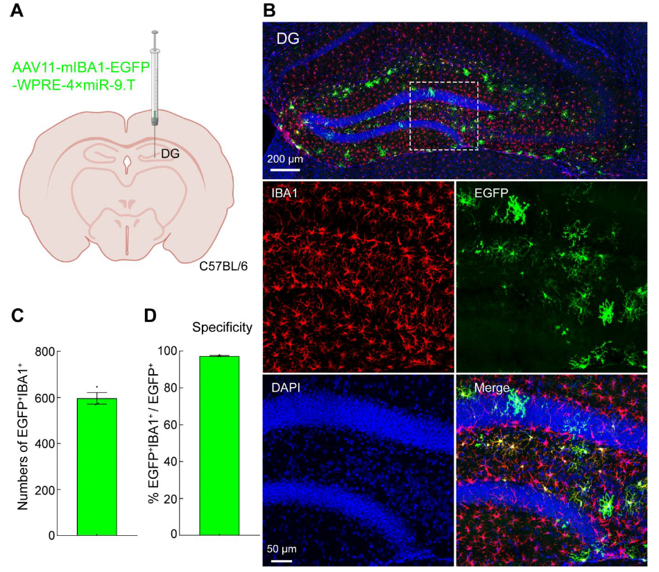
To verify whether AAV11 can efficiently transduce microglia in the DG region, AAV11 was injected at a dose of 2×10^9 VG (Figure 1A). Three weeks later, brain tissues were collected, sectioned, and subjected to immunofluorescence staining (Figure 1B). AAV11 demonstrated strong transduction capability in microglia within the hippocampal DG region (Figures 1B and 1C). Quantitative analysis of EGFP and IBA1 double-positive cells indicated that AAV11 primarily transduced microglia with approximately 97% specificity (Figure 1D).
Figure 1. AAV11 efficiently and specifically transduces microglia in the DG brain region
2. Substantia Nigra pars Reticulata (SNr) Region of the Mouse Brain
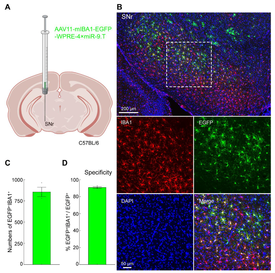
To verify the efficiency of AAV11 in transducing microglia in the SNr region, AAV11 was injected at a dose of 2×10^9 VG (Figure 2A). Three weeks later, brain tissues were collected, sectioned, and subjected to immunofluorescence staining (Figure 2B). In the SNr, most microglia were labeled with green fluorescence (Figures 2B and 2C). Fluorescence colocalization analysis showed that AAV11-mediated gene expression in SNr microglia had approximately 90% specificity (Figure 2D).
Figure 2. AAV11 efficiently and specifically transduces microglia in the SNr brain region
3. Spinal Cord of Mice
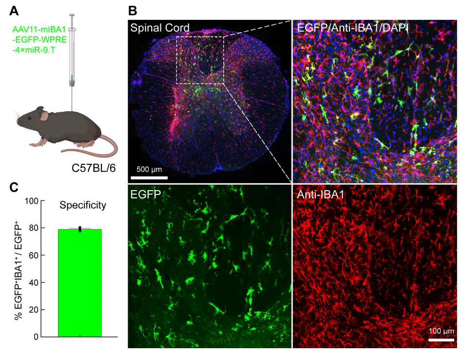
Spinal cord microglia play a crucial role in spinal cord injury repair, inflammation regulation, pain generation, and the development of potential therapeutic strategies. Therefore, developing a gene delivery system targeting spinal cord microglia is essential. The authors investigated whether AAV11 could efficiently transduce spinal cord microglia. AAV11 was injected into the lumbar segment at a dose of 5×10^9 VG (Figure 3A). Three weeks later, spinal cord tissues were collected, sectioned, and subjected to immunofluorescence staining (Figure 3B). Quantitative analysis of EGFP and IBA1 double-positive cells indicated that AAV11 exhibited strong transduction efficiency in spinal cord microglia, with approximately 80% specificity (Figures 3B and 3C).
Figure 3. AAV11 efficiently and specifically transduces microglia in the spinal cord
4. Sparse and Bright Labeling of Microglia by AAV11
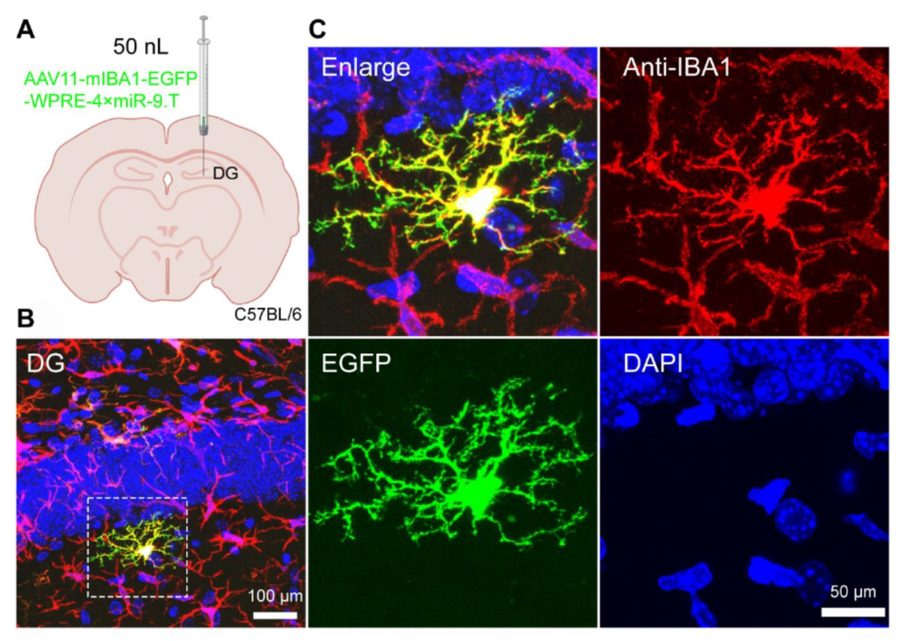
The complex morphology of microglia is critical for their function in the brain. Microglia possess intricate processes and can undergo morphological changes under different physiological or disease conditions. Labeling the fine morphology of microglia is essential for understanding their state-dependent changes and interactions with other neural cells. However, simple and universal methods for labeling microglial morphology in the mammalian brain are lacking.
In this study, by reducing the viral injection volume to 50 nL at a dose of 5×10^8 VG into the dentate gyrus (DG) of the hippocampus (Figure 4A), the authors found that AAV11 could sparsely label microglia in the DG region three weeks later (Figure 4B). Using laser confocal scanning, the fine structural details of microglia were clearly observed (Figure 4C). Therefore, AAV11 can be used for sparse labeling to analyze the detailed structure of microglia.
Figure 4. AAV11 sparsely labels microglia
By directly injecting AAV11 containing the IBA1 promoter and four tandem miR-9 targeting sequences, specific transduction of microglia in defined brain regions of adult mice was achieved. Additionally, AAV11 shows high transduction specificity for microglia across various brain regions and the spinal cord. Furthermore, by reducing the injection dose, AAV11 can be used for sparse labeling. This work provides a powerful viral tool for studying microglial structure and function. For researchers focused on glial biology, we are very interested in collaborating with your laboratory to expand the applications of this serotype. If you are interested, please feel free to contact us at kz.lin@siat.ac.cn orBD@ebraincase.com.
Service Type :
Select the service you'd like to purchase.
Order Information(Premade-AAVs)
Please provide us some information about the service you'd like to order.
Order Information(Custom AAV/Lentivirus)
Please provide us some information about the service you'd like to order.
Order Information(Others)
Please provide us some information about the service you'd like to order.