The basolateral amygdala (BLA) encodes stimulus salience through acetylcholine (ACh) signaling, with cholinergic inputs primarily originating from choline acetyltransferase (ChAT)-positive neurons in the substantia innominata (SI) of the basal forebrain. The nucleus accumbens (NAc) serves as a central hub integrating emotion, cognition, and motor functions and provides the major upstream input to basal forebrain ChAT neurons. However, the cell-type-specific circuit through which the NAc regulates BLA cholinergic signaling has remained unknown.
On June 26, 2026, research teams led by Prof. Xiao Xiong (Center for Excellence in Brain Science and Intelligence Technology, Chinese Academy of Sciences), Prof. Deng Hanfei (Fudan University), Prof. Xu Fuqiang (Shenzhen Institute of Advanced Technology, Chinese Academy of Sciences), and Prof. Xiao Lei (Fudan University) published their study in Nature Neuroscience, entitled :"Striatal control of amygdalar acetylcholine release during salience-associated processing."
The study demonstrates that D1- and D2-type medium spiny neurons (MSNs) in the NAc bidirectionally regulate ACh release in the BLA through SI ChAT neurons, thereby controlling stimulus salience representation and associative learning.
The study was co-first authored by Dr. Aixiao Chen, Yunjing Li, Hangfei Zhu, and Xiao Cui. Corresponding authors are Prof. Xiao Xiong, Prof. Deng Hanfei, Prof. Xu Fuqiang, and Prof. Xiao Lei.
🎯Dynamic Representation of Stimulus Salience by Acetylcholine Signaling in the BLA
To determine how behavioral context influences acetylcholine (ACh) signaling, two complementary behavioral paradigms were established (Figure 1). In the Go/No-go task, the reward-predicting cue (CSG) elicited robust ACh responses in the basolateral amygdala (BLA) after training, whereas the No-go cue (CSN) consistently evoked minimal cholinergic responses (Figure 1b–d).
The active avoidance task used the same auditory stimulus as the CSN, designated as the avoidance cue (CSA), while altering only the behavioral contingency: mice were required to actively lick to avoid punishment. Once the task was acquired, the same auditory cue evoked a markedly enhanced ACh response (Figure 1f–h).
The distinct cholinergic responses elicited by an identical auditory stimulus demonstrate that BLA acetylcholine signaling is determined not by the sensory stimulus itself, but by contextual factors, including task rules and behavioral demands (Figure 1i).
Physiological state further reshaped salience representation by BLA acetylcholine (Figure 1j–n). In water-deprived mice, reward-predicting cues elicited robust ACh responses accompanied by anticipatory licking behavior. Following water satiation, ACh responses to reward cues were markedly reduced, reflecting diminished motivational salience. In contrast, satiated mice exhibited significantly enhanced cholinergic responses to aversive foot shocks, indicating an increased behavioral priority assigned to threatening stimuli. Importantly, the contribution of licking behavior to ACh signaling was excluded throughout these experiments.
Collectively, these findings demonstrate that acetylcholine signaling in the BLA dynamically represents the perceived salience of environmental stimuli. The magnitude of cholinergic responses is flexibly regulated by learning, behavioral context, and physiological state, thereby closely matching the animal's level of motivational engagement.
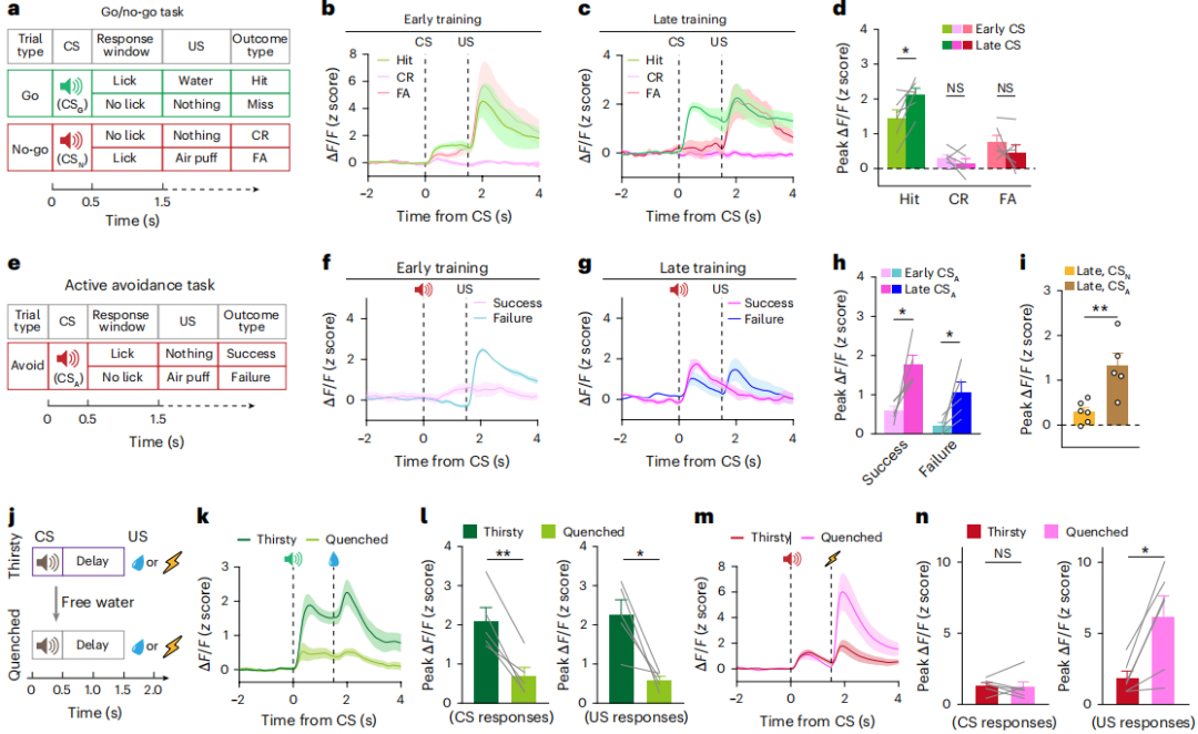
Figure 1 | Behavioral context and physiological state shape salience-related acetylcholine responses in the BLA
🎯Activation of the NAc→SI Pathway Does Not Immediately Affect Acetylcholine Release in the Cortex or Hippocampus
Anterograde tracing revealed that D1- and D2-type medium spiny neurons (MSNs) in the mouse nucleus accumbens (NAc) project extensively to the substantia innominata (SI). To determine the functional role of this pathway, ChR2 was expressed in NAc D1 or D2 neurons, while an acetylcholine (ACh) sensor was expressed in the basolateral amygdala (BLA). By delivering pathway-specific optical stimulation to the SI, they selectively manipulated the activity of the NAc→SI projection (Figure 2a).
Consistent with the effects observed following somatic activation, optogenetic activation of the NAc D1→SI pathway significantly increased ACh release in the BLA, whereas activation of the NAc D2→SI pathway markedly suppressed BLA acetylcholine levels (Figure 2b). These findings demonstrate that the two populations of NAc MSNs exert bidirectional and antagonistic regulation of BLA cholinergic signaling through the SI pathway.
In addition to innervating the BLA, ChAT-positive cholinergic neurons in the SI of the basal forebrain (BF) also project to multiple brain regions, including the medial prefrontal cortex (mPFC), auditory cortex (AUD), and hippocampal CA1. To determine whether the regulatory effects of the NAc→SI pathway were brain region-specific, the authors simultaneously monitored ACh dynamics across these regions (Figure 2c, e, g).
Both rewarding and aversive stimuli elicited robust increases in acetylcholine release in the cortex and hippocampus. However, these responses were not regulated by the NAc→SI pathway. Following activation of the NAc D1→SI pathway, only small, delayed fluctuations in cortical and hippocampal ACh signals were observed. These responses were substantially weaker than those detected in the BLA and lacked any immediate effect (Figure 2d, f, h–j), most likely resulting from weak activation of axon collaterals arising from a small subset of ChAT neurons.
By contrast, activation of the NAc D2→SI pathway produced no detectable changes in acetylcholine levels in either the cortex or hippocampus. The inhibitory effect mediated by D2 neurons was highly selective for the BLA. The weak, nonspecific signal fluctuations occasionally observed during prolonged stimulation most likely reflected changes in global arousal state rather than biologically meaningful circuit regulation.
Taken together, these findings demonstrate that the bidirectional regulation of acetylcholine by the NAc→SI pathway exhibits remarkable brain region specificity. Rather than broadly modulating cholinergic transmission throughout the forebrain, this circuit selectively and precisely regulates cholinergic signaling in the BLA while having little or no role in controlling acetylcholine dynamics in the cortex or hippocampus.
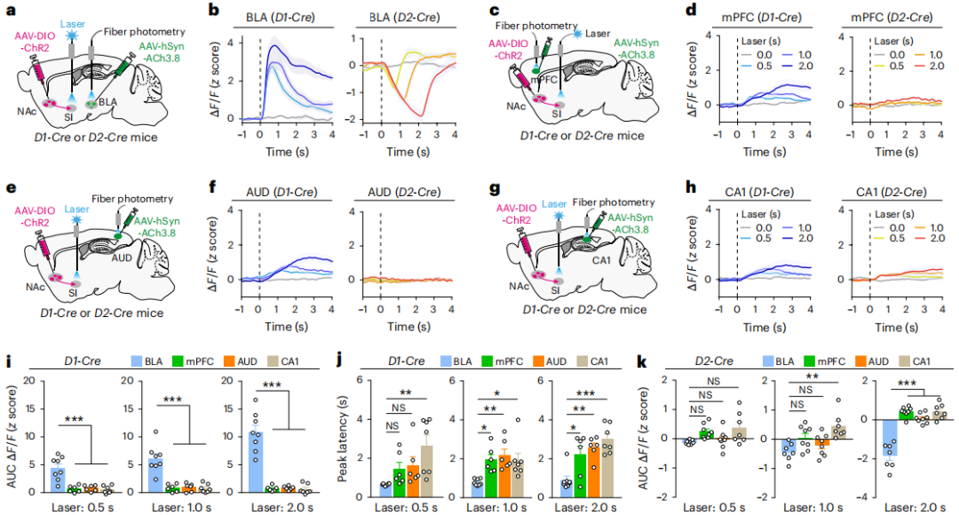
Figure 2 | Effects of NAc→SI pathway activation on acetylcholine release across multiple brain regions
🎯Differential Innervation of SI Cholinergic Neurons by NAc D1 and D2 Medium Spiny Neurons
Although both D1- and D2-type medium spiny neurons (MSNs) in the nucleus accumbens (NAc) are GABAergic inhibitory neurons, they exert completely opposite effects on acetylcholine (ACh) release in the basolateral amygdala (BLA). A circuit model was proposed in which D1 neurons indirectly activate SI cholinergic (ChAT) neurons by inhibiting local GABAergic interneurons within the substantia innominata (SI), whereas D2 neurons directly inhibit SI ChAT neurons.
To test this model, fluorescent synaptic labeling combined with high-resolution three-dimensional imaging was used to selectively label the presynaptic terminals of NAc neurons, allowing bona fide synaptic contacts to be distinguished from passing axons (Figure 3a–c).
Quantitative analysis revealed that D2 neurons formed significantly more synaptic contacts around the somata of SI ChAT neurons than D1 neurons, indicating that D2 neurons preferentially establish direct synaptic connections with SI ChAT neurons (Figure 3d–e).
Monosynaptic retrograde tracing together with in situ hybridization further confirmed that SI cholinergic neurons projecting to the BLA receive extensive inputs from the NAc. Moreover, the majority of their upstream presynaptic neurons were Drd2-expressing D2 MSNs, demonstrating that NAc D2 neurons directly and monosynaptically inhibit SI ChAT neurons (Figure 3f–j).
Slice electrophysiological recordings in transgenic mice further distinguished the functional connectivity of the two pathways. Optogenetic activation of NAc D1→SI axons predominantly evoked inhibitory postsynaptic currents (IPSCs) in local SI GABAergic neurons, while producing only minimal responses in ChAT neurons. In contrast, activation of NAc D2→SI axons preferentially elicited IPSCs in ChAT neurons, with little effect on local GABAergic interneurons. Together, these findings demonstrate that D1 neurons preferentially target local SI GABAergic interneurons, whereas D2 neurons directly innervate SI ChAT neurons.
In vivo calcium imaging further verified the functional organization of this circuit. Activation of the NAc D1→SI pathway significantly increased the activity of SI ChAT neurons, whereas activation of the NAc D2→SI pathway markedly suppressed their firing (Figure 3k–o).
Direct optogenetic inhibition of local SI GABAergic interneurons resulted in a robust increase in BLA acetylcholine release, with ACh signals remaining persistently above baseline, providing strong evidence for the disinhibitory mechanism underlying the D1 pathway (Figure 3p–s).
Collectively, these findings demonstrate that NAc D1 neurons inhibit local GABAergic interneurons within the SI, thereby relieving inhibitory control over ChAT neurons through a disinhibitory mechanism and promoting acetylcholine release in the BLA. In contrast, NAc D2 neurons directly and monosynaptically inhibit SI ChAT neurons, reducing their activity and consequently decreasing acetylcholine release in the BLA. Together, these complementary pathways constitute a bidirectional antagonistic circuit for cholinergic regulation.
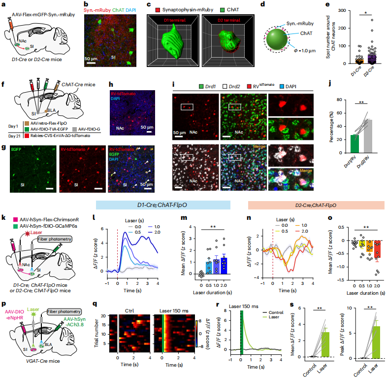
Figure 3 | Synaptic and functional connectivity between NAc MSNs and SI ChAT neurons
🎯Bidirectional Regulation of Behavior-Related Acetylcholine Signaling by Inhibition of the D1→SI and D2→SI Pathways
Previous optogenetic activation experiments demonstrated that NAc D1 and D2 neurons exert opposite effects on acetylcholine (ACh) release in the basolateral amygdala (BLA). However, whether these two pathways are required for physiological salience processing remained unclear. NpHR-mediated optogenetic inhibition was therefore used to selectively silence the NAc D1→SI and NAc D2→SI pathways, allowing their causal roles in salience encoding to be examined (Figure 4a).
Inhibition of the D1→SI pathway significantly attenuated BLA acetylcholine responses evoked by both rewarding and aversive stimuli, indicating that the D1 pathway is required for amplifying salience-related cholinergic signaling. By enhancing cholinergic transmission in the amygdala, this pathway increases the behavioral salience of sensory stimuli (Figure 4b, c, e).
Conversely, inhibition of the D2→SI pathway markedly enhanced stimulus-evoked ACh release, demonstrating that the D2 pathway continuously suppresses cholinergic signaling in the BLA under physiological conditions (Figure 4b, d, f).
Significant differences were observed between experimental and control trials for both pathways, indicating that D1 and D2 pathways act in concert to maintain the basal level of acetylcholine signaling. The relatively modest effects produced by inhibition of either pathway alone likely reflect the fact that SI cholinergic neurons integrate convergent inputs from multiple upstream brain regions, with the NAc representing only one component of this regulatory network.
This bidirectional antagonistic mode of regulation closely parallels the classical opposing functions of D1- and D2-expressing neurons within the basal ganglia circuitry.
Collectively, these findings demonstrate that the bidirectional NAc→SI circuit constitutes an essential neural pathway for regulating acetylcholine dynamics in the BLA and maintaining normal salience encoding of environmental stimuli.
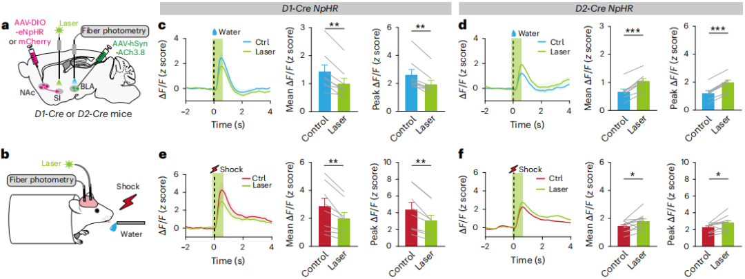
Figure 4 | Silencing the D1→SI pathway reduces, whereas silencing the D2→SI pathway enhances stimulus-evoked acetylcholine release
🎯Differential Salience Encoding by NAc D1→SI and D2→SI Medium Spiny Neurons Correlates with Amygdalar Acetylcholine Dynamics
Given the opposing effects of the NAc D1→SI and D2→SI pathways on acetylcholine (ACh) release in the basolateral amygdala (BLA), the salience-coding properties of these two neuronal populations were further characterized. Axon-targeted calcium indicators were expressed in D1 and D2 medium spiny neurons (MSNs), and fiber photometry recordings were performed in the substantia innominata (SI) to monitor neuronal responses to three categories of salient stimuli: reward, punishment, and novel noise (Figure 5a–c).
D1→SI neurons exhibited robust and reliable excitatory responses to all salient stimuli, indicating that they uniformly encode stimulus salience regardless of positive or negative valence. In contrast, D2→SI neurons predominantly displayed stimulus-evoked inhibitory responses, with highly heterogeneous response patterns and substantial variability across individual neurons (Figure 5d–l).
Simultaneous dual-channel recordings further showed that calcium activity in D1→SI neurons was strongly and positively correlated with BLA acetylcholine dynamics, whereas D2→SI neuronal activity exhibited a negative correlation with BLA acetylcholine levels, demonstrating two functionally opposing patterns of circuit activity (Figure 5m–o).
Pavlovian conditioning further revealed that the coupling between neuronal activity and BLA acetylcholine signaling was learning-dependent. Functional coupling was weak during the early stage of training but progressively strengthened as learning proceeded, with positive coupling between D1→SI activity and BLA ACh signals and negative coupling between D2→SI activity and BLA ACh signals becoming increasingly pronounced. These findings indicate that the functional association within this circuit is gradually reinforced during cue-reward learning (Figure 5p–s).
Collectively, these findings demonstrate that D1→SI and D2→SI neurons exhibit distinct salience-coding strategies and dynamically regulate BLA acetylcholine signaling through opposing activity patterns. Moreover, the functional coupling between these pathways and amygdalar cholinergic signaling is progressively strengthened as associative learning is acquired.
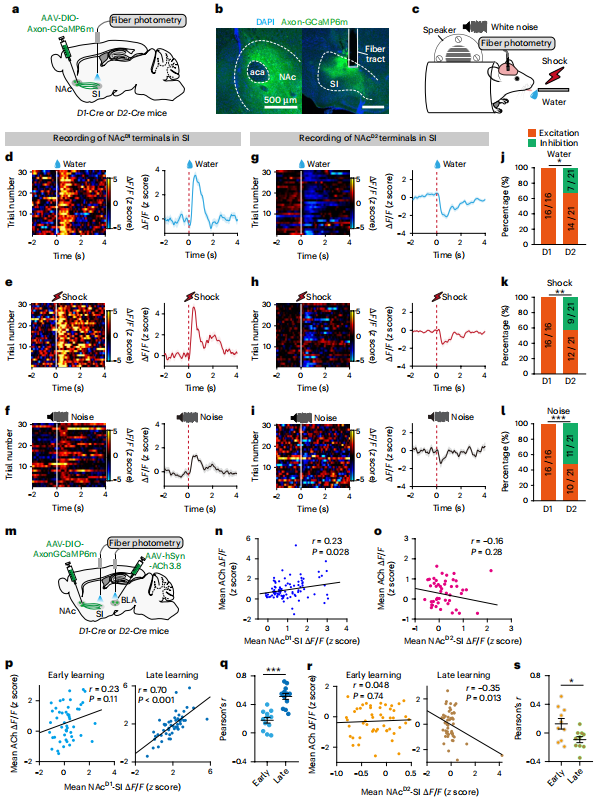
Figure 5 | Salience-related neural activity in D1→SI and D2→SI pathways and their correlations with BLA acetylcholine dynamics
🎯Closed-Loop Manipulation of the NAc→SI Pathway Bidirectionally Regulates Associative Learning
Cue–reward associative learning is fundamental to adaptive behavior. To determine how the NAc→SI pathway influences learning through the regulation of acetylcholine (ACh) signaling in the basolateral amygdala (BLA), an auditory cue–nose-poke reward paradigm was employed to examine the contribution of this pathway to associative learning efficiency (Figure 6a, b).
Behavioral recordings revealed a progressive reorganization of BLA acetylcholine dynamics throughout learning. During the pre-training stage, peak ACh responses occurred after reward delivery. As learning progressed, the cholinergic peak gradually shifted to the cue presentation period, accompanied by an increase in response amplitude and a reduction in response latency. Meanwhile, reward-evoked ACh signals progressively decreased and shifted earlier in time. These dynamic changes indicate that BLA cholinergic activity becomes tightly coupled to the formation of cue–reward associative memory (Figure 6c–h).
Closed-loop optogenetic manipulation further demonstrated the bidirectional regulatory function of the NAc→SI pathway. Activation of the NAc D1→SI pathway following correct behavioral responses increased BLA acetylcholine levels, significantly accelerated learning, shortened cue-response latency, and produced no detectable effects on locomotor activity or motivational state. In contrast, activation of the NAc D2→SI pathway suppressed cholinergic signaling, prolonged response latency, and impaired learning performance (Figure 6i–r).
Inhibition of the D1→SI pathway significantly impaired associative learning, whereas inhibition of the D2→SI pathway produced no obvious effect on learning performance.
Pharmacological blockade of muscarinic acetylcholine receptors (mAChRs) in the BLA by scopolamine completely abolished the learning-promoting effect of D1 pathway activation. Mice exhibited significantly prolonged cue-response latency without detectable changes in locomotor activity or motivation, demonstrating that the facilitation of learning mediated by the D1→SI pathway depends entirely on cholinergic receptor signaling within the BLA (Figure 6s–w).
Collectively, these findings demonstrate that NAc D1 and D2 neurons bidirectionally regulate cue–reward associative learning by oppositely controlling acetylcholine release in the BLA, thereby precisely modulating stimulus salience and learning-related plasticity.
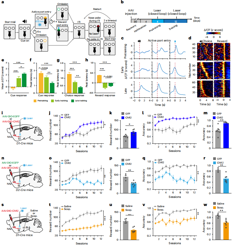
Figure 6 | Activation of D1→SI promotes, whereas activation of D2→SI suppresses reward-associated learning
🎯Summary
This work identifies a previously unrecognized striatum–basal forebrain–amygdala circuit, in which the nucleus accumbens (NAc) selectively regulates salience encoding in the basolateral amygdala (BLA) through the cholinergic system of the substantia innominata (SI). Distinct from the conventional cortical–thalamic salience network, this circuit establishes a complementary neural pathway linking the ventral striatum, basal forebrain, and amygdala for the regulation of stimulus salience.
Importantly, the SI→BLA cholinergic pathway selectively regulates stimulus salience without driving reward preference or aversive behavior, demonstrating that stimulus value (valence) and stimulus salience (behavioral priority) are encoded by two functionally distinct neural systems.
These findings further extend the classical antagonistic D1/D2 striatal circuitry to the cholinergic regulation of attention and salience. D1 neurons enhance salience encoding, whereas D2 neurons suppress it, together providing a dynamic mechanism for prioritizing behaviorally relevant environmental cues.
All viral vectors used in this study are available from BrainCase: