E-mail:BD@ebraincase.com
Tel:+8618971215294
English 中文版
Pioneering Innovation in Gene Vector Research - BrainCasePioneering Innovation in Gene Vector Research - BrainCase
  • Home
  • Pre-made AAV Library
    CRISPRRNAiHSV-helperRV-helperNeurotropic virusCalcium SensorsOptogenetics activationOptogenetics inhibitionChemical geneticsSparse labelingFluorescent proteinBiosensorsRecombinaseApoptosis & AutophagyDisease ModelNeurotoxicityOther
  • Products & Service

    Product Center

    Virus

    Vesicular Stomatitis Virus-VSV
    RCAS-TVA
    Lentivirus
    Rabies Virus-CVS Strain-RABV
    Herpes simplex virus-HSV
    Pseudorabies virus-PRV
    Adeno-Associated Virus-AAV
    Neurotropic virus list
    Baculovirus

    Kits & Regents

    EndoFree Plasmid Maxi Kit
    Lentivirus Concentration Kit
    VESSL Exosome Solutions
    AAV Rainbow Combo™

    Plasmid Construction & Vector cloning

    Lentiviral Vector
    AAV Vector
    Plasmid
    Plasmid Library

    Popular Applications

    Gene Regulation

    Gene Overexpression-Brain Case
    RNA interference(RNAi)-siRNA-Brain Case
    Gene Editing - CRSIPR cloning - BrainCase

    Neural Circuit Function Research

    Optogenetics - BrainCase
    Chemical genetics-DREADDs-Brain Case
    Calcium signal recording-Gels- Brain Case
    GRAB Neurotransmitter Fluorescent Sensor- Brain Case
    Functional Magnetic Resonance Imaging Technology- Brain Case

    Research on the structure of neural circuits

    Direct Input and Output-viral vectors- Brain Case
    Anterograde Mono-synaptic Tracing -HSV- Brain Case
    Antrograde Muti-synaptic Tracing-HSV & VSV-Brain Case
    Retrograde Mono-synaptic Tracing-Rabies Virus(CVS Strain)-Brain Case
    Retrograde Muti-synaptic Tracing-PRV-Brain Case

    Featured Services

    Experiment Service

    Research Services | Neural Circuit Tracing + Targeted Spatial Omics + Neuronal Classification Analysi
    Research Service | Cell and Tissue Patch-Clamp Recording and Analysis Services
    BrainCase Biotech| Whole-Brain Transparency Imaging and Analysis Services

    AAV Serotype Screening

    AAV Serotypes screening-gene therapy

    Collaboration Products

    Kiryl Piatkevich lab
    Yulong Li lab
    Zhifei Fu lab
    Woo-ping Ge lab

    Oncolytic Virus Modification

    Oncolytic virus-for cancer therapy-Brain Case
    Herpes Virus Vector-anti-tumor- BrainCase
    Vesicular stomatitis virus-killing tumor cells-Brain Case
  • News
    Corporate News New Product Launch Test Report Investor News
  • Support
    Literature interpretation Customer article FAQs Blog Legal
  • About Us
    Pre-made AAV Library Products & Service News Support About Us Contact
  • Contact
    Contact Us Join us
  • 中文
    English 中文版
  • Home
  • Support
  • Customer article
  • Support
  • Literature interpretation
  • Customer article
  • FAQs
  • Blog
  • Legal

Client Article | Advanced Science | Team led by Changgeng Peng, Rougang Xie, Xin Yu, Xiafeng Shen, et al. Reveal: From Single Target to Dual Blockade—Discovery of Nav1.7/1.8 Supramolecular Complex Reshapes Chronic Pain Treatment Strategy

Release time:2026-07-06 11:04:20
Neuropathic pain (NP) affects 7%–10% of the global population, while current medications show limited efficacy. Voltage-gated sodium channels Nav1.7 and Nav1.8 are key ion channels in dorsal root ganglion (DRG) neurons closely associated with neuropathic pain. However, single-target drugs against either channel have shown unsatisfactory therapeutic effects, suggesting a potential pathological synergistic mechanism between the two.

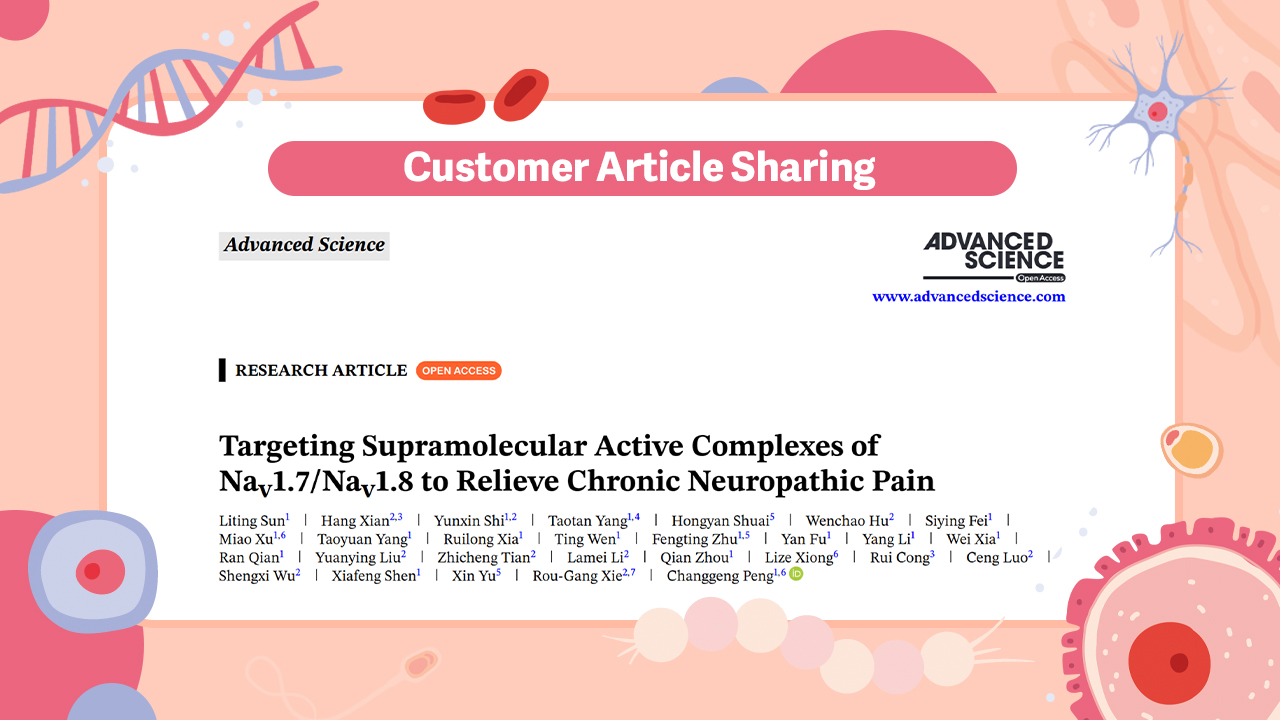
Recently, a collaborative team from Tongji University-affiliated Shanghai Fourth People’s Hospital (Changgeng Peng team), Air Force Medical University (Rougang Xie team), Dali University (Xin Yu), and Shanghai First Rehabilitation Hospital (Xiafeng Shen team) published a paper in Advanced Science titled “Targeting Supramolecular Active Complexes of Nav1.7/Nav1.8 to Relieve Chronic Neuropathic Pain.”

The study demonstrates that a hallmark of chronic neuropathic pain is the Nav1.7/Nav1.8/TrkB supramolecular active complex (SMAC). Its assembly is regulated by the TrkB/CREB signaling pathway and jointly controlled by five cytoskeletal proteins, among which SPTAN1 and DSP are core molecules maintaining its structure and function.

SMAC promotes sodium ion enrichment and amplifies sodium currents, leading to hyperexcitability and repetitive firing of DRG neurons. This is a fundamental mechanism underlying persistent and refractory neuropathic pain.

Dual blockade of Nav1.7 and Nav1.8 represents an effective therapeutic strategy for this condition. In addition, cytoskeletal proteins and the TrkB–DSP interaction interface may serve as promising novel drug targets.

https://doi.org/10.1002/advs.202522185


1. Formation and Structural Characteristics of Nav1.7/Nav1.8/TrkB SMAC in Dorsal Root Ganglion (DRG) Neurons of Neuropathic Pain Mice

Previous studies have shown that miR-96 knockout induces pain-related behaviors accompanied by coordinated upregulation of Nav1.7 and Nav1.8, suggesting that the proportion of neurons co-expressing these two sodium channels may be remodeled under nerve injury conditions.

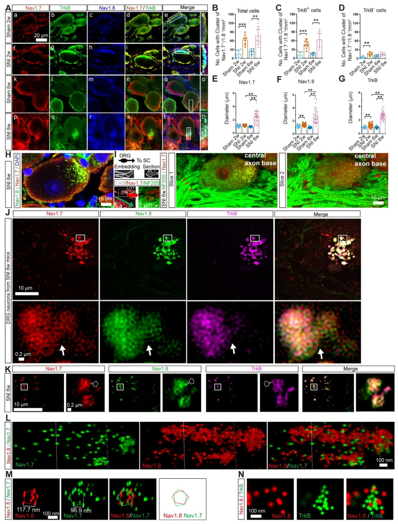
In this study, we first examined expression changes of Nav1.7 and Nav1.8 in dorsal root ganglion (DRG) neurons of mice subjected to sciatic nerve branch injury (SNI). Imaging at 60× magnification revealed that at 2 weeks post-SNI, Nav1.7/Nav1.8 co-expressing DRG neurons exhibited membrane clustering. By 6 weeks post-injury, Nav1.7/Nav1.8/TrkB SMAC structures had further formed, with a significantly higher number of positive neurons compared with the sham group. These structures were predominantly distributed in TrkB (tropomyosin receptor kinase B)-positive neurons (Fig. 1A–D). At 6 weeks post-surgery, SMAC size was significantly increased (Fig. 1E–G) and was localized to the axonal initial segment, further confirmed by Dil staining (Fig. 1H, I). Only a few small aggregates were observed in the sham group, suggesting a potential association with post-surgical allodynia or neuropathic pain during recovery.

Fig. 1. Nerve injury induces formation of Nav1.7/Nav1.8 SMAC in mouse DRG neurons
 
Using 60 nm super-resolution imaging, SMACs in DRG neurons 6 weeks after SNI were observed as polygonal columnar lattice structures composed of Nav1.7, Nav1.8, and TrkB. The lattice had edge lengths of approximately 160–250 nm, with proteins interconnected to form complete clustered units (Fig. 1J, K).

Stochastic optical reconstruction microscopy (STORM, ~20 nm resolution) further revealed a gradient distribution of the three proteins along the longitudinal axis of the lattice. Quantitative analysis showed that the average molecular densities of Nav1.7 and Nav1.8 were approximately 340 molecules/μm³ and 902 molecules/μm³, respectively. The inter-molecular spacing between heterologous proteins in the core region of the complex was approximately 30 nm, while the spacing between homologous proteins was approximately 70 nm, with many molecules arranged in near-contact proximity (Fig. 1L–N).

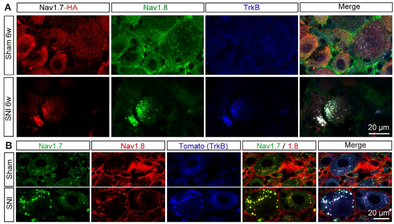
Using Scn9a^HA/+ mice (which display normal mechanical pain behavior), the HA tag was shown to efficiently label Nav1.7-positive neurons. Labeled Nav1.7 was clearly localized within SMACs, and Nav1.7 expression exhibited cellular heterogeneity (Fig. 2A).

Tracing in Trk2A-Tomato/+ reporter mice further demonstrated that SMACs primarily form in TrkB-positive low-threshold mechanoreceptor neurons (Fig. 2B).

Fig. 2. Distribution of Nav1.7 and TrkB within SMACs in DRG neurons of SNI mice
 

2. SMAC Formation in Dorsal Root Ganglion Neurons of Patients with Neuropathic Pain

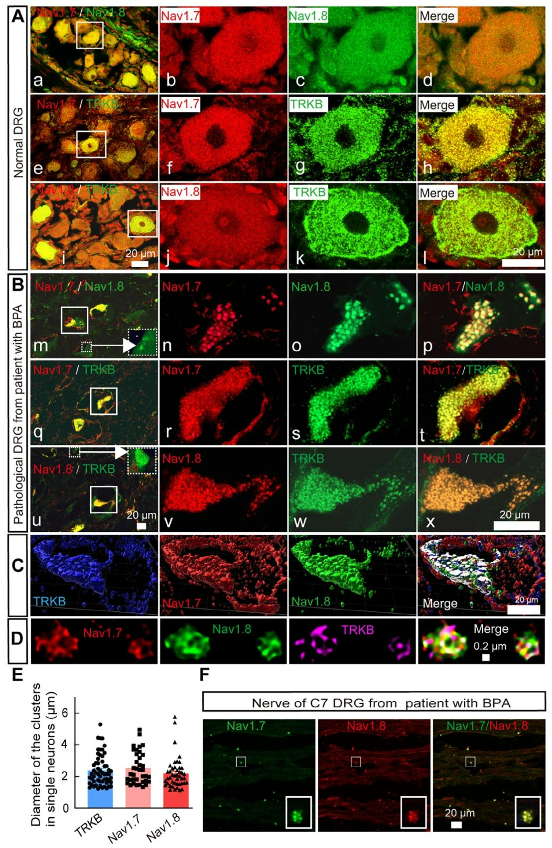
Consistent with the findings in mouse models, dorsal root ganglion (DRG) neurons from patients with neuropathic pain induced by brachial plexus avulsion (BPA) commonly form large Nav1.7/Nav1.8/TrkB SMAC structures. Such structures are absent in normal tissues. The complexes are mainly localized on the cell membrane and are widely distributed in neurons with diameters of 30–120 μm, with large neurons (>60 μm in diameter) accounting for over 73% (Fig. 3A–C).

Structured illumination microscopy (SIM) imaging confirmed that human SMACs also form polygonal lattice structures with edge lengths of 160–250 nm, assembled into clusters with an average diameter of approximately 2 μm. Some clusters contain only Nav1.8 or TrkB. These complexes are also abundantly distributed along DRG nerve fibers (Fig. 3D, F). In summary, Nav1.7/Nav1.8/TrkB SMAC represents a characteristic pathological marker of severe chronic neuropathic pain.

Fig. 3. Formation of Nav1.7/Nav1.8/TrkB SMAC in DRG neurons of BPA-induced neuropathic pain patients


3. Synergistic Relief of Neuropathic Pain by Combined Nav1.7 and Nav1.8 Blockade in Mouse Models

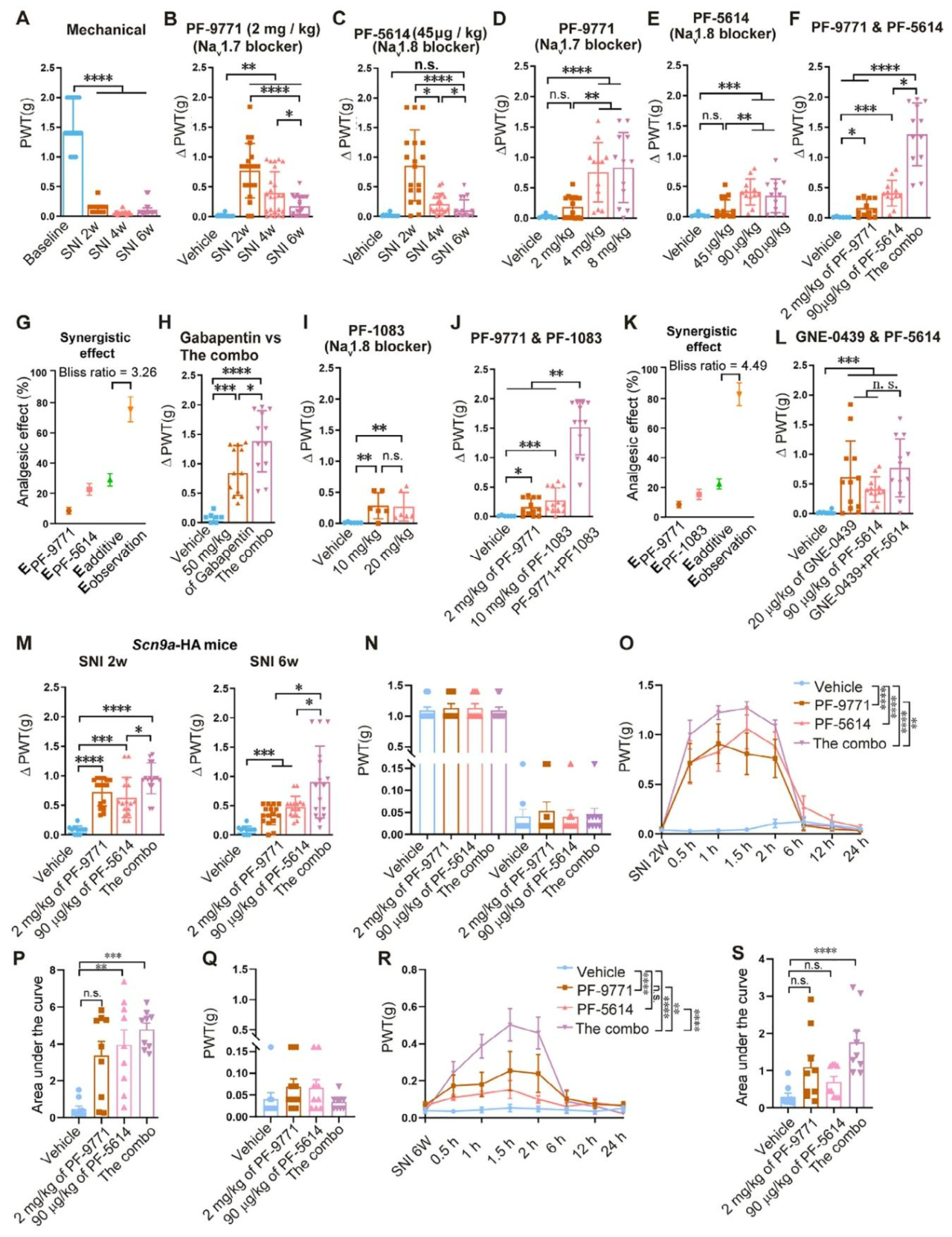
Based on the synergistic pathogenic role of the two sodium channels within SMAC, pharmacological intervention experiments were conducted. As SMAC assembly matured following SNI (2–6 weeks post-injury), the analgesic efficacy of either the Nav1.7 blocker (PF-05089771) or the Nav1.8 blocker (PF-04885614) alone progressively declined. By 6 weeks post-surgery, even increased dosing failed to alleviate mechanical allodynia (Fig. 4A–E).

In contrast, combined administration of the two inhibitors produced a strong synergistic effect, with significantly better efficacy than monotherapy or gabapentin (100 mg/kg), and without the sedative side effects associated with gabapentin (Fig. 4F–H).

When PF-05089771 was combined with an alternative Nav1.8 blocker (PF-04531083), the synergistic effect was maintained. However, combining GNE-0439 (Nav1.7 blocker) with PF-04885614 showed no efficacy. This synergistic effect was further validated in Scn9a^HA/+ mice (Fig. 4I–M).

Twenty-four-hour pharmacodynamic monitoring showed that at 2 weeks post-surgery, both monotherapy and combination therapy provided partial relief, whereas at 6 weeks post-surgery, only the combination therapy group achieved significant pain relief (Fig. 4N–S).

Fig. 4. Combined inhibition of Nav1.7 and Nav1.8 effectively alleviates mechanical allodynia in SNI mice


4. Involvement of Nav1.7/Nav1.8 Supramolecular Active Complexes in the Development of Diabetic Neuropathic Pain

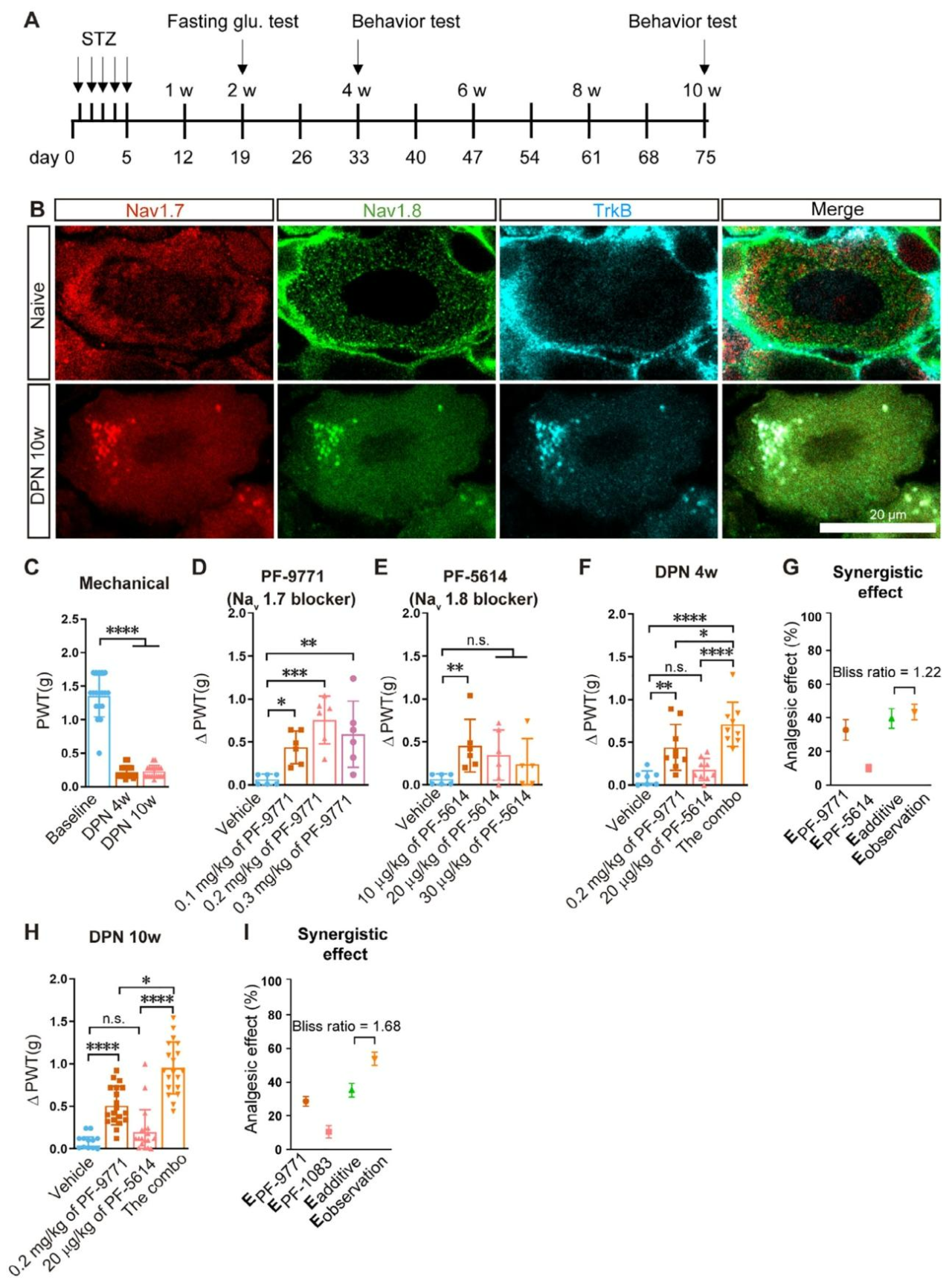
To investigate whether SMAC is involved in diabetic neuropathic pain, a streptozotocin (STZ)-induced diabetic mouse model was established via intraperitoneal injection. Two weeks after the final administration, fasting blood glucose levels exceeded 13 mM, confirming successful model induction (Fig. 5A).

Morphological analysis revealed no typical SMAC structures in DRG neurons of healthy mice. In contrast, abundant Nav1.7/Nav1.8/TrkB supramolecular active complexes were observed in DRG neurons of diabetic mice 10 weeks after STZ treatment.

Behavioral assessments showed that mice developed significant mechanical allodynia starting at 4 weeks post-STZ administration, which persisted up to 10 weeks (Fig. 5B, C).

Pharmacological intervention demonstrated that combined administration of the Nav1.7 blocker PF-05089771 and Nav1.8 blocker PF-04885614 significantly alleviated neuropathic pain both at early (4 weeks) and late (10 weeks) stages of diabetes. The combination therapy showed markedly superior efficacy compared with either monotherapy, indicating a clear synergistic analgesic effect (Fig. 5D–I).

Fig. 5. Nav1.7/Nav1.8 SMAC participates in the development of diabetic neuropathic pain in mice


5. Activation of Tyrosine Kinase Receptor B (TrkB) Promotes SMAC Formation

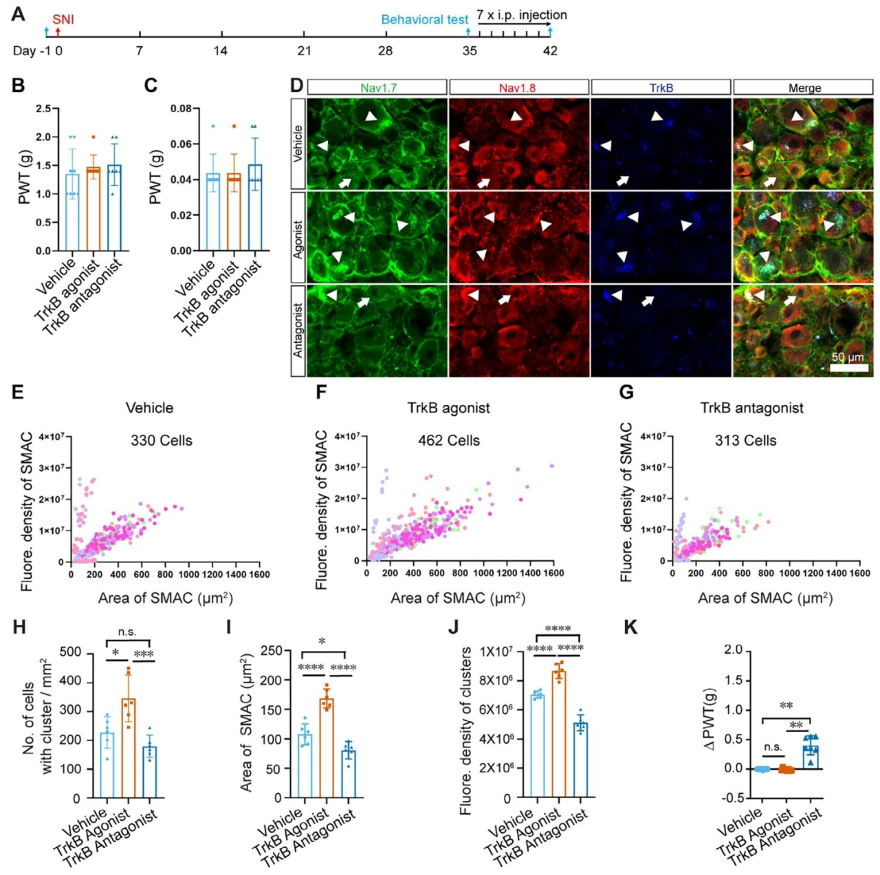
To clarify the regulatory role of TrkB in SMAC formation, from day 35 after SNI surgery, mice were administered either a TrkB agonist (7,8-dihydroxyflavone) or antagonist (ANA-12) for 7 consecutive days (Fig. 6A–C).

The results showed that the TrkB agonist significantly increased the number of DRG neurons co-expressing Nav1.7/Nav1.8/TrkB, and enlarged SMAC area and fluorescence intensity. In contrast, the antagonist markedly reduced SMAC size and decreased cluster density (Fig. 6D–J).

Behaviorally, the antagonist partially alleviated mechanical pain. Although the agonist promoted SMAC maturation and reduced pain thresholds in 5/8 mice, no statistically significant differences were observed between groups due to the low baseline pain threshold in the model (Fig. 6K).

Fig. 6. TrkB activation promotes SMAC formation

6. Cytoskeletal Proteins Promote SMAC Formation Under Pathological Conditions
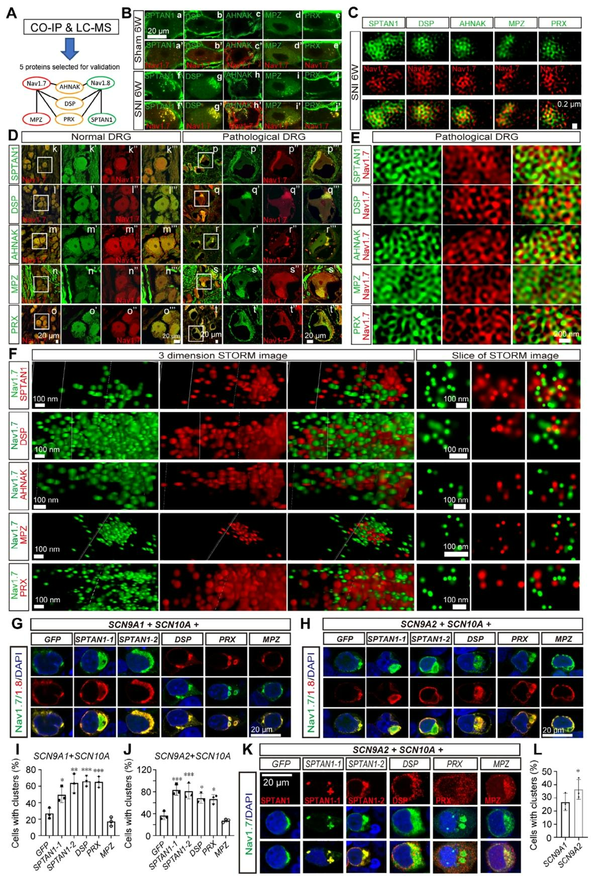
To identify proteins involved in SMAC assembly, Nav1.7/Nav1.8 affinity purification followed by mass spectrometry was performed on DRG tissues from mice 6 weeks after SNI. Multiple interacting proteins were identified, and five cytoskeletal proteins—SPTAN1, DSP, AHNAK, MPZ, and PRX—were selected for validation (Fig. 7A).

Immunostaining showed that all five proteins co-localized with Nav1.7 within SMACs in DRG neurons of SNI mice, whereas only small clusters were observed in the sham group, with no detectable MPZ signal (Fig. 7B).

SIM imaging confirmed that these proteins form polygonal lattice structures that connect Nav1.7 clusters, thereby facilitating SMAC assembly (Fig. 7C).

Consistent results were observed in DRG tissues from BPA-induced neuropathic pain patients. Super-resolution imaging revealed that SPTAN1 links multiple Nav1.7 clusters, and STORM imaging confirmed a highly intermingled distribution of the five proteins with Nav1.7 (Fig. 7D–F).

HEK293 overexpression experiments showed that a single cytoskeletal protein promotes Nav1.7/Nav1.8 clustering, while co-expression of multiple proteins leads to the formation of larger channel clusters. Two SPTAN1 isoforms fully co-localized with Nav1.7, whereas DSP, PRX, and MPZ showed partial co-localization. Long Nav1.7 isoforms exhibited a stronger tendency to aggregate (Fig. 7G–L).

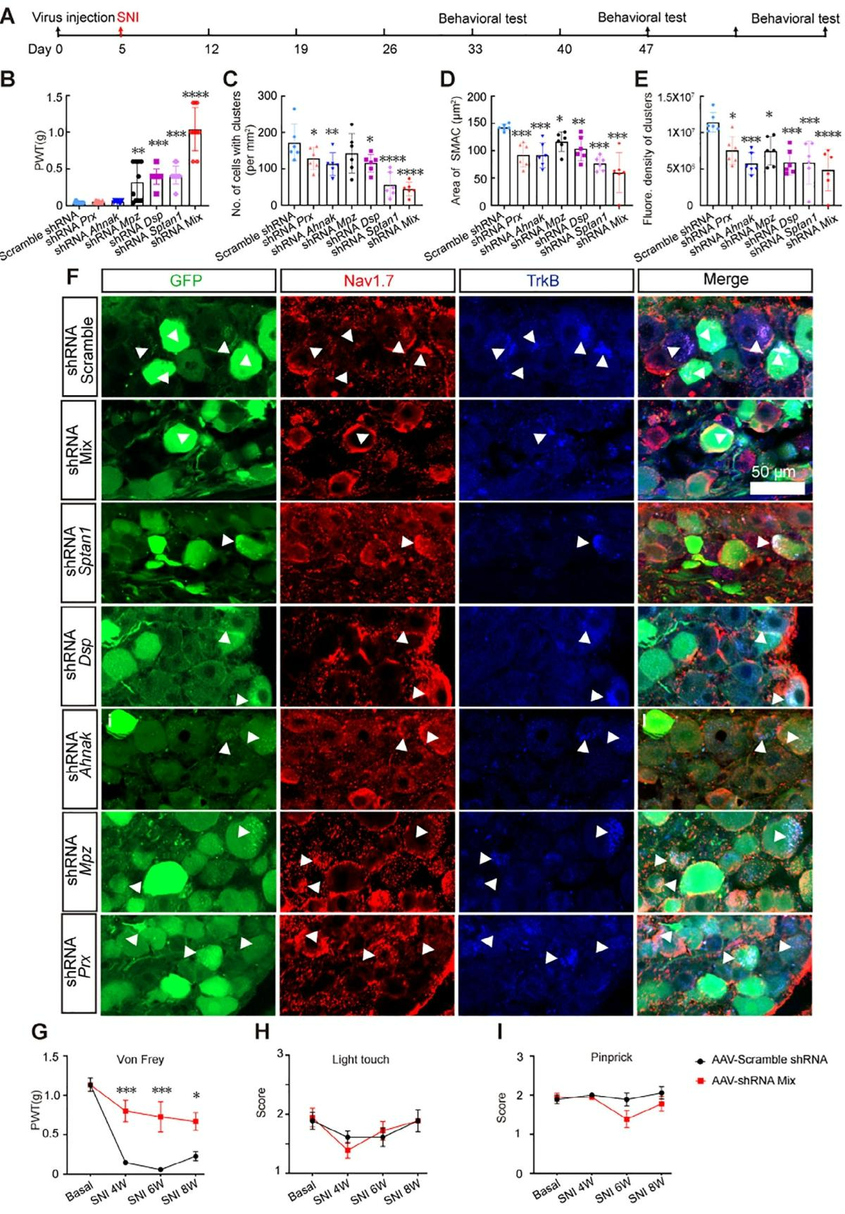
In vivo knockdown experiments demonstrated that injection of mixed shRNA-expressing viral vectors into the L4 and L5 DRG 5 days before SNI completely prevented pain development, reduced the number of SMAC-positive neurons, decreased SMAC size, and lowered molecular density (Fig. 8A–F).

Knockdown of SPTAN1 or DSP alone suppressed SMAC formation and alleviated pain. Knockdown of AHNAK reduced SMAC number but did not affect pain behavior. Knockdown of MPZ increased pain thresholds (Fig. 8B–C).

Long-term monitoring showed that AAV-shRNA-mediated knockdown produced sustained analgesia from 2 to 8 weeks post-surgery, without affecting normal touch or acute nociception (Fig. 8G–I).

Fig. 7. Cytoskeletal proteins promote Nav1.7 and Nav1.8 clustering


Fig. 7. Cytoskeletal proteins promote Nav1.7 and Nav1.8 clustering
 

7. Construction of the SMAC Scaffold via Specific Interactions Among Five Cytoskeletal Proteins

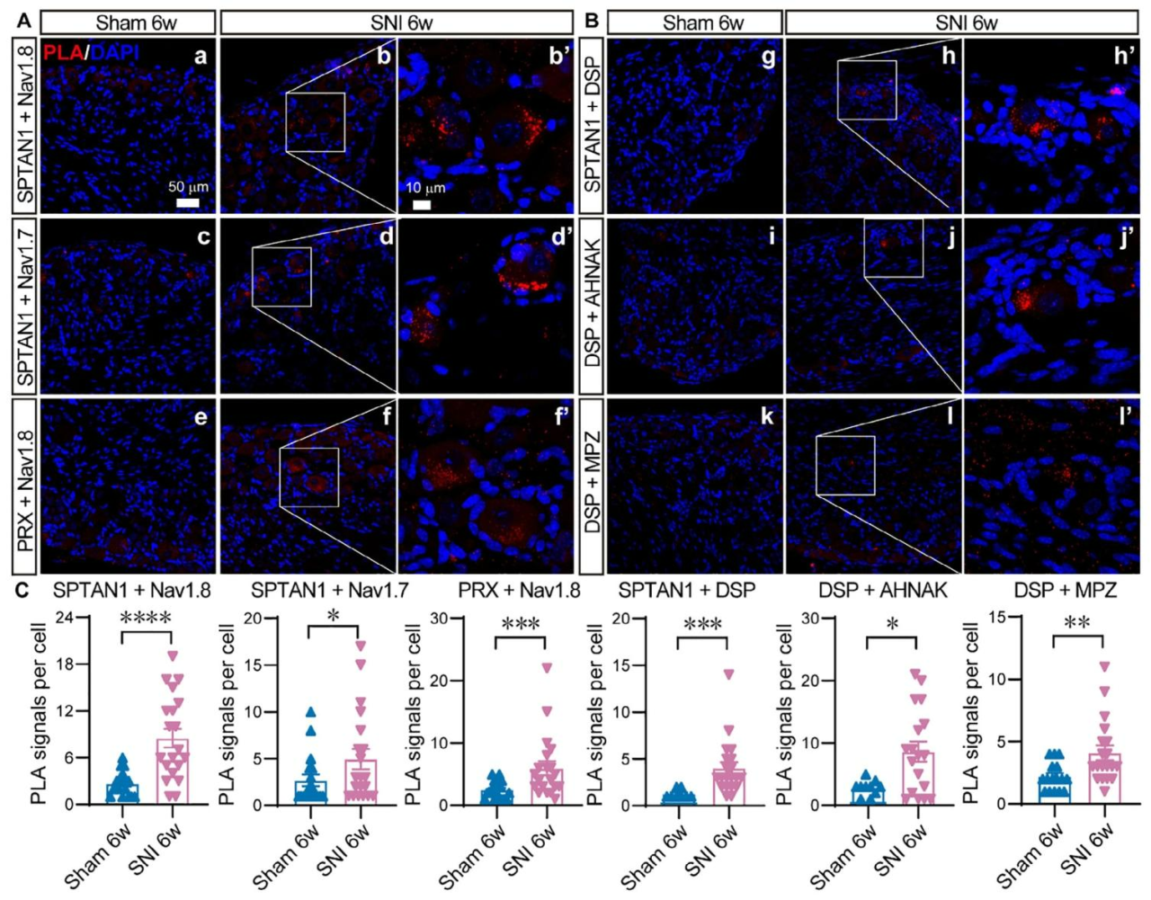
Using molecular docking (HADDOCK 2.4) and proximity ligation assays (PLA), direct interactions between sodium channels and five cytoskeletal proteins were examined in DRG tissues from mice 6 weeks after SNI.

The results showed that both Nav1.7 and Nav1.8 directly bind to SPTAN1, while Nav1.8 can also interact with PRX. DSP interacts with SPTAN1, AHNAK, and MPZ, respectively (Fig. 9A, B).

PLA fluorescence signals were significantly higher in the SNI group than in the sham group, indicating enhanced protein–protein interactions under pathological conditions (Fig. 9C).

Based on these findings, a SMAC assembly model was proposed: DSP acts as a central hub, interacting with SPTAN1, AHNAK, and MPZ to form the core scaffold. SPTAN1 recruits Nav1.7 and Nav1.8, while Nav1.8 additionally binds PRX, collectively facilitating SMAC assembly.

Fig. 9. Direct interactions between Nav1.7, Nav1.8, and five cytoskeletal proteins


8. Regulation of Four Cytoskeletal Proteins by the TrkB/CREB Signaling Pathway and Direct Interaction Between TrkB and DSP

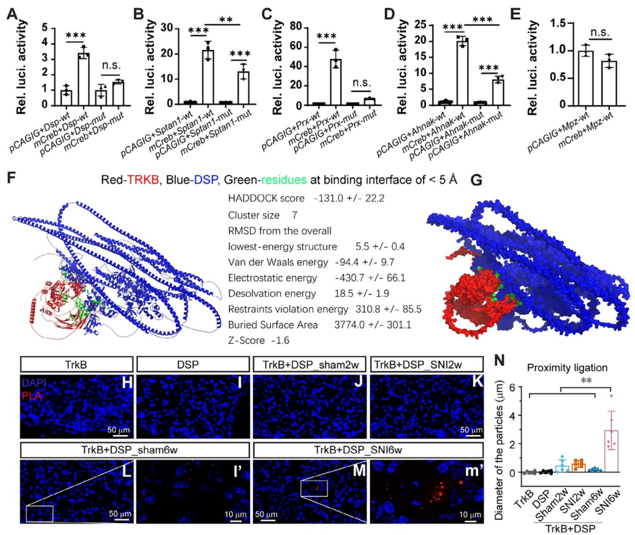
It was found that the promoter regions of four cytoskeletal protein genes (Dsp, Sptan1, Prx, and Ahnak) contain CREB-binding sites.

Dual-luciferase reporter assays demonstrated that CREB overexpression significantly enhanced promoter activity of these genes, while mutation of the binding sites attenuated or abolished this transcriptional activation, indicating that the BDNF/TrkB/CREB signaling pathway upregulates these proteins and promotes SMAC formation (Fig. 10A–E).

Molecular docking using HADDOCK predicted binding affinities between TrkB and the five cytoskeletal proteins, showing the strongest affinity between TrkB and DSP (score −131.0 ± 22.2, interface distance <5 Å) (Fig. 10F, G).

Proximity ligation assays further confirmed that, compared with the sham group, TrkB–DSP interaction signals were significantly increased in DRG neurons at both 2 and 6 weeks after SNI, indicating enhanced binding under pathological conditions (Fig. 10H–N).

Fig. 10. BDNF/TrkB/CREB signaling activates expression of four cytoskeletal proteins, and TrkB directly interacts with DSP


9. Combined Blockade of Nav1.7 and Nav1.8 Suppresses Action Potentials

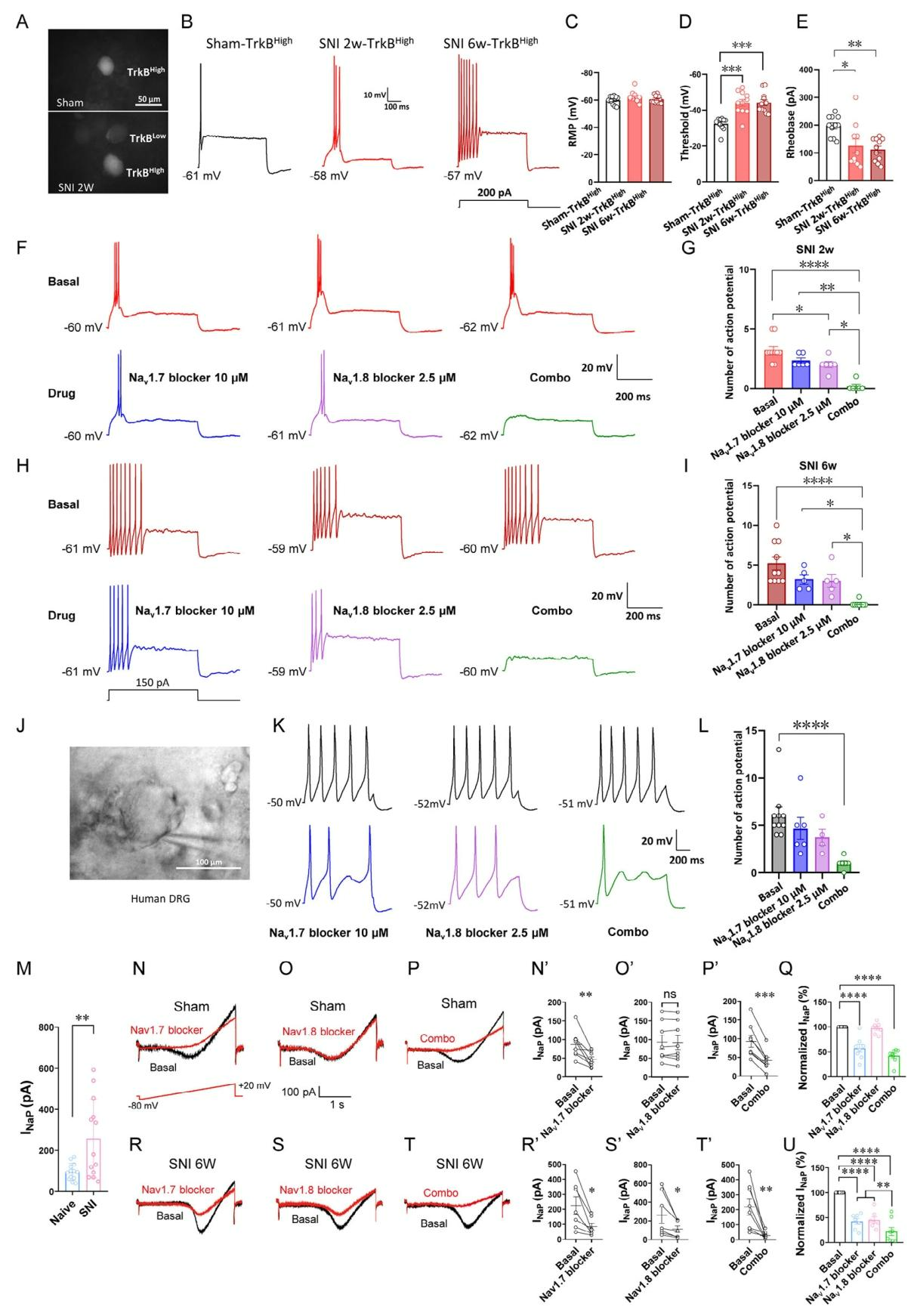
Whole-cell patch-clamp recordings showed that after SNI, TrkB-positive low-threshold mechanoreceptor neurons exhibited reduced activation thresholds, a shift from single firing to repetitive firing, and progressively increasing firing frequency over time (Fig. 11A–E).

At 2 weeks post-injury, either Nav1.7 or Nav1.8 blockade alone partially inhibited action potentials, with combination therapy showing superior efficacy. By 6 weeks, neurons became resistant to monotherapy, whereas only the combined treatment effectively and reversibly suppressed repetitive firing (Fig. 11F–I). Similar results were observed in human pathological DRG neurons (Fig. 11J–L).

Electrophysiological analysis showed that combined inhibition reduced total sodium current by 71%. Persistent sodium current in diseased neurons was 2.7-fold higher than in normal neurons.

In normal neurons, inhibition rates of persistent sodium current were 54% (PF-05089771), 0% (PF-04885614), and 62% for combination therapy, respectively. In diseased neurons, the corresponding values were 65%, 61%, and 91% (Fig. 11M–U).

These results suggest that Nav1.7 is the primary carrier of persistent sodium current within SMAC, while Nav1.8 contributes to its regulation. Their synergistic interaction enhances neuronal excitability, providing the molecular basis for the strong analgesic effect of dual-target therapy.

Fig. 11. Combined inhibition of Nav1.7 and Nav1.8 blocks action potentials in hyperexcitable DRG neurons
 

10. SMAC Regulates Neuronal Repetitive Firing

Using a combination of fluorescent ion indicators, two-photon imaging, and patch-clamp electrophysiology, it was observed that during action potential generation, the membrane potential dynamics within the SMAC region differ from other cellular regions. Local sodium ions accumulate and are retained within the SMAC, and outward sodium flux can trigger secondary action potentials, thereby driving repetitive neuronal firing (Fig. 12A–E).

Combined administration of Nav1.7 and Nav1.8 blockers rapidly suppressed sodium influx and prevented secondary firing, confirming that SMAC functions in a manner analogous to a biological transistor by concentrating sodium ions and amplifying sodium currents (Fig. 12F, G).

TrkB agonists reduced action potential threshold and baseline firing intensity, whereas antagonists produced the opposite effect. Neither had a significant impact on resting membrane potential, suggesting that TrkB cooperates with sodium channels to regulate neuronal excitability (Fig. 12H–M).

Knockdown of the five cytoskeletal proteins resulted in most neurons failing to generate action potentials. Individual knockdown of SPTAN1 or DSP increased firing thresholds and reduced spike amplitude, while all knockdown conditions increased baseline excitability (Fig. 12N–S).

In summary, SMAC exacerbates pain by altering electrophysiological properties, and SPTAN1 and DSP are key scaffold proteins essential for maintaining its structure and function.

Fig. 12. Nav1.7/Nav1.8/TrkB SMAC regulates neuronal repetitive firing


Summary

This study is the first to identify and validate the existence of Nav1.7/Nav1.8 SMAC in chronic neuropathic pain and to demonstrate its central pathogenic role, providing a new perspective for understanding the underlying mechanisms of this disorder.

Based on this discovery, a dual-target blockade strategy against Nav1.7 and Nav1.8 shows superior analgesic efficacy and safety compared with existing drugs, offering a strong theoretical and experimental foundation for the development of first-in-class small-molecule therapeutics and future combination clinical therapies.

The viral tools used in this study are available from Brain Case Biotech
Product Category No. Product Name
Gene Regulation BC-1856 rAAV-U6-shRNA(mAhnak)-CMV-EGFP
BC-1787 rAAV-U6-shRNA(mDsp)-CMV-EGFP
BC-1795 rAAV-U6-shRNA(mMpz)-CMV-EGFP
BC-1783 rAAV-U6-shRNA(mSptanl)-CMV-EGFP
BC-1790 rAAV-U6-shRNA1(mPrx)-CMV-EGFP
BC-0186 rAAV-U6-shRNA(Scramble)-CMV-EGFP

Brain Case Biotech also provides various customized services. For more information,
please contact:
bd@ebraincase.com

Service Type :

Select the service you'd like to purchase.

Order Information(Premade-AAVs)

Please provide us some information about the service you'd like to order.

How did you hear about BrainCase Biotech or our products?(Fill in this field to get a discount coupon)

Detailed requirements:

scroll

Order Information(Custom AAV/Lentivirus)

Please provide us some information about the service you'd like to order.

Gene ID or gene information:

Selection of the reporting gene:

Special Instructions:

scroll

Order Information(Others)

Please provide us some information about the service you'd like to order.

Virus name/Detailed requirements:

scroll

Related products

Client Article | Advanced Science | Team led by Changgeng Peng, Rougang Xie, Xin Yu, Xiafeng Shen, et al. Reveal: From Single Target to Dual Blockade—Discovery of Nav1.7/1.8 Supramolecular Complex Reshapes Chronic Pain Treatment Strategy

Client Article | Advanced Science | Team led by Changgeng Peng, Rougang Xie, Xin Yu, Xiafeng Shen, et al. Reveal: From Single Target to Dual Blockade—Discovery of Nav1.7/1.8 Supramolecular Complex Reshapes Chronic Pain Treatment Strategy

Client Articles | Neuron | Can’t sleep well under stress? Air Force Medical University team (Hailong Dong, Guangchao Zhao) reveals VTA–mPFC pathway drives sleep architecture disruption under chronic stress

Client Articles | Neuron | Can’t sleep well under stress? Air Force Medical University team (Hailong Dong, Guangchao Zhao) reveals VTA–mPFC pathway drives sleep architecture disruption under chronic stress

Customer Article | Sci. Adv. | Jinhao Sun’s Team at Shandong University Unveils: How Does the Brain “Remember” Gastric Inflammation? Hcn1-Dependent PVN Engram Neurons Are the Key!

Customer Article | Sci. Adv. | Jinhao Sun’s Team at Shandong University Unveils: How Does the Brain “Remember” Gastric Inflammation? Hcn1-Dependent PVN Engram Neurons Are the Key!

Sci. Adv. | How Does the Brain Recognize Familiar Individuals? Hao Wang / Gao Chen / Hongbin Yang / Libiao Pan Reveal VGluT2 Neurons Precisely Encode Conspecific Identity, and Exclusive Memories Can Be Reactivated

Sci. Adv. | How Does the Brain Recognize Familiar Individuals? Hao Wang / Gao Chen / Hongbin Yang / Libiao Pan Reveal VGluT2 Neurons Precisely Encode Conspecific Identity, and Exclusive Memories Can Be Reactivated

map
{dede:global.cfg_webname/}

Pre-made AAV Library

CRISPR
RNAi
Neurotropic virus
Optogenetics activation
Biosensors

News

Corporate News
New Product Launch
Test Report
Investor News

Support

Literature interpretation
Customer article
FAQs
Blog
Legal
微信

WhatsApp Business Account

Tel: +8618971215294
E-mail: BD@ebraincase.com

Address:-

Address:-

  • Copyright © 2024 Brain Case All Rights Reserved.