E-mail:BD@ebraincase.com
Tel:+8618971215294
English 中文版
Pioneering Innovation in Gene Vector Research - BrainCasePioneering Innovation in Gene Vector Research - BrainCase
  • Home
  • Pre-made AAV Library
    CRISPRRNAiHSV-helperRV-helperNeurotropic virusCalcium SensorsOptogenetics activationOptogenetics inhibitionChemical geneticsSparse labelingFluorescent proteinBiosensorsRecombinaseApoptosis & AutophagyDisease ModelNeurotoxicityOther
  • Products & Service

    Product Center

    Virus

    Vesicular Stomatitis Virus-VSV
    RCAS-TVA
    Lentivirus
    Rabies Virus-CVS Strain-RABV
    Herpes simplex virus-HSV
    Pseudorabies virus-PRV
    Adeno-Associated Virus-AAV
    Neurotropic virus list
    Baculovirus

    Kits & Regents

    EndoFree Plasmid Maxi Kit
    Lentivirus Concentration Kit
    VESSL Exosome Solutions
    AAV Rainbow Combo™

    Plasmid Construction & Vector cloning

    Lentiviral Vector
    AAV Vector
    Plasmid
    Plasmid Library

    Popular Applications

    Gene Regulation

    Gene Overexpression-Brain Case
    RNA interference(RNAi)-siRNA-Brain Case
    Gene Editing - CRSIPR cloning - BrainCase

    Neural Circuit Function Research

    Optogenetics - BrainCase
    Chemical genetics-DREADDs-Brain Case
    Calcium signal recording-Gels- Brain Case
    GRAB Neurotransmitter Fluorescent Sensor- Brain Case
    Functional Magnetic Resonance Imaging Technology- Brain Case

    Research on the structure of neural circuits

    Direct Input and Output-viral vectors- Brain Case
    Anterograde Mono-synaptic Tracing -HSV- Brain Case
    Antrograde Muti-synaptic Tracing-HSV & VSV-Brain Case
    Retrograde Mono-synaptic Tracing-Rabies Virus(CVS Strain)-Brain Case
    Retrograde Muti-synaptic Tracing-PRV-Brain Case

    Featured Services

    Experiment Service

    Research Services | Neural Circuit Tracing + Targeted Spatial Omics + Neuronal Classification Analysi
    Research Service | Cell and Tissue Patch-Clamp Recording and Analysis Services
    BrainCase Biotech| Whole-Brain Transparency Imaging and Analysis Services

    AAV Serotype Screening

    AAV Serotypes screening-gene therapy

    Collaboration Products

    Kiryl Piatkevich lab
    Yulong Li lab
    Zhifei Fu lab
    Woo-ping Ge lab

    Oncolytic Virus Modification

    Oncolytic virus-for cancer therapy-Brain Case
    Herpes Virus Vector-anti-tumor- BrainCase
    Vesicular stomatitis virus-killing tumor cells-Brain Case
  • News
    Corporate News New Product Launch Test Report Investor News
  • Support
    Literature interpretation Customer article FAQs Blog Legal
  • About Us
    Pre-made AAV Library Products & Service News Support About Us Contact
  • Contact
    Contact Us Join us
  • 中文
    English 中文版
  • Home
  • Support
  • Customer article
  • Support
  • Literature interpretation
  • Customer article
  • FAQs
  • Blog
  • Legal

Sci. Adv.丨Could Intranasal Oxytocin Help Police Officers Prevent PTSD? Xiao Lei’s Team from Fudan Institute of Brain Science Reveals the Precise Neural Circuit Underlying Oxytocin-Mediated Suppression of Fear Learning

Release time:2026-07-08 10:05:42
The medial prefrontal cortex (mPFC) is a key brain region involved in threat evaluation and fear regulation. Within the mPFC, the prelimbic cortex (PL) primarily governs fear expression. Somatostatin-positive (SST) interneurons in the PL contribute to fear memory encoding and express oxytocin receptors (OxtR). However, the cellular and circuit mechanisms through which oxytocin signaling in the PL regulates fear learning remain unclear.

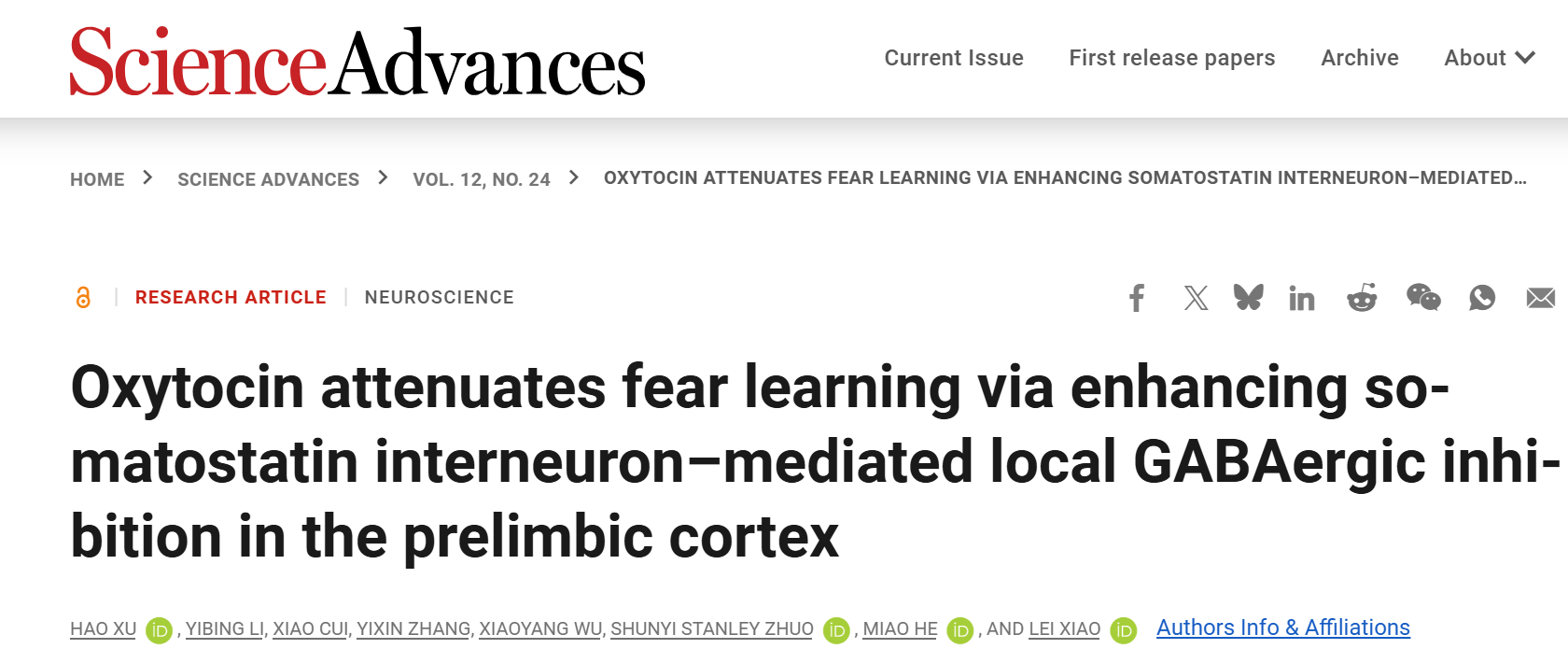
On June 12, 2026, Lei Xiao’s research group from the Institute of Brain Science at Fudan University / National Key Laboratory of Brain Function and Disease published a study entitled “Oxytocin attenuates fear learning via enhancing somatostatin interneurons-mediated local GABAergic inhibition in the prelimbic cortex” in Science Advances.

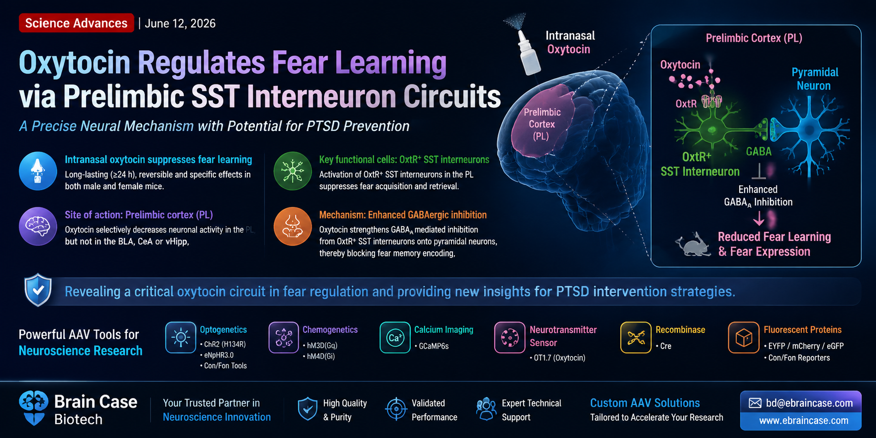
The study demonstrated that intranasal administration of oxytocin can produce long-lasting, reversible, and specific suppression of fear learning, with comparable effects observed in both male and female subjects, suggesting potential applications in the prevention of post-traumatic stress disorder (PTSD).

Mechanistically, oxytocin activates oxytocin receptor-expressing somatostatin interneurons in the prelimbic cortex, enhancing their GABAergic inhibitory control over pyramidal neurons and subsequently disrupting the encoding of fear memories. This work reveals a precise neural circuit mechanism through which oxytocin regulates fear learning and provides new insights into potential therapeutic strategies for stress-related disorders.

https://doi.org/10.1126/sciadv.aef8400
 

1. Intranasal Oxytocin Produces Long-Lasting and Reversible Suppression of Fear Learning Through Regulation of the Prelimbic Cortex

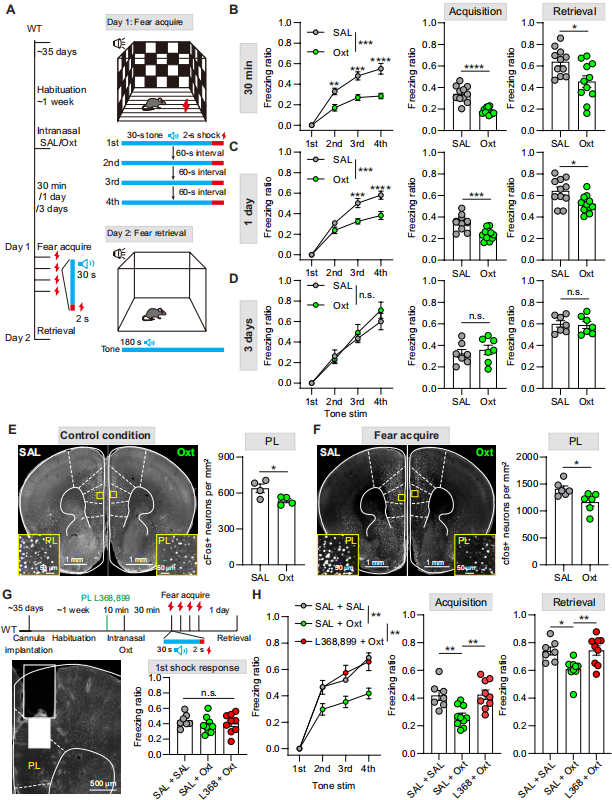
In this experiment, mice received intranasal administration of oxytocin or saline, followed by a cued fear conditioning test (Fig. 1A). The results showed that mice treated with oxytocin exhibited significantly reduced freezing responses and impaired fear memory retrieval (Fig. 1B). This effect was consistent in both male and female mice.

A time-course analysis of oxytocin efficacy revealed that the suppressive effect on fear learning remained significant one day after administration (Fig. 1C) but completely disappeared after three days (Fig. 1D). The effects observed 30 minutes and one day after administration were significantly stronger than those observed after three days. The platform-mediated avoidance (PMA) test further demonstrated that oxytocin reduced active avoidance learning, confirming that its inhibitory effect on fear learning persisted for more than 24 hours.

The basolateral amygdala (BLA), prelimbic cortex (PL), and ventral hippocampus (vHipp) are key brain regions involved in fear regulation. cFos immunostaining revealed that oxytocin selectively reduced neuronal activity in the PL, while exerting no significant effects on the BLA, central amygdala (CeA), or vHipp (Fig. 1E-F). These findings further support previous evidence linking PL hyperactivation to fear-related behaviors.

Following local infusion of the oxytocin receptor (OxtR) antagonist L-368,899 into the PL, the fear-suppressing effect of oxytocin was completely abolished (Fig. 1G-H). Collectively, these findings demonstrate that intranasal oxytocin achieves long-lasting suppression of fear learning by activating oxytocin receptor signaling pathways within the prelimbic cortex.

Figure 1. Intranasal administration of oxytocin suppresses fear learning and reduces neuronal activity in the PL.
 

2. Modulation of Oxytocin Signaling in the Prelimbic Cortex Alters Fear Learning and Memory Retrieval

Previous studies have established that the PL plays a critical role in regulating learned fear. This study further demonstrated that intranasal oxytocin suppresses fear learning through the PL (Fig. 1).

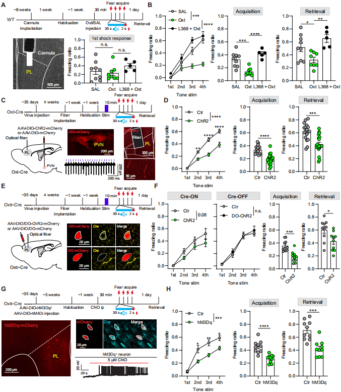
Researchers locally injected oxytocin into the PL (Fig. 2A), producing results consistent with intranasal administration: this manipulation did not affect mice’s responses to foot shocks but significantly impaired fear learning and memory retrieval (Fig. 2B). Blocking OxtR signaling in the PL with L-368,899 completely eliminated the effects of oxytocin, whereas receptor blockade alone produced no significant behavioral changes.

Previous anterograde tracing studies have shown that oxytocinergic neurons in the paraventricular nucleus of the hypothalamus (PVN) directly project to the PL. Using optogenetic approaches to activate these endogenous oxytocinergic fibers (Fig. 2C), researchers found that fear acquisition and retrieval were significantly reduced (Fig. 2D).
Using OxtR-Cre mice, selective optogenetic activation of OxtR-expressing neurons in the PL markedly suppressed fear-related behaviors, whereas activation of OxtR-negative neurons had no effect (Fig. 2E-F). This effect persisted for more than 24 hours. Similarly, chemogenetic activation of PL OxtR-positive neurons also weakened fear acquisition (Fig. 2G-H).

Unlike the sex-dependent effects previously reported for oxytocin signaling in the medial prefrontal cortex, oxytocin-related interventions targeting the PL produced comparable effects in both male and female mice.

Together, these findings demonstrate that enhancing oxytocin signaling within the prelimbic cortex can significantly suppress fear learning and fear expression.

Figure 2. Enhancement of oxytocin signaling in the prelimbic cortex attenuates fear acquisition and memory retrieval.
 

3. Dynamic Changes in Prelimbic Cortex Oxytocin Signaling During Fear Learning

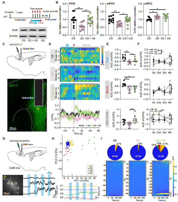
Previous studies have demonstrated that stress can alter the expression levels of oxytocin and its receptors in the brain. In this study, three experimental groups were established: tone-only stimulation (CS), foot shock-only stimulation (US), and paired tone–shock stimulation (CS+US), to investigate changes in oxytocin signaling during fear conditioning (Fig. 3A).
Given that the half-life of oxytocin is approximately 20–30 minutes and that the prelimbic cortex (PL) and infralimbic cortex (IL) lack a clear anatomical boundary, the study combined mPFC and paraventricular nucleus of the hypothalamus (PVN) tissues for analysis. The results showed that oxytocin levels in the US group were significantly reduced compared with the CS group, whereas no significant change was observed in the CS+US group. In addition, OxtR expression levels in both the CS and CS+US groups were higher than those in the US group (Fig. 3B).

Using fiber photometry combined with the oxytocin sensor AAV-OXT1.7, researchers monitored real-time oxytocin release in the PL (Fig. 3C). Tone stimulation increased oxytocin levels, whereas foot shock reduced them. After shock termination, oxytocin levels rapidly recovered in the CS+US group, while recovery was slower in the US group (Fig. 3D-E). Data from multiple rounds of experiments showed that oxytocin levels gradually increased in the CS group, whereas the CS+US group exhibited a transient decrease after shock exposure but had recovered to levels close to those of the CS group by the fourth trial, consistent with tissue-level measurements (Fig. 3F).

Using GCaMP6s calcium imaging to monitor the activity of OxtR-expressing neurons in the PL (Fig. 3G), researchers identified four distinct activity patterns through clustering analysis: weak slow activation (C1), stable activity (C2), rapid strong activation (C3), and activity attenuation (C4) (Fig. 3H).

The CS and US groups contained only C1 and C2 neurons, whereas the CS+US group involved in associative fear learning exhibited all four neuronal activity patterns. Among these, C3 neurons showed significantly increased activity following foot shock, while C4 neurons displayed sustained decreases in activity (Fig. 3I). The researchers proposed that C3 neurons may represent fear-learning-activated somatostatin interneurons, which exert inhibitory effects on C4 neurons.

Collectively, these findings indicate that fear learning induces multilayered plasticity within the PL oxytocin circuit through regulation of receptor expression, neuropeptide release, and neuronal activation patterns, enabling adaptive encoding of threat-related signals.

Figure 3. Changes in prelimbic cortex oxytocin signaling during tone–shock fear conditioning.
 

4. Fear Conditioning Differentially Regulates the Activity of Two Neuronal Populations in the Prelimbic Cortex

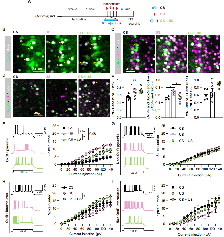
Oxytocin exerts distinct effects on PL pyramidal neurons and GABAergic interneurons. In this experiment, Oxtr-Cre; Ai3 mice were divided into three groups: tone-only stimulation (CS), foot shock-only stimulation (US), and paired tone–shock stimulation (CS+US) (Fig. 4A).

cFos staining revealed that the activation level of oxytocin receptor-positive (OxtR+) neurons in the PL was higher in both the US and CS+US groups compared with the CS group. Among these populations, OxtR+ pyramidal neurons exhibited the highest activation in the US group, whereas OxtR+ somatostatin interneurons showed the strongest activation in the CS+US group (Fig. 4B-E).

Ex vivo electrophysiological recordings further supported these findings. OxtR+ pyramidal neurons displayed increased excitability in the US group but reduced excitability in the CS+US group. In contrast, OxtR+ interneurons exhibited the highest excitability in the CS+US group (Fig. 4F-H). No significant differences were observed in the excitability of OxtR-negative neurons among the three groups (Fig. 4G, 4I).

Additionally, the two neuronal populations in the US group showed opposite changes in resting membrane potential characteristics. Together, these findings demonstrate that compared with non-associative foot shock exposure, associative fear learning selectively activates OxtR-positive somatostatin interneurons in the PL while suppressing OxtR-positive pyramidal neurons.

Figure 4. Differential effects of tone–shock fear conditioning on neuronal excitability in the prelimbic cortex.
 

5. Prelimbic OxtR-Positive Somatostatin Interneurons Mediate Oxytocin-Dependent Regulation of Fear Learning

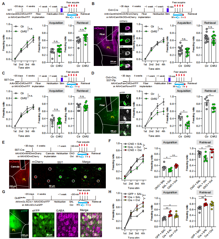
Both PL pyramidal neurons and somatostatin (SST) interneurons in the prelimbic cortex express oxytocin receptors (OxtR). In this study, researchers used optogenetic approaches to selectively activate pyramidal neurons, parvalbumin (PV) interneurons, and SST interneurons in the medial prefrontal cortex. None of these manipulations altered fear learning or memory retrieval in mice (Fig. 5A, C).

Further investigation targeting specific OxtR-expressing cell populations revealed that optogenetic activation of OxtR-positive pyramidal neurons in the PL produced no significant behavioral effects (Fig. 5B). In contrast, selective activation of PL OxtR-positive SST interneurons significantly suppressed fear acquisition and memory retrieval (Fig. 5D), whereas activation of OxtR-positive PV neurons had no effect.

Chemogenetic silencing of SST neurons in the PL reversed the fear-suppressing effects of oxytocin (Fig. 5E-F). Furthermore, conditional deletion of OxtR from PL GABAergic neurons enhanced fear-related behaviors and completely abolished the effects of oxytocin (Fig. 5G-H).

Together, these findings demonstrate that OxtR-expressing SST interneurons in the PL are indispensable functional mediators for oxytocin-dependent regulation of fear learning.

Figure 5. PL OxtR-positive SST interneurons mediate oxytocin regulation of fear learning.
 

6. Oxytocin Enhances GABAergic Inhibitory Effects of PL OxtR-Positive SST Interneurons

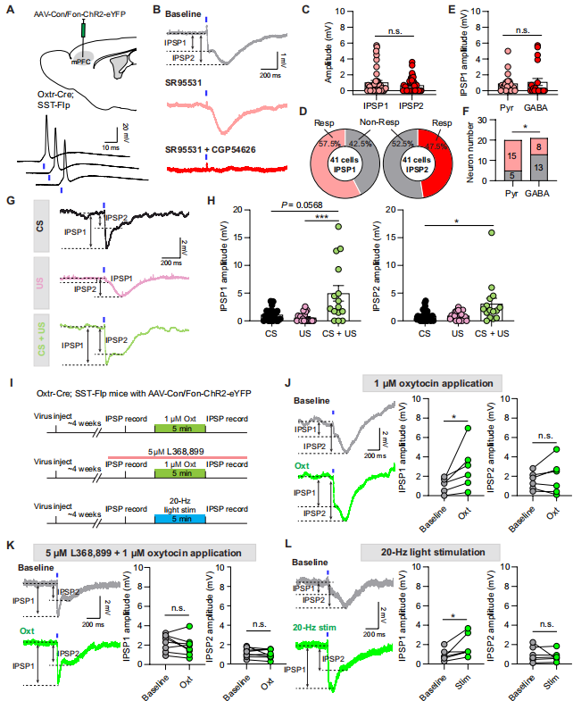
SST interneurons in the prelimbic cortex are known to bidirectionally regulate pyramidal neuron activity. In this study, researchers used Oxtr-Cre; SST mice to label OxtR-positive SST neurons. Optogenetic stimulation of these neurons induced two types of inhibitory postsynaptic potentials (IPSPs): IPSP1 (GABAA receptor-mediated) and IPSP2 (GABAB receptor-mediated) (Fig. 6A-B).

These neurons preferentially targeted pyramidal neurons, with a projection rate of approximately 75%, significantly higher than that of general GABAergic interneurons (38%) (Fig. 6C-F).

Consistent with the finding that fear learning activates OxtR-positive SST neurons (Fig. 4), synaptic analysis revealed that the paired tone–shock conditioning group (CS+US) exhibited significantly increased IPSP1 amplitude, indicating that fear learning strengthens this inhibitory pathway. No significant differences were observed in the basic electrophysiological properties of neurons among groups (Fig. 6G-H).

Application of oxytocin selectively increased the amplitude of GABAA-mediated IPSP1, and this effect was blocked by an oxytocin receptor antagonist (Fig. 6I-K). Similarly, 20 Hz optical pre-activation of OxtR-positive SST neurons also enhanced IPSP1 responses (Fig. 6I, L).

Collectively, these results demonstrate that oxytocin regulates fear learning by strengthening monosynaptic GABAergic inhibition from OxtR-positive SST interneurons onto pyramidal neurons in the prelimbic cortex.

Figure 6. Oxytocin signaling enhances GABAergic inhibition of pyramidal neurons mediated by PL OxtR-positive SST interneurons.
 

Conclusion

By integrating optogenetic manipulation, in vivo calcium imaging, and cell-type-specific electrophysiological recording, this study demonstrates that oxytocin signaling in the prelimbic cortex regulates fear processing by enhancing local inhibitory control from SST interneurons onto pyramidal neurons. These findings reveal a precise neural circuit mechanism through which oxytocin modulates fear-related behaviors and provide new insights into potential therapeutic strategies for stress-related disorders such as post-traumatic stress disorder (PTSD).


Viral Tools Used in This Study Provided by Brain Case Biotech

Brain Case Biotech provides a comprehensive range of viral vectors supporting optogenetics, chemogenetics, recombinase systems, neurotransmitter monitoring, calcium imaging, and fluorescent labeling applications.
Product Category Product ID Product Name
Optogenetics BC-0108 rAAV-EF1α-DIO-hChR2(H134R)-mCherry
BC-0819 rAAV-hSyn-DO-hChR2(H134R)-EGFP
BC-0125 rAAV-EF1α-DIO-eNpHR3.0-EYFP
BC-0100 rAAV-CaMKIIα-hChR2(H134R)-EYFP
BC-0708 rAAV-CaMKIIα-DIO-hChR2(H134R)-mCherry
BC-0356 rAAV-hSyn-Con Fon-hChR2(H134R)-EYFP
Chemogenetics BC-0143 rAAV-hSyn-DIO-hM3D(Gq)-mCherry
BC-0153 rAAV-hSyn-DIO-hM4D(Gi)-mCherry
Recombinase BC-0322 rAAV-mDlx-SV40 NLS-Cre
Neurotransmitter Sensor BC-0757 rAAV-hSyn-OT1.7
Calcium Imaging BC-0238 rAAV-hSyn-DIO-GCaMP6s
Fluorescent Proteins BC-0029 rAAV-CaMKIIα-EYFP
BC-0242 rAAV-EF1α-DIO-EYFP
BC-0025 rAAV-hSyn-DIO-mCherry
BC-0468 rAAV-CaMKIIα-DIO-mCherry
BC-0063 rAAV-hSyn-Con Fon-EYFP
BC-0936 rAAV-mDLX-eGFP

Brain Case Biotech also provides a wide range of customized viral vector services.
For more information, please contact bd@ebraincase.com


Service Type :

Select the service you'd like to purchase.

Order Information(Premade-AAVs)

Please provide us some information about the service you'd like to order.

How did you hear about BrainCase Biotech or our products?(Fill in this field to get a discount coupon)

Detailed requirements:

scroll

Order Information(Custom AAV/Lentivirus)

Please provide us some information about the service you'd like to order.

Gene ID or gene information:

Selection of the reporting gene:

Special Instructions:

scroll

Order Information(Others)

Please provide us some information about the service you'd like to order.

Virus name/Detailed requirements:

scroll

Related products

Sci. Adv.丨Could Intranasal Oxytocin Help Police Officers Prevent PTSD? Xiao Lei’s Team from Fudan Institute of Brain Science Reveals the Precise Neural Circuit Underlying Oxytocin-Mediated Suppression of Fear Learning

Sci. Adv.丨Could Intranasal Oxytocin Help Police Officers Prevent PTSD? Xiao Lei’s Team from Fudan Institute of Brain Science Reveals the Precise Neural Circuit Underlying Oxytocin-Mediated Suppression of Fear Learning

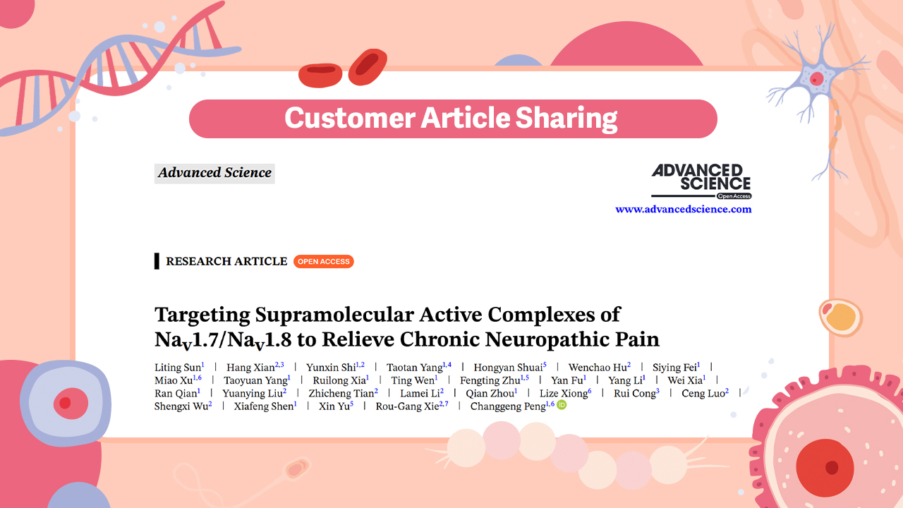
Client Article | Advanced Science | Team led by Changgeng Peng, Rougang Xie, Xin Yu, Xiafeng Shen, et al. Reveal: From Single Target to Dual Blockade—Discovery of Nav1.7/1.8 Supramolecular Complex Reshapes Chronic Pain Treatment Strategy

Client Article | Advanced Science | Team led by Changgeng Peng, Rougang Xie, Xin Yu, Xiafeng Shen, et al. Reveal: From Single Target to Dual Blockade—Discovery of Nav1.7/1.8 Supramolecular Complex Reshapes Chronic Pain Treatment Strategy

Client Articles | Neuron | Can’t sleep well under stress? Air Force Medical University team (Hailong Dong, Guangchao Zhao) reveals VTA–mPFC pathway drives sleep architecture disruption under chronic stress

Client Articles | Neuron | Can’t sleep well under stress? Air Force Medical University team (Hailong Dong, Guangchao Zhao) reveals VTA–mPFC pathway drives sleep architecture disruption under chronic stress

Customer Article | Sci. Adv. | Jinhao Sun’s Team at Shandong University Unveils: How Does the Brain “Remember” Gastric Inflammation? Hcn1-Dependent PVN Engram Neurons Are the Key!

Customer Article | Sci. Adv. | Jinhao Sun’s Team at Shandong University Unveils: How Does the Brain “Remember” Gastric Inflammation? Hcn1-Dependent PVN Engram Neurons Are the Key!

map
{dede:global.cfg_webname/}

Pre-made AAV Library

CRISPR
RNAi
Neurotropic virus
Optogenetics activation
Biosensors

News

Corporate News
New Product Launch
Test Report
Investor News

Support

Literature interpretation
Customer article
FAQs
Blog
Legal
微信

WhatsApp Business Account

Tel: +8618971215294
E-mail: BD@ebraincase.com

Address:-

Address:-

  • Copyright © 2024 Brain Case All Rights Reserved.