E-mail:BD@ebraincase.com
Tel:+8618971215294
English 中文版
Pioneering Innovation in Gene Vector Research - BrainCasePioneering Innovation in Gene Vector Research - BrainCase
  • Home
  • Pre-made AAV Library
    CRISPRRNAiHSV-helperRV-helperNeurotropic virusCalcium SensorsOptogenetics activationOptogenetics inhibitionChemical geneticsSparse labelingFluorescent proteinBiosensorsRecombinaseApoptosis & AutophagyDisease ModelNeurotoxicityOther
  • Products & Service

    Product Center

    Virus

    Vesicular Stomatitis Virus-VSV
    RCAS-TVA
    Lentivirus
    Rabies Virus-CVS Strain-RABV
    Herpes simplex virus-HSV
    Pseudorabies virus-PRV
    Adeno-Associated Virus-AAV
    Neurotropic virus list
    Baculovirus

    Kits & Regents

    EndoFree Plasmid Maxi Kit
    Lentivirus Concentration Kit
    VESSL Exosome Solutions
    AAV Rainbow Combo™

    Plasmid Construction & Vector cloning

    Lentiviral Vector
    AAV Vector
    Plasmid
    Plasmid Library

    Popular Applications

    Gene Regulation

    Gene Overexpression-Brain Case
    RNA interference(RNAi)-siRNA-Brain Case
    Gene Editing - CRSIPR cloning - BrainCase

    Neural Circuit Function Research

    Optogenetics - BrainCase
    Chemical genetics-DREADDs-Brain Case
    Calcium signal recording-Gels- Brain Case
    GRAB Neurotransmitter Fluorescent Sensor- Brain Case
    Functional Magnetic Resonance Imaging Technology- Brain Case

    Research on the structure of neural circuits

    Direct Input and Output-viral vectors- Brain Case
    Anterograde Mono-synaptic Tracing -HSV- Brain Case
    Antrograde Muti-synaptic Tracing-HSV & VSV-Brain Case
    Retrograde Mono-synaptic Tracing-Rabies Virus(CVS Strain)-Brain Case
    Retrograde Muti-synaptic Tracing-PRV-Brain Case

    Featured Services

    Experiment Service

    Research Services | Neural Circuit Tracing + Targeted Spatial Omics + Neuronal Classification Analysi
    Research Service | Cell and Tissue Patch-Clamp Recording and Analysis Services
    BrainCase Biotech| Whole-Brain Transparency Imaging and Analysis Services

    AAV Serotype Screening

    AAV Serotypes screening-gene therapy

    Collaboration Products

    Kiryl Piatkevich lab
    Yulong Li lab
    Zhifei Fu lab
    Woo-ping Ge lab

    Oncolytic Virus Modification

    Oncolytic virus-for cancer therapy-Brain Case
    Herpes Virus Vector-anti-tumor- BrainCase
    Vesicular stomatitis virus-killing tumor cells-Brain Case
  • News
    Corporate News New Product Launch Test Report Investor News
  • Support
    Literature interpretation Customer article FAQs Blog Legal
  • About Us
    Pre-made AAV Library Products & Service News Support About Us Contact
  • Contact
    Contact Us Join us
  • 中文
    English 中文版
  • Home
  • News
  • New Product Launch
  • News
  • Corporate News
  • New Product Launch
  • Test Report
  • Investor News

PRV Tracers for Peripheral Neural Circuit Tracing | Representative Applications Across Multiple Organs

Time:2026-07-03 20:44:58
Neural circuit tracing is a cornerstone technique for elucidating the mechanisms underlying nervous system function. Owing to its unique capability for retrograde transsynaptic tracing across multiple synaptic orders, pseudorabies virus (PRV) has become a gold-standard tool for mapping bidirectional neural connectivity between peripheral organs and the central nervous system, including the brain and spinal cord.Compared with intracerebral injection, PRV delivery into peripheral target tissues more closely preserves physiological neural connectivity, enabling comprehensive mapping of both afferent and efferent pathways innervating visceral organs, skin, skeletal muscle, glands, and other peripheral targets.

This article showcases a collection of representative applications of Brain Case PRV tracers for peripheral neural circuit mapping, covering a wide range of target organs, animal models, and experimental strategies.
 

I. Key Advantages of PRV-Based Peripheral Neural Circuit Tracing


▶  Highly specific retrograde transsynaptic labeling
PRV is transported exclusively in the retrograde direction along neuronal pathways, enabling sequential labeling of upstream neurons and precise identification of central nuclei innervating peripheral target organs.

▶  Broad compatibility with peripheral tissues
PRV can be efficiently delivered into a wide range of peripheral tissues, including skeletal muscle, visceral organs, skin, joints, spleen, intestine, and heart, providing robust and reproducible infection efficiency.

▶  Multiple fluorescent reporter options
PRV tracers expressing EGFP or mCherry are available, facilitating immunofluorescence co-immunostaining and colocalization analyses.

▶  Compatible with multiple animal models
PRV tracing systems have been successfully applied in commonly used laboratory animals, including mice, rats, and guinea pigs.
 

II. Representative Applications of PRV Tracing in Peripheral Tissues


Part 1. Mammary Tumor | Brain–Tumor Neural Circuit

Published: The Journal  of Clinical Investigation (2023)
Research group: Guangyan Wu / Yi Zhang, Army Medical University
Virus: BC-PRV-531 (PRV-CAG-EGFP, green fluorescence)
Experimental procedure:
PRV was injected directly into 4T1 mammary tumors (total volume: 1 μL; 2 × 10⁸ PFU/mL). Six days after injection, whole-brain sections revealed PRV-EGFP-positive neurons distributed throughout multiple central regions, including the intermediolateral cell column (IML) of the spinal cord, periaqueductal gray (PAG), gigantocellular reticular nucleus (PGi), paraventricular nucleus of the hypothalamus (PVN), and the medial subdivision of the central amygdala (CeM), delineating a complete neural pathway linking peripheral tumors with the brain. Immunofluorescence co-immunostaining further demonstrated that the vast majority of PRV-labeled neurons within the CeM co-expressed corticotropin-releasing hormone (CRH).
Figure 1. Retrograde PRV tracing of newly established sympathetic innervation in 4T1 mammary tumors
Original link: https://www.jci.org/articles/view/167725

Part 2. Spleen | Dorsal Root Ganglion–Spleen Neuroimmune Circuit

Published: Cell (2024)
Research group: Huiyan Li / Yucheng Zhang / Tao Zhou, Academy of Military Medical Sciences
Virus: BC-PRV-531 (PRV-CAG-EGFP, green fluorescence)
Experimental procedure:
PRV was microinjected into multiple sites of the mouse spleen (total volume: 3 μL; 5 × 10⁹ PFU/mL). Following tissue collection, whole-mount immunostaining was performed on the dorsal root ganglia (DRGs) and nodose ganglia (NGs). Retrograde tracing demonstrated that TRPV1-positive nociceptive neurons innervating the spleen were predominantly localized to the left T8–T13 DRGs. Most PRV-positive neurons also expressed calcitonin gene-related peptide (CGRP), identifying the spinal sensory origin of splenic sensory innervation.
Figure 2. Splenic nociceptive sensory neurons predominantly originate from the left T8–T13 dorsal root ganglia
Original link: https://www.sciencedirect.com/science/article/pii/S0092867424004537

Part 3. Heart | Superior Cervical Ganglion–Cardiac Sympathetic Circuit

Published: Journal of Controlled Release (2025)
Research group: Chao Xie; Hanbing Wang
Virus: BC-PRV-531 (PRV-CAG-EGFP, green fluorescence)
Experimental procedure:
PRV was microinjected into multiple regions of the mouse heart, including the apex, the free wall of the left ventricle, the anterior and posterior walls of the left ventricle, and the interventricular septum (100 nL per injection site). Retrograde transsynaptic tracing demonstrated a sympathetic neural circuit connecting the heart and the superior cervical ganglion (SCG). PRV-positive sympathetic neurons were readily detected within the SCG, providing direct evidence that the SCG serves as a critical peripheral sympathetic relay mediating the exacerbation of myocardial ischemia–reperfusion injury under chronic pain conditions.
Figure 3. Retrograde multisynaptic PRV tracing of cardiac sympathetic circuits
Original link: https://www.sciencedirect.com/science/article/abs/pii/S0168365925000306
 

Part 4. Femoral Bone Marrow | Brain–Bone Stress Neural Circuit

Published: Science Advances (2025)
Research group: Air Force Medical University; Xueyu Hu / Yulin Dong
Virus: BC-PRV-531 (PRV-CAG-EGFP, green fluorescence)
Experimental procedure:
PRV was microinjected into the femoral bone marrow cavity of mice (total volume: 2 μL; ≥2 × 10⁸ PFU/mL). Following injection, the bone entry site was sealed with bone wax. Whole-brain sections were collected for analysis 6 days after injection. Retrograde transsynaptic tracing identified abundant PRV-positive neurons in multiple brain regions, including the locus coeruleus (LC), central amygdala (CeA), and paraventricular nucleus of the hypothalamus (PVN). PRV-labeled neurons in the LC colocalized with tyrosine hydroxylase (TH), whereas a large proportion of labeled neurons in the CeA co-expressed corticotropin-releasing hormone (CRH). These findings delineated the central-to-peripheral sympathetic circuit involving the CeA–LC–PVN axis that regulates skeletal tissue.

Figure 4. Retrograde PRV tracing of sympathetic neural circuits
Original link: https://www.science.org/doi/10.1126/sciadv.adz9329


Part 5. Kidney | Brainstem–Kidney Sympathetic Circuit Regulating Blood Pressure

Published: Advanced Science (2025)
Research group: Nanjing University; Jingning Zhu / Xiaoyang Zhang
Virus: BC-PRV-531 (PRV-CAG-EGFP, green fluorescence)
Experimental procedure:
PRV was microinjected into multiple sites within the left renal parenchyma of rats, including the rostral, middle, and caudal regions (0.8 μL per injection site). Brainstem sections were collected 4 days after injection following transcardial perfusion. Retrograde tracing labeled GAD65/67-positive inhibitory presympathetic neurons in the rostral ventromedial medulla (RVMM). A large proportion of PRV-positive neurons also expressed histamine H4 receptor (H4R), providing direct evidence that GABAergic neurons in the RVMM serve as a key upstream sympathetic center regulating renal sympathetic outflow and mediating antihypertensive effects.

Figure 5. Retrograde tracing of inhibitory and excitatory sympathetic efferent pathways from the RVMM to the kidney
Original link: https://advanced.onlinelibrary.wiley.com/doi/10.1002/advs.202508176


Part 6. Lacrimal Gland | Locus Coeruleus–Superior Cervical Ganglion–Lacrimal Gland Sympathetic Circuit

Published:  Nature Communications (2025)
Research group: Qingdao Eye Hospital, Shandong First Medical University; Lixin Xie / Qingjun Zhou
Viruses:
         ▶   BC-PRV-531 (PRV-CAG-EGFP, green fluorescence)
         ▶   BC-PRV-724 (PRV-CAG-mRFP, red fluorescence)
Experimental procedure:
Green fluorescent PRV was microinjected into multiple sites of the unilateral lacrimal gland (total volume: 1 μL; 5 × 10⁹ PFU/mL), while red fluorescent PRV was simultaneously injected into the superior cervical ganglion (SCG) (total volume: 1 μL; 1 × 10⁹ PFU/mL). Animals were perfused at different time points between 1 and 4 days after injection, followed by whole-brain sectioning. Dual-fluorescence colocalization signals were predominantly detected in the locus coeruleus (LC), nucleus tractus solitarius (NTS), and paraventricular nucleus of the hypothalamus (PVN), whereas neurons labeled exclusively with GFP were observed in the superior salivatory nucleus (SSN). These results demonstrated that the LC serves as a major upstream sympathetic center regulating lacrimal gland innervation. Most PRV-labeled neurons were also positive for TH.

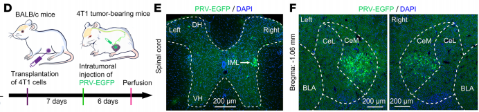
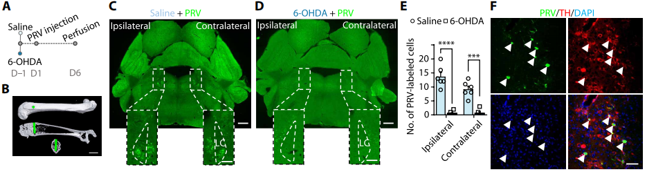
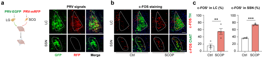
Figure 6. Brain regions providing noradrenergic projections to the lacrimal gland
Original link: 
https://www.nature.com/articles/s41467-025-60476-z

Part 7. Lung | Hypothalamic PVN–Lung Sympathetic Circuit in Acute Lung Injury

Published: Nature Communications (2025)
Research group: The Second Xiangya Hospital, Central South University; Ruping Dai / Hui Li

Virus: BC-PRV-531 (PRV-CAG-EGFP, green fluorescence)
Experimental procedure:
PRV was microinjected into multiple sites of the left lung lobe in mice (total volume:
1 μL). Six days after injection, spinal cord and whole-brain sections were collected following transcardial perfusion. 
Retrograde tracing identified abundant CRH-expressing PRV-positive neurons in the paraventricular nucleus of the hypothalamus (PVN). PRV-positive neurons were also detected throughout the spinal cord and brainstem, providing direct anatomical evidence for a multisynaptic central sympathetic pathway projecting from the PVN to the lung.


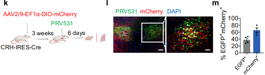
Figure 7. Retrograde PRV tracing in Crh-IRES-Cre mice
Original link: https://www.nature.com/articles/s41467-025-63953-7

Part 8. Small Intestine | Brain–Vagus–Gut Neural Circuit

Published: Cell Stem Cell (2025)
Research group: Shanghai Institute of Nutrition and Health, Chinese Academy of Sciences; Jun Qin; Shanghai Jiao Tong University; Qi Han; Tongji University; Moubin Lin; Shanghai Institute of Immunity and Infection, Chinese Academy of Sciences; Xiao Su

Virus: BC-PRV-531 (PRV-CAG-EGFP, green fluorescence)
Experimental procedure:
PRV was microinjected into three sites of the mouse duodenum (total volume:
2 μL). Seventy-two hours after injection, tissues were collected following transcardial perfusion. Sections of the intestine, celiac ganglion, spinal cord, and whole brain were prepared for analysis. 
Retrograde transsynaptic tracing labeled multiple orders of upstream neurons innervating the intestine. Numerous PRV-positive neurons were identified in the dorsal motor nucleus of the vagus (DMV), intermediolateral cell column (IML) of the spinal cord, and celiac ganglion, delineating the complete brain–vagus–gut neural circuit underlying chronic stress–mediated regulation of intestinal stem cells.


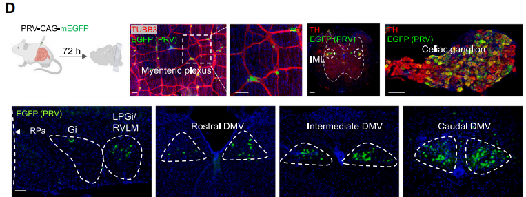
Figure 8. Retrograde transsynaptic PRV tracing delineates the neural circuitry connecting the brain and the intestine
Original link: 
https://www.sciencedirect.com/science/article/abs/pii/S1934590925000840

Part 9. Tibialis Anterior and Gastrocnemius Muscles | Hypothalamus–Pons–Spinal Cord Motor Circuit

Published: Nature Communications (2025)
Research group: Institute of Neuroscience, Chinese Academy of Sciences; Yi Li; Nanjing Medical University; Weihua Cai

Virus: BC-PRV-531 (PRV-CAG-EGFP, green fluorescence)
Experimental procedure:
PRV was microinjected into multiple sites of the
tibialis anterior and gastrocnemius muscles in mice. Three injection sites were established within each muscle at a depth of 0.5–1.0 mm, with 1 μL administered per site. Whole-brain clearing and imaging were performed 5.5 days after injection. 
PRV-EGFP-positive neurons were highly enriched in the caudal lateral hypothalamic area (cLHA) and were broadly distributed throughout the oral pontine reticular nucleus (PnO) and the intermediolateral cell column (IML) of the spinal cord. Retrograde tracing reconstructed the multisynaptic motor circuit extending from peripheral hindlimb muscles to the hypothalamus. Three-dimensional reconstruction further demonstrated that glutamatergic neurons in the cLHA constitute a major upstream neuronal population controlling hindlimb motor function.


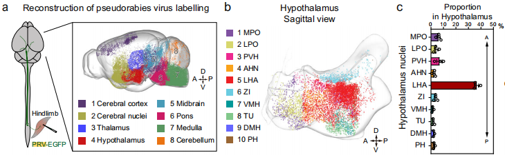
Figure 9. Retrograde labeling of upstream neurons innervating the tibialis anterior and gastrocnemius muscles
Original link: 
https://www.nature.com/articles/s41467-025-67133-5


Part 10. Tibialis Anterior Muscle | Reconstruction of Motor Circuits Following Thoracic Spinal Cord Organoid Transplantation

Published: Nature Biomedical Engineering (2025)
Research group: Tongji University School of Medicine; Rongrong Zhu / Liming Cheng
Virus: 
BC-PRV-803 (PRV-CAG-3Ms, red fluorescence)
Experimental procedure:
PRV was microinjected into four sites of the mouse tibialis anterior muscle (0.75 μL per site; 2 × 10⁹ PFU/mL). Spinal cord and whole-brain sections were collected 7 days after injection. PRV-positive signals were detected both within the transplanted engineered thoracic spinal cord organoids (enTsOrg) and in the host thoracic spinal cord ventral horn, demonstrating that the transplanted organoids established functional retrograde motor circuits with the hindlimb skeletal muscles. Combined with anterograde biotinylated dextran amine (BDA) tracing, these findings further confirmed reconstruction of a three-tier neural pathway connecting the host brain, transplanted organoids, and peripheral skeletal muscles.


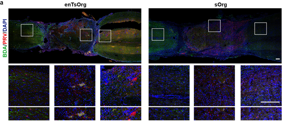
Figure 10. Neuronal tracing using combined BDA anterograde tracing and PRV retrograde tracing
Original link: 
https://www.nature.com/articles/s41551-025-01549-8

Part 11. Knee Synovium and Articular Cartilage | Brain–Joint Neural Circuit

Published: Annals of the Rheumatic Diseases (2025)
Research group: Bone and Joint Rehabilitation Center, Yangzhi Rehabilitation Hospital, Tongji University; Jian Luo
Virus: 
BC-PRV-531 (PRV-CAG-EGFP, green fluorescence)
Experimental procedure:
PRV was microinjected into the mouse knee joint cavity (2 μL; 7 × 10⁹ PFU/mL) at a depth of 1.0–2.0 mm. Whole-brain and spinal cord sections were collected 96 hours after injection. PRV-EGFP-positive neurons were highly enriched in the paraventricular nucleus of the hypothalamus (PVN) and were also distributed throughout multiple upstream nuclei in the brainstem and spinal cord. Immunofluorescence co-immunostaining demonstrated extensive colocalization of PRV-labeled neurons with CRH in the PVN, providing direct anatomical evidence for a multisynaptic sympathetic pathway connecting PVN CRH-expressing neurons with the knee joint synovium.


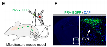
Figure 11. Retrograde transsynaptic labeling of upstream neurons innervating the knee joint
Original link: 
https://ard.eular.org/article/S0003-4967(25)04597-2/abstract

Contact Us
At the same time, Brain Case Biotech also offers a range of customized services. Please contact 
bd@ebraincase.com for more information.

Service Type :

Select the service you'd like to purchase.

Order Information(Premade-AAVs)

Please provide us some information about the service you'd like to order.

How did you hear about BrainCase Biotech or our products?(Fill in this field to get a discount coupon)

Detailed requirements:

scroll

Order Information(Custom AAV/Lentivirus)

Please provide us some information about the service you'd like to order.

Gene ID or gene information:

Selection of the reporting gene:

Special Instructions:

scroll

Order Information(Others)

Please provide us some information about the service you'd like to order.

Virus name/Detailed requirements:

scroll

Related products

PRV Tracers for Peripheral Neural Circuit Tracing | Representative Applications Across Multiple Organs

PRV Tracers for Peripheral Neural Circuit Tracing | Representative Applications Across Multiple Organs

Nat. Neurosci.: First Red Fluorescent Acetylcholine Sensor Enables Multiplex In Vivo Imaging of Neurotransmitters

Nat. Neurosci.: First Red Fluorescent Acetylcholine Sensor Enables Multiplex In Vivo Imaging of Neurotransmitters

Fluorescent “superheroes”! mBaoJin + mScarlet3-H (also known as mYongHong) Tool Viruses Empower Super-Resolution and High-Efficiency Imaging

Fluorescent “superheroes”! mBaoJin + mScarlet3-H (also known as mYongHong) Tool Viruses Empower Super-Resolution and High-Efficiency Imaging

Nat Biomed Eng | Woo-ping Ge and Wenzhi Sun’s teams develop the rAAV-miniBEND system: Breaking through the challenge of brain endothelial cell gene delivery and enabling cerebrovascular malformation modeling  !

Nat Biomed Eng | Woo-ping Ge and Wenzhi Sun’s teams develop the rAAV-miniBEND system: Breaking through the challenge of brain endothelial cell gene delivery and enabling cerebrovascular malformation modeling !

map
{dede:global.cfg_webname/}

Pre-made AAV Library

CRISPR
RNAi
Neurotropic virus
Optogenetics activation
Biosensors

News

Corporate News
New Product Launch
Test Report
Investor News

Support

Literature interpretation
Customer article
FAQs
Blog
Legal
微信

WhatsApp Business Account

Tel: +8618971215294
E-mail: BD@ebraincase.com

Address:-

Address:-

  • Copyright © 2024 Brain Case All Rights Reserved.