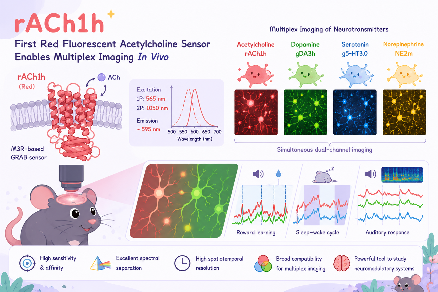
Nat. Neurosci.: First Red Fluorescent Acetylcholine Sensor Enables Multiplex In Vivo Imaging of Neurotransmitters
Time:2026-07-01 15:38:30
Acetylcholine (ACh), the first neurotransmitter ever identified, plays indispensable roles in both the central and peripheral nervous systems. It is critically involved in regulating attention, motor control, learning and memory, and the sleep–wake cycle, while also contributing to higher-order brain functions such as motivation, reward processing, decision-making, and emotion. In recent years, several green fluorescent genetically encoded ACh sensors—including GRABACh2.0, GRABACh3.0, and iAChSnFR—have been developed and widely adopted, allowing researchers to visualize ACh dynamics in real time. However, accumulating evidence suggests that ACh rarely functions in isolation. Instead, it acts as an integral component of the brain's neuromodulatory network, often operating in concert with other neurotransmitters such as dopamine (DA) and serotonin (5-HT). Understanding the dynamic interplay between ACh and other neurochemical signals therefore requires multicolor fluorescent sensors capable of simultaneously monitoring multiple neurotransmitters. Unfortunately, all currently available ACh sensors are based on green fluorescent proteins. At the same time, most widely used genetically encoded sensors for dopamine (DA), serotonin (5-HT), norepinephrine (NE), and calcium also emit within the green spectral range. This spectral overlap has long prevented researchers from simultaneously monitoring ACh together with other neurochemical signals in the same brain region of the same animal at the same time, significantly limiting investigations into the coordinated actions of multiple neuromodulatory systems.
On June 16, 2026, a collaborative team led by Prof. Yulong Lifrom the School of Life Sciences, Peking University, the IDG/McGovern Institute for Brain Research at Peking University, and the PKU–THU Center for Life Sciences, together with Prof. Changwei Weifrom Beijing Chaoyang Hospital, Capital Medical University, published their study entitled "Red-shifted GRAB acetylcholine sensors for multiplex imaging in vivo" in Nature Neuroscience.
In this work, the researchers developed the first high-performance red fluorescent genetically encoded ACh sensor, enabling simultaneous dual-channel in vivo imaging of ACh together with dopamine (DA), serotonin (5-HT), norepinephrine (NE), and other neuromodulatory signals across multiple spatial scales. This breakthrough provides a powerful new tool for dissecting the coordinated interactions among neuromodulatory systems in the living brain.
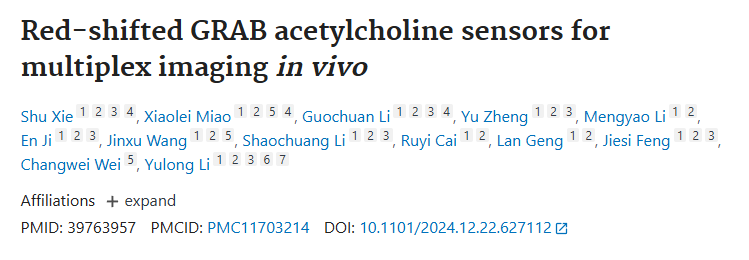
Building upon their previously established GRAB (GPCR-Activation-Based) sensor platform, Li and colleagues engineered a chimeric scaffold by combining human and mouse M3 muscarinic acetylcholine receptors (M3Rs). A circularly permuted red fluorescent protein (cpmApple) was inserted into the receptor's third intracellular loop (ICL3). Through systematic optimization of the insertion site, receptor scaffold, and fluorescent module via extensive mutagenesis and screening, the team successfully developed a high-affinity red fluorescent ACh sensor, designated rACh1h.
Optically, rACh1h exhibits a one-photon excitation peak at 565 nm and two-photon excitation peaks at 1050 nm, providing excellent spectral separation from commonly used green fluorescent sensors. In cultured cells, rACh1h demonstrated efficient plasma membrane localization and produced a fluorescence response to 100 μM ACh that was five times greater than that of gACh3.0, with an apparent affinity of approximately 400 nM.
Compared with existing green fluorescent ACh sensors, rACh1h delivers superior fluorescence signal-to-noise ratio and higher affinity, making it a high-performance tool for simultaneously monitoring the dynamics of multiple neurotransmitters in vivo.
Figure 1. Development of the rACh1h Red Fluorescent Acetylcholine Sensor
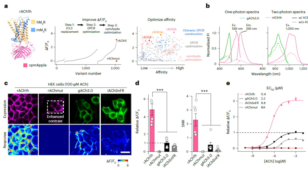
To evaluate whether rACh1h could reliably detect endogenous ACh release in vivo, the researchers combined the sensor with optogenetic stimulation and successfully recorded endogenous ACh dynamics in the mouse brain. The specificity of the fluorescence signals was further confirmed through pharmacological validation.
Benefiting from its excellent sensitivity and robust performance, rACh1h not only enabled reliable detection of evoked ACh release, but also provided sensitive and stable monitoring of spontaneous cholinergic activity in freely behaving mice.
These results demonstrate that rACh1h is capable of faithfully reporting endogenous acetylcholine dynamics under both experimentally evoked and physiological conditions, establishing it as a powerful tool for monitoring cholinergic signaling in the living brain.
Figure 2. rACh1h Enables Detection of Endogenous Acetylcholine Release In Vivo
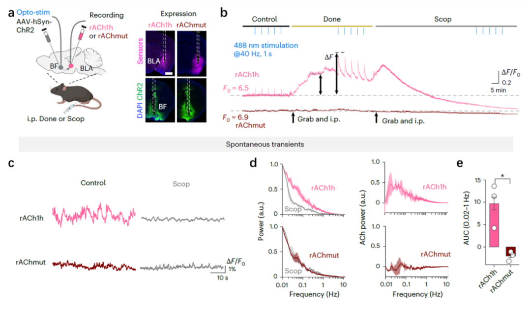
The most significant advance offered by rACh1h is its ability to enable simultaneous imaging of ACh alongside other neurotransmitters. To demonstrate this capability, the researchers co-expressed the red fluorescent rACh1h sensor with the green fluorescent dopamine sensor gDA3h in the basolateral amygdala (BLA) of mice and simultaneously monitored ACh and DA dynamics during reward learning.
The recordings revealed that the release patterns of both neurotransmitters evolved as learning progressed. Initially, ACh and DA responses were primarily evoked by the delivery of the sucrose reward itself. As learning was acquired, however, both signals gradually shifted to respond to the auditory cue predicting reward, reflecting the neural encoding of reward-predictive stimuli.
The researchers further combined rACh1h with the green fluorescent serotonin sensor g5-HT3.0 to perform long-term simultaneous recordings of ACh and serotonin throughout the sleep–wake cycle.The results showed that the two neuromodulators exhibited similar activity patterns during wakefulness and non-rapid eye movement (NREM) sleep. In contrast, during rapid eye movement (REM) sleep, their dynamics diverged markedly: ACh levels increased, whereas serotonin levels decreased.
These findings suggest that distinct neuromodulatory systems play different functional roles during transitions between sleep states. More importantly, they highlight the unique capability of dual-color imaging for dissecting the coordinated dynamics of multiple neurotransmitter systems in the living brain.
Figure 3. Simultaneous Imaging of Acetylcholine and Other Neurotransmitters Using rACh1h
In addition to its outstanding performance in fiber photometry, rACh1h also demonstrated excellent compatibility with wide-field imaging, enabling high spatiotemporal resolution monitoring of ACh dynamics across multiple brain regions.
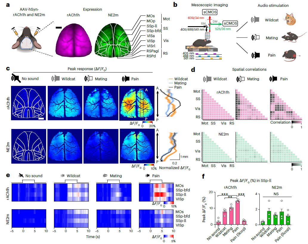
To evaluate its performance in large-scale imaging applications, the researchers co-expressed rACh1h with the green fluorescent norepinephrine sensor NE2m in the mouse dorsal cortex. Combined with a wide-field fluorescence imaging system, this approach enabled real-time visualization of ACh and norepinephrine (NE) dynamics across multiple cortical areas simultaneously.
Using an auditory stimulation paradigm, mice were presented with recordings of cat vocalizations, mouse mating calls, and mouse distress vocalizations. Distinct spatial activity patterns and dynamic response profiles of ACh and NE were observed across different cortical regions in response to these behaviorally relevant sounds.
These results demonstrate that rACh1h, together with NE2m, enables reliable dual-color imaging of neurotransmitter dynamics across the cortex, providing a powerful approach for investigating how multiple neuromodulatory systems coordinate sensory information processing in the brain.
Figure 4. Dual-Color Imaging with rACh1h and NE2m Reveals Cortical Neurotransmitter Dynamics
In summary, the Li laboratory has successfully expanded the spectral repertoire of genetically encoded acetylcholine sensors by developing the first red fluorescent ACh sensor, rACh1h. The sensor enables highly sensitive, highly specific, and high spatiotemporal resolution monitoring of ACh dynamics in vivo.
By overcoming the spectral limitations of existing green fluorescent ACh sensors, rACh1h can be readily combined with a broad range of green fluorescent sensors to simultaneously monitor the dynamics of ACh alongside other neurochemical signals. This capability provides researchers with a powerful platform for investigating how multiple neuromodulatory systems interact to shape brain function and behavior.
As a versatile tool for multiplex neurochemical imaging, rACh1h opens new opportunities for dissecting the coordinated actions of neurotransmitter systems across diverse physiological and behavioral processes, advancing our understanding of the neural mechanisms underlying complex brain functions.
For technical consultation, custom vector design, or viral packaging services,
please contact:bd@ebraincase.com
Service Type :
Select the service you'd like to purchase.
Order Information(Premade-AAVs)
Please provide us some information about the service you'd like to order.
Order Information(Custom AAV/Lentivirus)
Please provide us some information about the service you'd like to order.
Order Information(Others)
Please provide us some information about the service you'd like to order.