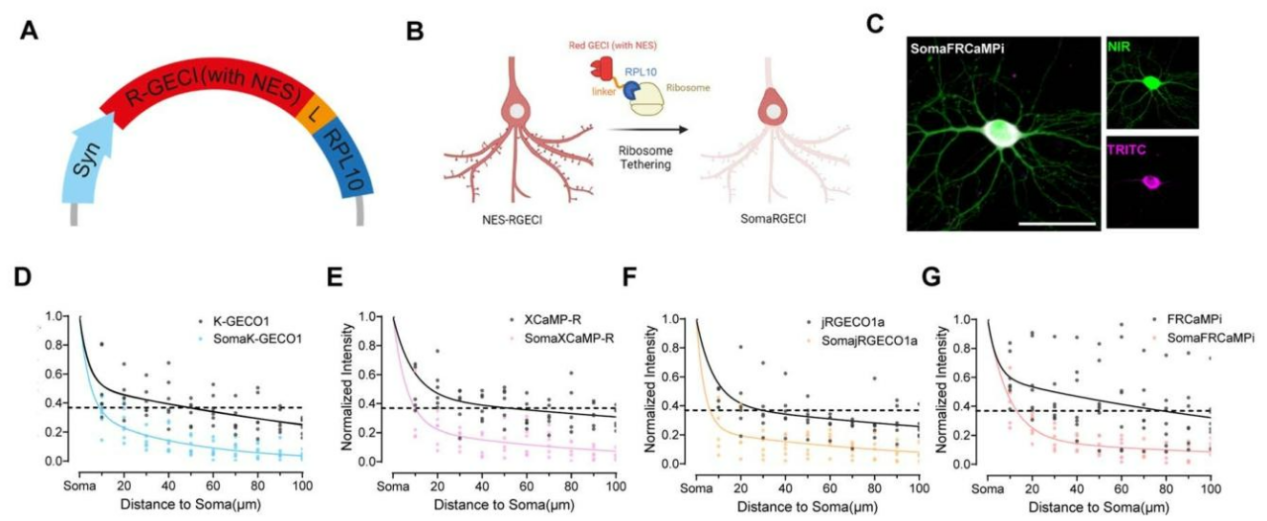
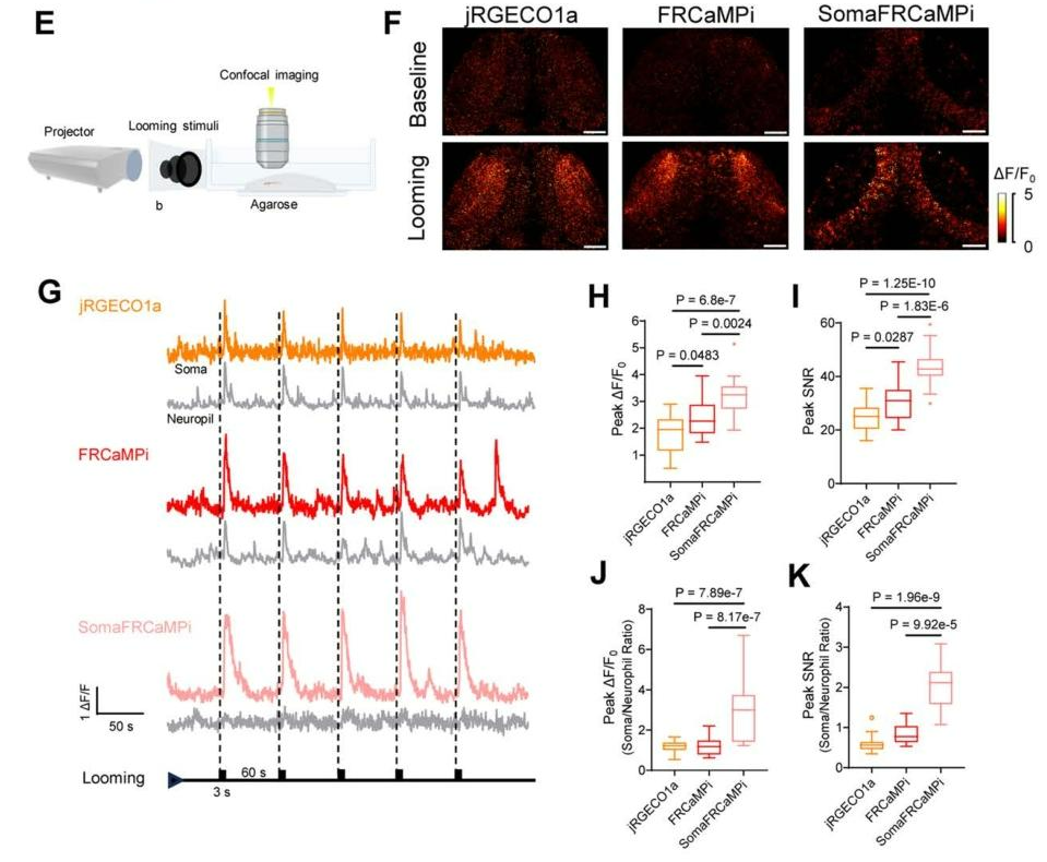
✅ Superior Long-Term Stability
When expressed in the mouse brain for up to four months, SomaFRCaMPi exhibited substantially less signal decay than jRGECO1a, demonstrating excellent long-term stability for chronic in vivo imaging experiments.
✅ Compatible with Dual-Color Imaging
SomaFRCaMPi can be readily combined with the green soma-targeted calcium indicator RiboL1-GCaMP8s for dual-color imaging. The two indicators exhibit comparable Z-score signal-to-noise ratios, while SomaFRCaMPi provides lower baseline noise, enabling more reliable simultaneous imaging of distinct neuronal populations.
✅ Optimized for Multiple Imaging Modalities
SomaFRCaMPi is compatible with a wide range of imaging platforms, including widefield microscopy, two-photon microscopy, and fiber photometry, making it suitable for diverse neuroscience applications.
Notably, in densely labeled brain regions where 80–90% of neurons express the indicator, SomaFRCaMPi demonstrated remarkable performance improvements over FRCaMPi:
🌟 3.3-fold more detectable neuronal ROIs
🌟 50% increase in detected calcium events
🌟 Up to 40% higher signal-to-noise ratio
🌟 30% reduction in long-range fluorescence crosstalk
These improvements enable more accurate extraction of neuronal activity from densely populated neural circuits.
Recommended Applications
🌟 High-density, single-cell calcium imaging in complex brain regions
🌟 Dual-color calcium imaging
🌟 Long-term in vivo recording of neuronal circuit activity
Current Limitation
Because SomaFRCaMPi is derived from the mApple fluorescent protein scaffold, it remains susceptible to blue-light excitation. Therefore, its compatibility with blue-light optogenetic actuators requires further validation before broad application in all-optical experimental paradigms.
Breakthrough II: PinkyCaMP — An Ultra-Bright, Photoswitching-Free Red Calcium Indicator
On April 24, 2026, researchers from the University of Cologne, in collaboration with the University of Tokyo, Ruhr University Bochum, and several other institutions, published "PinkyCaMP: an mScarlet-based calcium sensor with enhanced brightness, photostability and multiplexing capabilities" in Nature Methods.
Built on the mScarlet fluorescent protein scaffold, PinkyCaMP is a next-generation red genetically encoded calcium indicator that delivers exceptional brightness, superior photostability, and photoswitching-free performance, with overall imaging capabilities approaching those of state-of-the-art GCaMP indicators.
Key Innovation
PinkyCaMP is the first red genetically encoded calcium indicator engineered from the mScarlet fluorescent protein. By leveraging the intrinsic properties of mScarlet, the sensor combines exceptional molecular brightness, minimal protein aggregation, and complete resistance to blue-light-induced photoswitching, overcoming several longstanding limitations of previous red GECIs.
Key Advantages
✅ Exceptional Brightness
PinkyCaMP delivers outstanding fluorescence brightness, exceeding that of existing red GECIs. Its baseline fluorescence is more than twice as bright as jRGECO1a and RCaMP3, and approximately 14-fold brighter than jRCaMP1a, providing significantly improved image quality, particularly for deep-tissue and low-signal imaging applications.
✅ Superior Photostability
PinkyCaMP exhibits remarkable resistance to photobleaching, with a photobleaching half-life more than twice that of RCaMP3. This enhanced photostability enables prolonged imaging sessions while maintaining consistent fluorescence intensity, making it well suited for long-term in vivo imaging experiments.
✅ Photoswitching-Free Performance
Unlike many mApple-based red calcium indicators, PinkyCaMP does not undergo blue-light-induced photoswitching. This unique property allows seamless integration with blue-light optogenetic actuators, including CoChR and stGtACR2, without introducing spectral crosstalk or imaging artifacts.
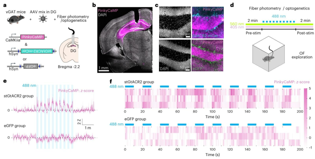 Figure 3. PinkyCaMP enables seamless integration with blue-light optogenetic tools.
✅ Excellent Compatibility for Dual-Color Imaging
PinkyCaMP is fully compatible with multiple green fluorescent indicators, including GCaMP8s and sDarken, enabling simultaneous monitoring of neuronal activity and complementary physiological signals. This compatibility supports neuronal subtype identification as well as synchronized calcium and neurotransmitter imaging within the same preparation.
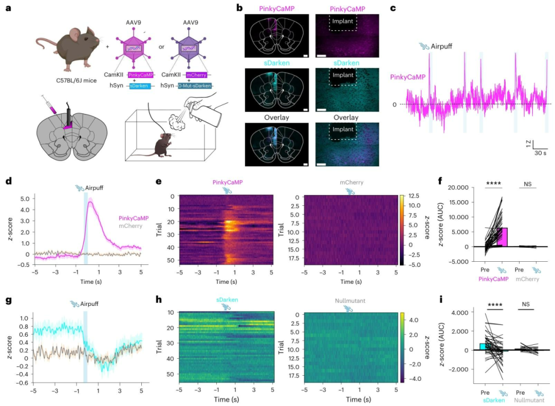 Figure 4. Simultaneous monitoring of neuronal activity and serotonin dynamics using PinkyCaMP and the green fluorescent serotonin sensor sDarken.
✅ Broad Compatibility Across Imaging Modalities
PinkyCaMP performs robustly across multiple imaging platforms, including fiber photometry, one-photon miniature microscopy (miniscopes), and two-photon imaging in awake animals. Its exceptional brightness is particularly advantageous for deep-tissue imaging, where photon collection is inherently limited.
✅ Safe for Long-Term Expression
PinkyCaMP exhibits excellent biocompatibility, with no detectable lysosomal accumulation, no Ca²⁺-dependent puncta formation, and no observable cytotoxicity after at least 90 days of expression, supporting its use in long-term functional imaging studies.
Recommended Applications
🌟 All-optical experiments combining calcium imaging and optogenetics
🌟 Dual-color functional imaging
🌟 Deep-brain and long-term in vivo imaging
🌟 Fiber photometry and other multimodal imaging applications
Current Limitation
Compared with green GECIs such as GCaMP, PinkyCaMP displays relatively slower kinetics, with a half-rise time of approximately 670 ms and a half-decay time of approximately 5.6 s. Consequently, it is less suitable for resolving high-frequency action potential firing with high temporal precision.
PinkyCaMP vs. SomaFRCaMPi: Which Red GECI Is Right for Your Research?
| Feature |
PinkyCaMP |
SomaFRCaMPi |
| Fluorescent Protein Scaffold |
mScarlet |
mApple |
| Excitation / Emission Peak |
568 nm / 600 nm |
560 nm / 590 nm |
| Ca²⁺ Affinity (Kd) |
54 nM |
81 nM |
| Dynamic Range (ΔF/F) |
15.1-fold |
16.3-fold |
| Single-AP Response (ΔF/F₀) |
~0.4 |
1.74 |
| Baseline Brightness |
High (≈14× brighter than jRCaMP1a) |
Moderate (comparable to jRGECO1a) |
| Soma Targeting |
No |
Red soma-targeted |
| Neuropil Contamination |
Present |
Significantly reduced |
| Photostability |
Excellent (>2× longer than RCaMP3) |
Moderate (~2× photobleaching rate of jRGECO1a) |
| Photoswitching |
No blue-light photoswitching |
Present (potential risk associated with the mApple scaffold) |
| Kinetics (Half-decay Time) |
Slow (~5.6 s) |
Moderate (~1.95 s) |
| Optimal Activity Range |
Low-frequency / sustained activity |
Low- to medium-frequency activity |
| High-Frequency Spike Resolution |
Not recommended |
Capable of resolving spike onset |
| Signal-to-Noise Ratio (SNR) |
Significantly improved over jRGECO1a |
10–40% higher sensitivity than FRCaMPi under dense labeling |
| Responsive Neurons Detected |
— |
~50% more responsive neurons than jRGECO1a |
| Compatibility with Blue-Light Optogenetics |
Fully compatible |
Limited; suitable for multicolor imaging |
| Recommended Applications |
Multicolor imaging, all-optical experiments, long-term imaging, maximum brightness |
Soma-specific imaging, single-cell segmentation, applications requiring minimal neuropil contamination |
🎯Choosing the Right Red GECI
Choose PinkyCaMP if you need:
💎 Maximum fluorescence brightness
💎 Outstanding photostability for long-term imaging
💎 Seamless compatibility with blue-light optogenetics
💎 Dual-color imaging with green fluorescent sensors
💎 Deep-tissue imaging and multimodal imaging applications
Choose SomaFRCaMPi if you need:
💎 Soma-specific labeling with minimal neuropil contamination
💎 High-precision single-cell segmentation in densely labeled brain regions
💎 Higher sensitivity for detecting neuronal activity
💎 Chronic in vivo imaging of neuronal populations with improved cellular resolution
Looking for Viral Vectors Expressing These Next-Generation Red GECIs?
At eBrainCase, we provide high-quality AAV tool viruses for cutting-edge neuroscience research. Viral vectors expressing PinkyCaMP, SomaFRCaMPi, and other genetically encoded indicators are available to support a broad range of applications, including in vivo calcium imaging, fiber photometry, two-photon imaging, multicolor imaging, and optogenetics.
Whether your research requires ultra-bright fluorescence, soma-specific labeling, or advanced all-optical experimental designs, our viral vector solutions are designed to accelerate your neuroscience discoveries.
The viral tools used in this study are available from Brain Case Biotech
| Product Category |
No. |
Product Name |
| PinkyCaMP |
BC-4412 |
rAAV-hSyn-DIO-PinkyCaMP-WPRE-hGH polyA |
| BC-4411 |
rAAV-hSyn-PinkyCaMP-WPRE-hGH polyA |
| SomaFRCaMPi |
BC-4389 |
rAAV-hSyn-DIO-NES-SomaFRCaMPi |
| BC-4183 |
rAAV-CAG-DIO-NES-SomaFRCaMPi |
| BC-4182 |
rAAV-hSyn-NES-SomaFRCaMPi |
For technical consultation, custom vector design, or viral packaging services,
please contact:bd@ebraincase.com