- E-mail:BD@ebraincase.com
- Tel:+8618971215294

Optic neuropathies are characterized by the progressive loss of retinal ganglion cells (RGCs) and degeneration of the optic nerve. They represent an important cause of irreversible vision loss and include conditions associated with glaucoma, inflammation, ischemia, trauma, and genetic abnormalities.
Adeno-associated virus (AAV) vectors provide a versatile platform for manipulating gene expression in RGCs. However, commonly used broad promoters such as CAG and CMV may drive expression in multiple retinal cell types. For applications requiring selective manipulation of RGCs, a promoter capable of restricting transgene expression to the target cell population is therefore highly desirable.
A study published in The Journal of Neuroscience identified the mouse γ-synuclein (mSncg) promoter as a potent RGC-specific promoter. When combined with AAV2 and intravitreal administration, mSncg enabled strong and sustained transgene expression in mouse RGCs and was also functional in human RGCs.
Since then, mSncg-based AAV tools have been applied to a growing range of applications, including gene overexpression, CRISPR/Cas9-mediated gene editing, longitudinal calcium imaging, neuroprotection, and optic nerve regeneration.
To identify an effective RGC-specific promoter, researchers compared multiple neuronal and RGC-associated promoters using an AAV2-EGFP reporter system.
Following intravitreal injection at 3 × 10⁹ vg/eye, broad promoters produced reporter expression across multiple retinal cell populations. In contrast, expression driven by the mSncg promoter was predominantly localized to the ganglion cell layer (GCL). The original study therefore identified mSncg as a promoter capable of driving potent and selective transgene expression in mouse RGCs.
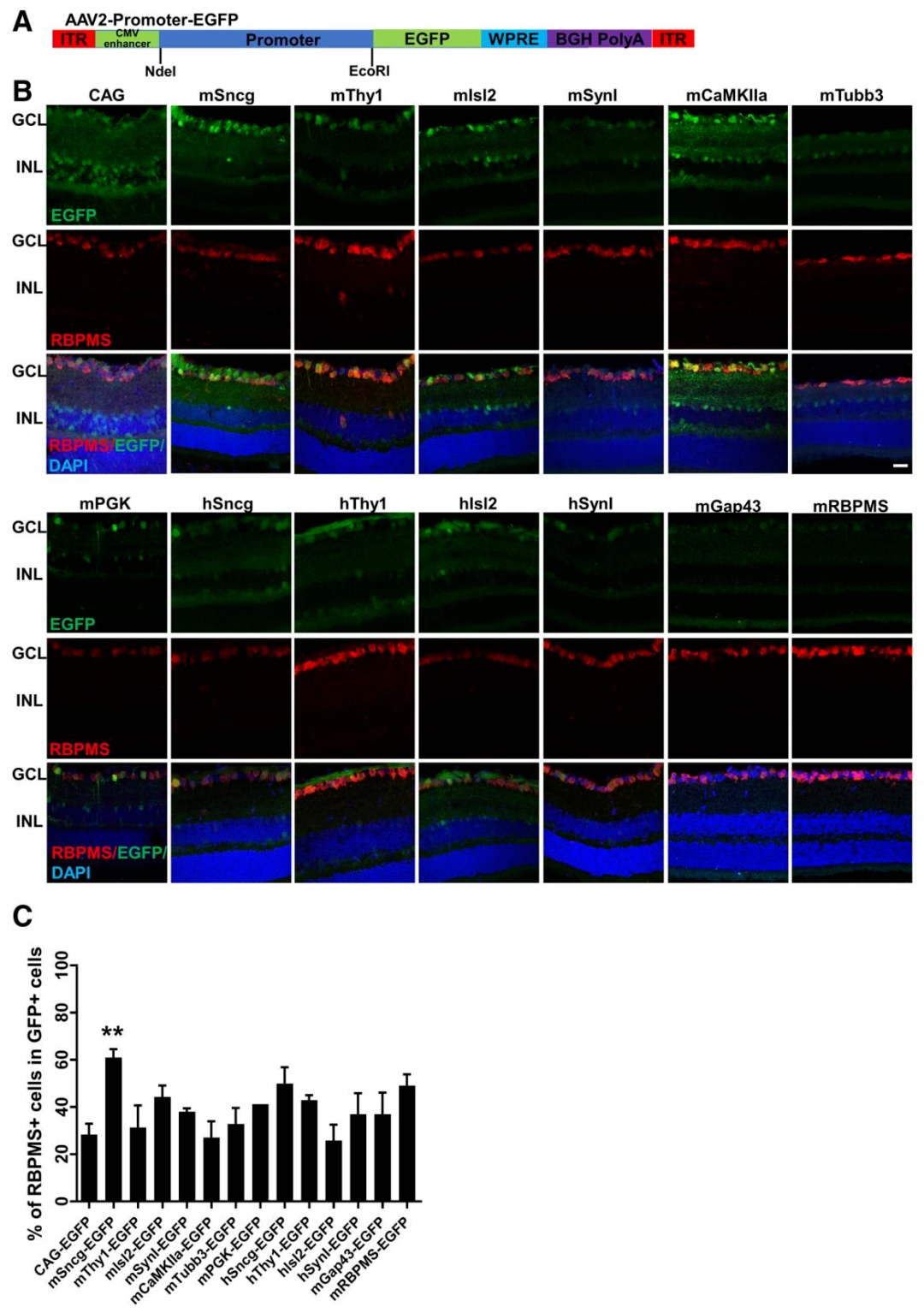
Figure 1. Layer-specific EGFP expression driven by different promoters in the mouse retina.
Additional characterization using RBPMS immunostaining and retrograde labeling further demonstrated efficient targeting of RGCs. In the study summarized in the original BrainCase article, approximately 78% of RGCs were transduced, 85% of reporter-positive cells were RGCs, and approximately 75% of projection RGCs were labeled.
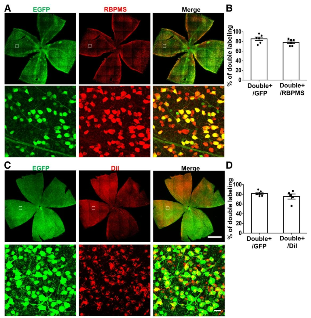
Figure 2. Identification of AAV-mSncg-EGFP-positive RGCs using RBPMS immunostaining and DiI retrograde tracing.
Importantly, the activity of the mouse Sncg promoter is not limited to mouse retinal cells.
The study evaluated mSncg in human RGC models, including RGCs differentiated from human pluripotent stem cells and primary human RGCs. In these models, mSncg showed stronger transgene expression than the corresponding human Sncg promoter (hSncg).
The original paper likewise concluded that the promoter functions in human RGCs, supporting its utility as an experimental tool for RGC-targeted gene manipulation.
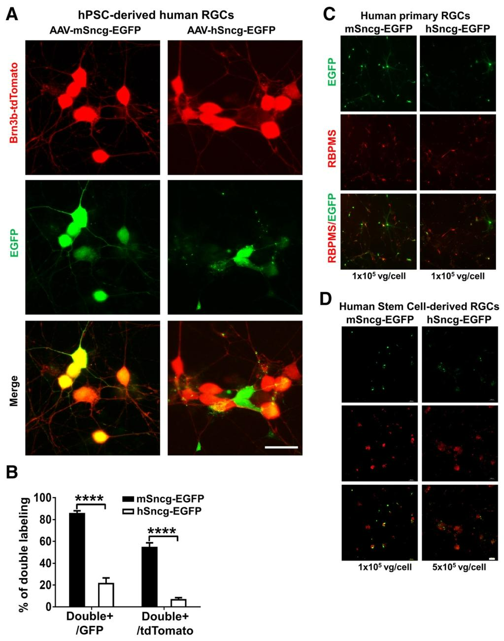
Figure 3. Transduction efficiency of AAV-mSncg-EGFP in human RGCs from different sources.
For longitudinal studies and experimental gene therapy, sustained expression is often as important as cell-type specificity.
Using scanning laser ophthalmoscopy (SLO) to monitor reporter expression over time, researchers evaluated the long-term activity of the mSncg promoter.
Expression driven by mSncg reached a high level approximately four weeks after injection. Following an initial decline, expression increased again from approximately three months and remained robust for up to 12 months in the study.
These findings indicate that the mSncg promoter can support long-term transgene expression in RGCs, making it particularly useful for longitudinal retinal and optic nerve studies.
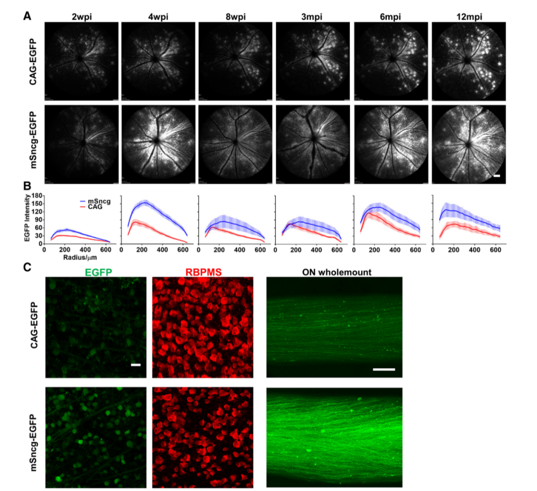
Figure 4. Long-term EGFP expression driven by the mSncg or CAG promoter in mouse RGCs.
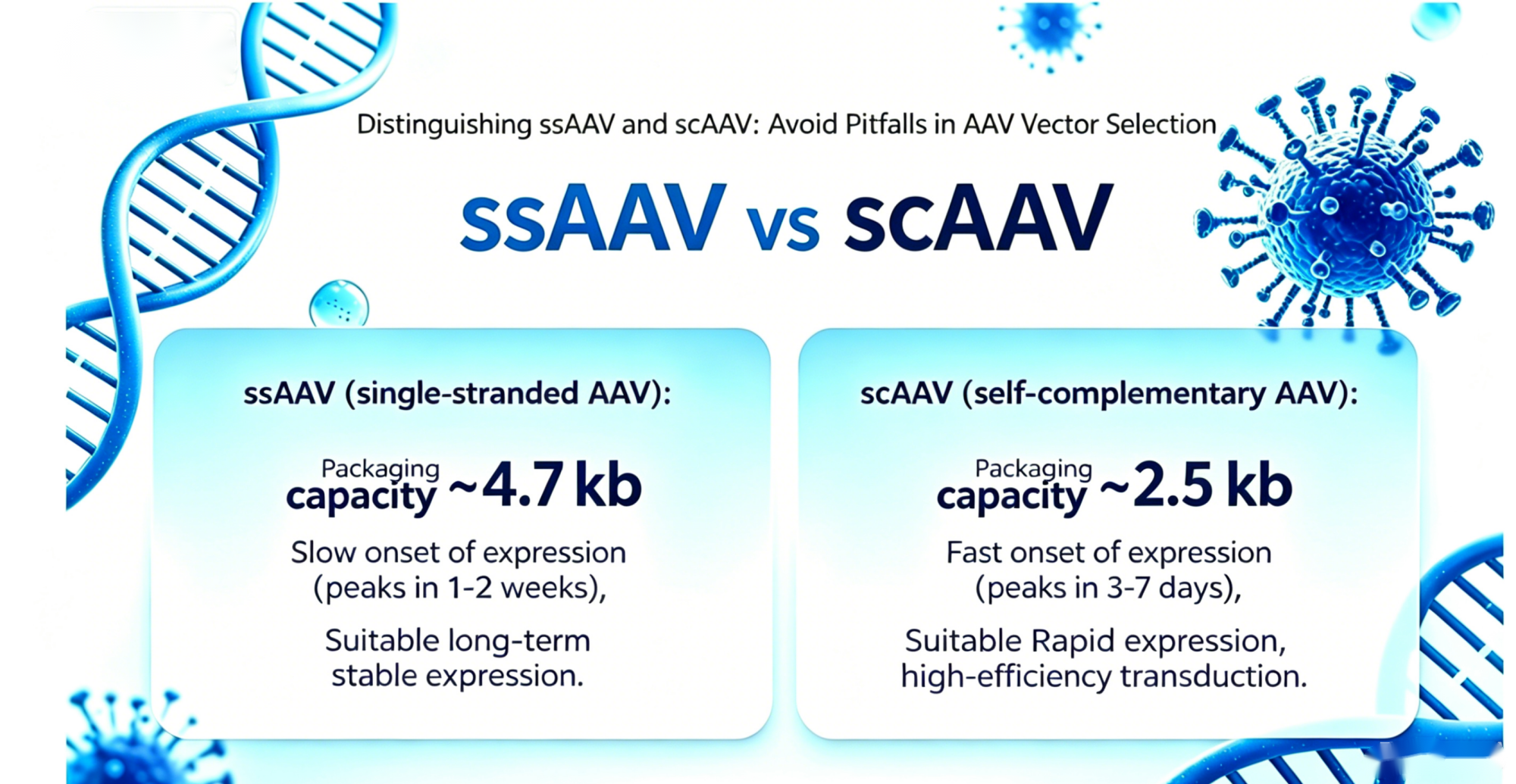
One limitation of AAV vectors is their packaging capacity of approximately 4.7 kb. For large payloads such as CRISPR/Cas9 components, reducing promoter size can provide valuable additional space.
The full-length mSncg promoter used in the study is approximately 1.45 kb. Researchers therefore generated three truncated variants:
1.03 kb → 0.66 kb → 0.27 kb
All three truncated promoters retained substantial RGC promoter activity. However, increasing truncation was associated with greater nonspecific reporter expression in the inner nuclear layer, indicating a trade-off between promoter size and RGC specificity.
The shortest 0.27-kb mSncg variant provides sufficient space to accommodate larger genetic components and was subsequently used for AAV-mediated CRISPR/Cas9 experiments.
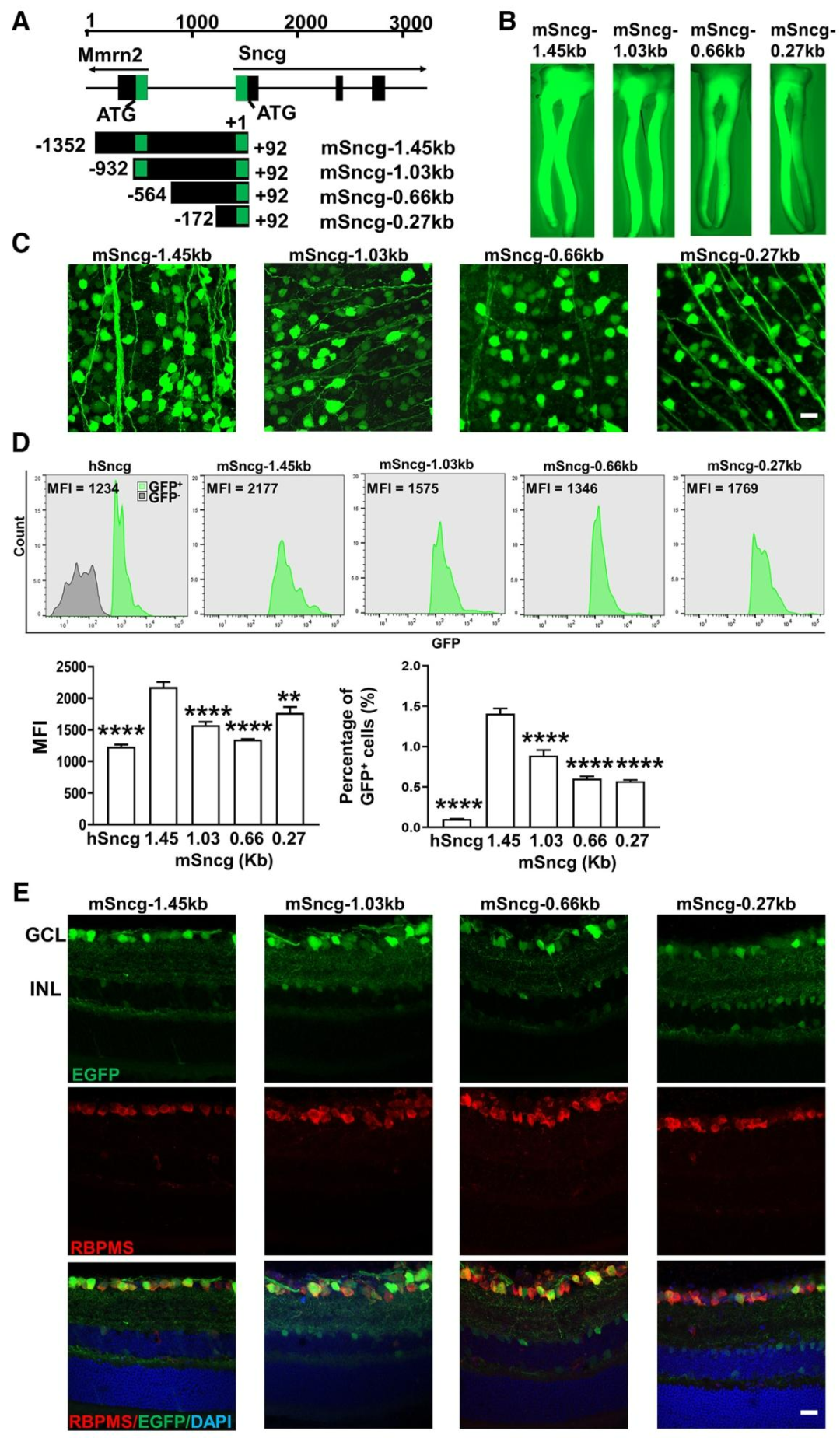
Figure 5. Specificity and activity of truncated mSncg promoter variants in mouse RGCs.
The mSncg promoter can also be combined with CRISPR/Cas9 to manipulate endogenous genes involved in RGC and optic nerve degeneration.
Two genes were selected as targets:
Ddit3 (CHOP) is associated with endoplasmic-reticulum-stress-mediated RGC soma degeneration, whereas Sarm1 plays an important role in axonal degeneration.
Researchers used the 0.27-kb truncated mSncg promoter to drive SpCas9, while the full-length mSncg promoter was also evaluated in a Cre-dependent Cas9 mouse system. Combined with gRNAs targeting Ddit3 and Sarm1, the system enabled gene knockdown specifically in RGCs.
The original J Neurosci study confirmed significant knockdown of endogenous Ddit3 and Sarm1 and demonstrated preservation of injured RGC somata and axons in an optic nerve crush model.
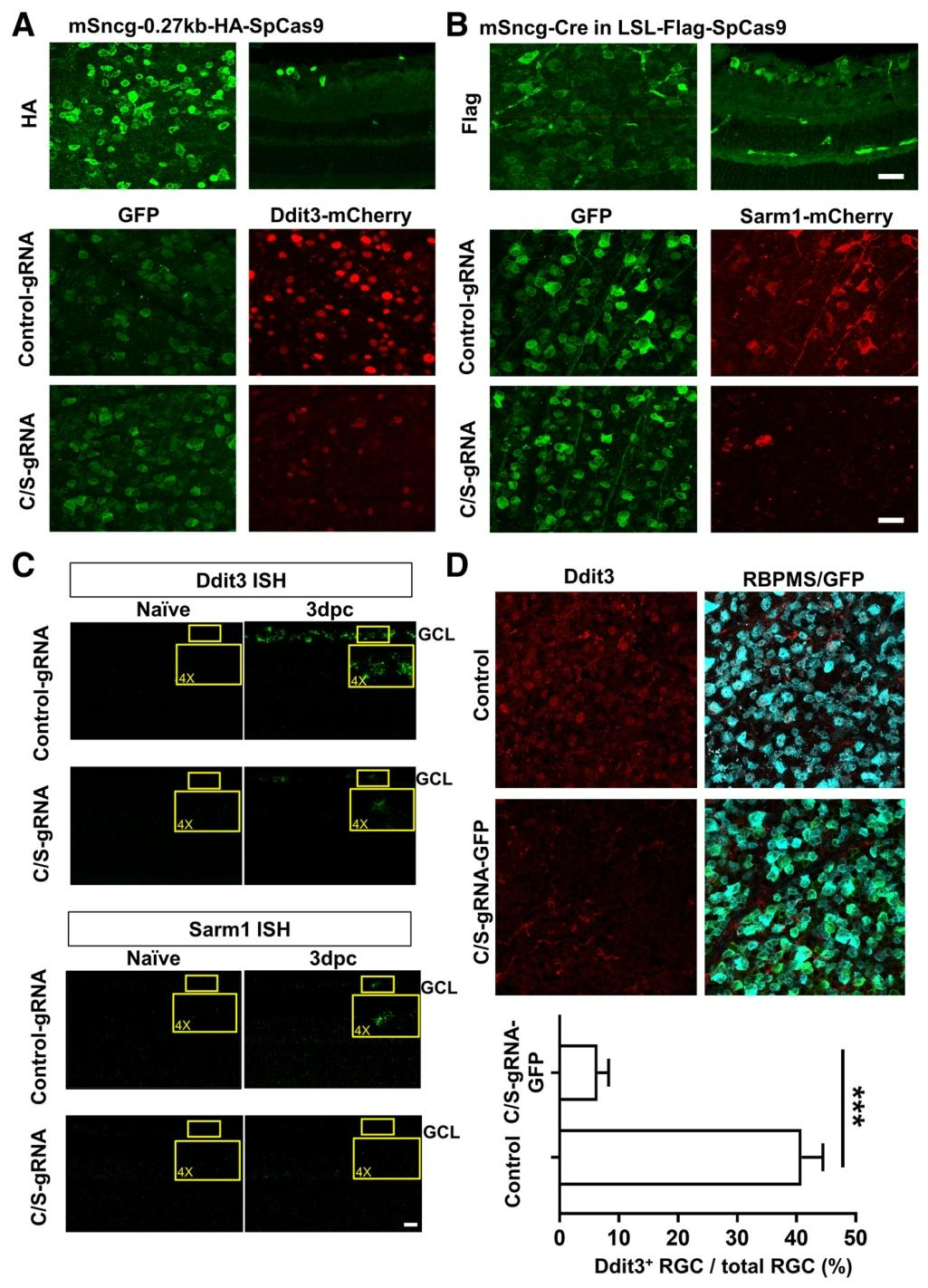
Figure 6. AAV-mSncg-CRISPR/Cas9-mediated targeting of Ddit3 and Sarm1 in mouse RGCs.
The system was further evaluated in an acute mouse optic nerve crush (ONC) model.
Following injury, untreated animals showed substantial thinning of the ganglion cell complex (GCC), RGC loss, and optic nerve axonal degeneration. In contrast, simultaneous targeting of Ddit3 and Sarm1 improved preservation of RGC somata, GCC thickness, and optic nerve axons.
These experiments provide a proof of concept for combining an RGC-specific promoter with AAV-mediated CRISPR/Cas9 to investigate neurodegenerative pathways and potential neuroprotective strategies.
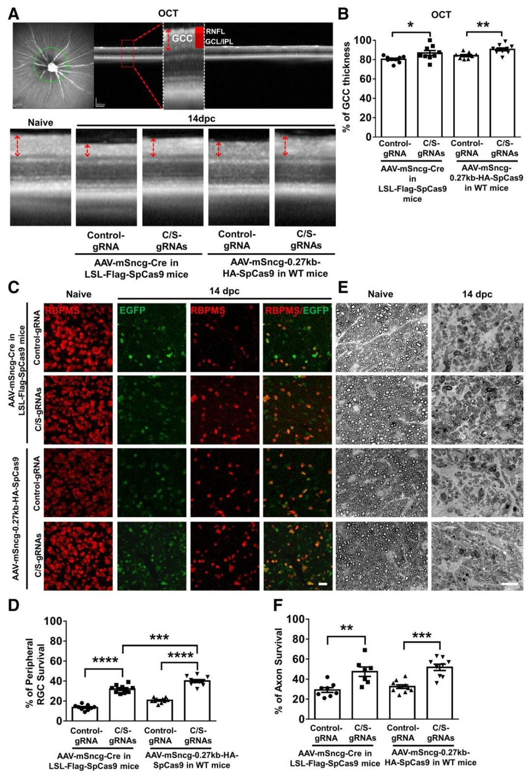
Figure 7. RGC soma and optic nerve axon preservation following AAV-mSncg-CRISPR/Cas9-mediated targeting of Ddit3 and Sarm1.
Beyond the initial characterization, mSncg-based AAV vectors have subsequently been used in several RGC research applications.
PNAS, 2022 — Liang Li, Yang Hu and colleagues
An AAV2-mSncg-jGCaMP7s vector was used to selectively express a genetically encoded calcium indicator in RGCs.
Combined with confocal scanning laser ophthalmoscopy, this approach enabled longitudinal monitoring of light-evoked calcium responses from large populations of individual RGCs in living mice.
Rather than evaluating only structural changes or cell survival, longitudinal calcium imaging enabled researchers to characterize functional changes occurring in RGCs during disease progression, including in optic nerve crush and glaucoma models.
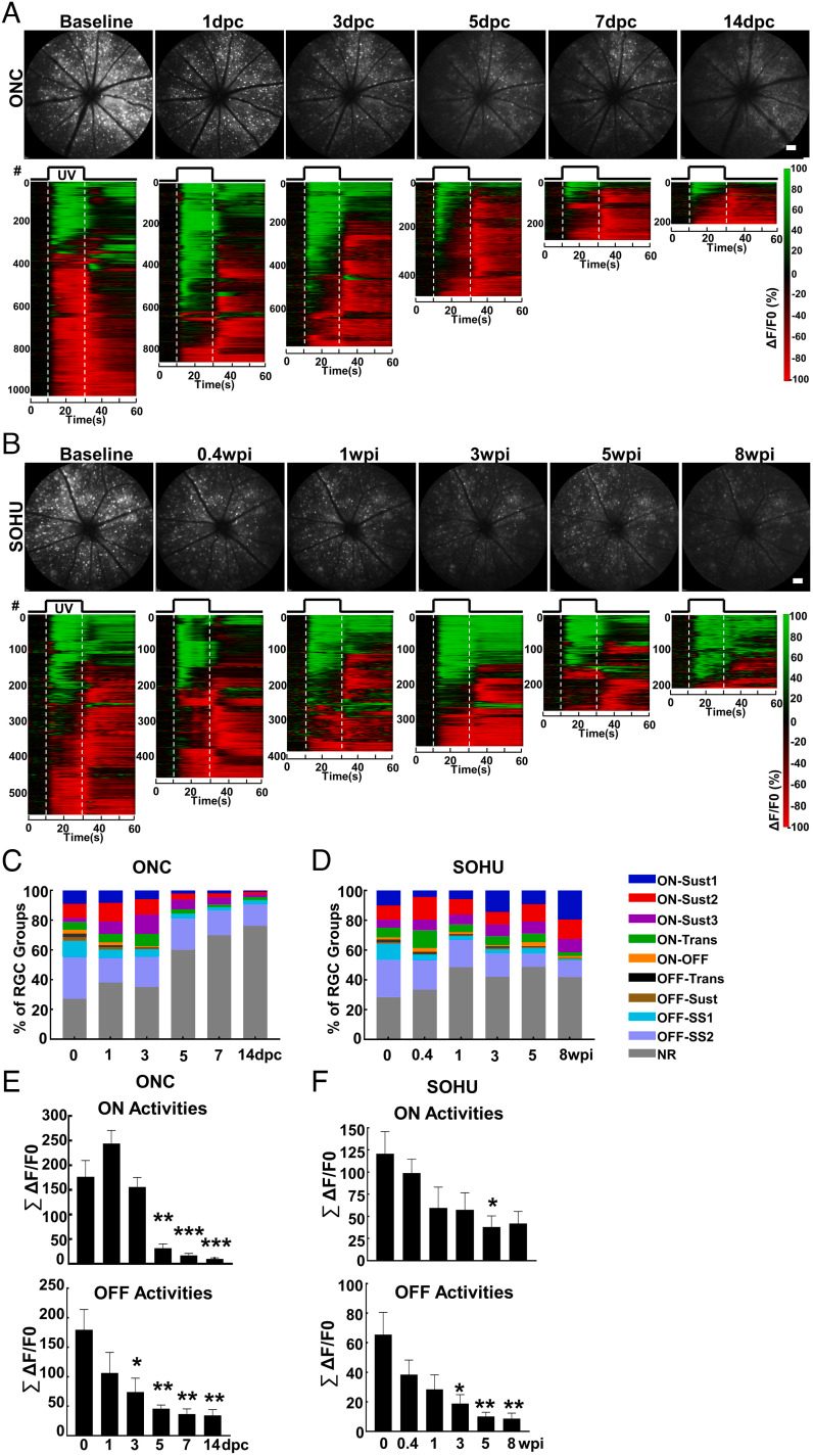
Figure 8. Longitudinal Ca²⁺ imaging reveals dynamic changes in RGC activity in ONC and SOHU glaucoma models.
Molecular Therapy, 2022 — Fang Fang, Yang Hu and colleagues
NMNAT2 is an important regulator of neuronal survival and axonal integrity and was found to be downregulated in glaucomatous RGCs.
Researchers used AAV2-mSncg-NMNAT2 Δex6 delivered by intravitreal injection to selectively overexpress a longer-lived NMNAT2 variant in RGCs.
The treatment increased NAD⁺ levels and was associated with protection of RGC somata and optic nerve axons and preservation of visual function in experimental models of glaucoma and optic nerve injury.
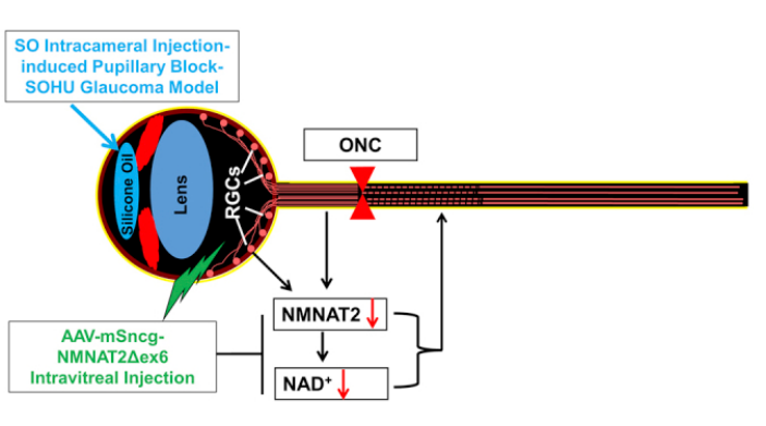
Figure 9. Proposed mechanism underlying NMNAT2-mediated neuroprotection in glaucoma.
Cell, 2021 — Xinzheng Guo, Bo Chen and colleagues
Following RGC soma or optic nerve axonal injury, reduced phosphorylation of CaMKIIα at T286 can impair CaMKII-CREB signaling.
Expression of the constitutively active CaMKIIα-T286D mutant was shown to protect RGC somata and long-range axonal projections.
Using the AAV2-mSncg promoter enables RGC-selective delivery of CaMKIIα-T286D, providing a strategy to investigate CaMKII-mediated neuroprotection while limiting expression in non-target retinal cells.
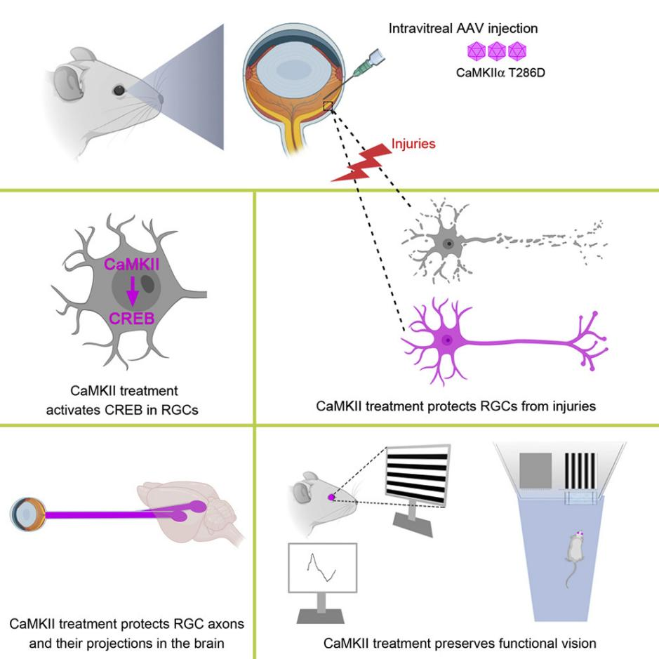
Figure 10. CaMKII-mediated protection of retinal ganglion cells and preservation of visual function.
Neuron, 2022 — Liang Li, Yang Hu and colleagues
Single-cell transcriptomic analysis comparing regenerating RGCs with surviving but non-regenerating RGCs identified several candidate neural repair genes, including Anxa2, ILK, and Mpp1.
RGC-specific expression using AAV2-mSncg provided a means to evaluate these candidates in vivo and investigate their effects on RGC survival, axonal regeneration, and visual function in experimental glaucoma models.
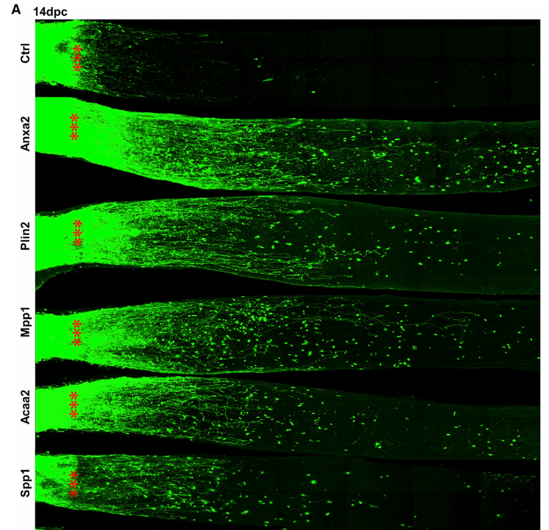
Figure 11. Anxa2 promotes optic nerve regeneration following optic nerve crush.
Science Translational Medicine, 2026 — Liang Li, Yang Hu and colleagues
More recently, gelsolin (Gsn) was investigated as a key regeneration-associated gene upregulated in regenerating RGCs.
Gelsolin promotes actin depolymerization by regulating filamentous actin (F-actin). RGC-specific AAV-mediated expression of Gsn and related actin-regulatory genes promoted optic nerve regeneration and RGC protection in mouse models of optic nerve crush and ocular-hypertension glaucoma. These effects were associated with decreased axonal F-actin and improved mitochondrial transport.
The Chinese BrainCase article describes the construct used in this context as AAV-CS265 (truncated mSncg)-Gsn.
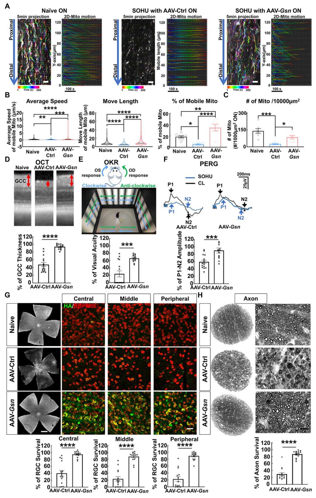
Figure 12. RGC-specific Gsn overexpression improves mitochondrial transport, visual function, and RGC and optic nerve survival in a mouse glaucoma model.
The mSncg promoter provides a useful platform for RGC-selective transgene expression and has now been applied across a broad range of experimental applications:
RGC-specific expression · Gene overexpression · CRISPR/Cas9 gene editing · Calcium imaging · Neuroprotection · Optic nerve regeneration · Glaucoma research
Its combination of cell-type specificity, sustained expression, compatibility with truncated promoter designs, and suitability for intravitreal AAV delivery makes mSncg a valuable tool for studying retinal ganglion cell biology and developing experimental strategies for optic nerve repair.
BrainCase provides customized AAV-mSncg vector construction and virus packaging services for different experimental applications.
Available designs can be customized according to your gene of interest, reporter, regulatory elements, AAV serotype, and experimental application.
Contact us to discuss your RGC-targeting AAV design and experimental requirements.


WhatsApp Business Account
Address:-
Address:-