AAV-ROOT: A New Retrograde Vector Reveals How Sensory Neurons Regulate Adipose Tissue
Release time:2026-07-13 16:18:13
For decades, adipose tissue has been viewed primarily as an endocrine organ. Hormones such as leptin and adiponectin carry metabolic information from fat to the brain, while sympathetic nerves transmit signals from the brain back to adipose tissue, together maintaining whole-body energy homeostasis.
But is hormonal signaling the entire story?
Recent evidence suggests that adipose tissue is also directly innervated by sensory neurons. However, studying these neurons has been challenging because researchers lacked tools capable of specifically labeling and manipulating organ-innervating sensory circuits.
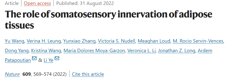
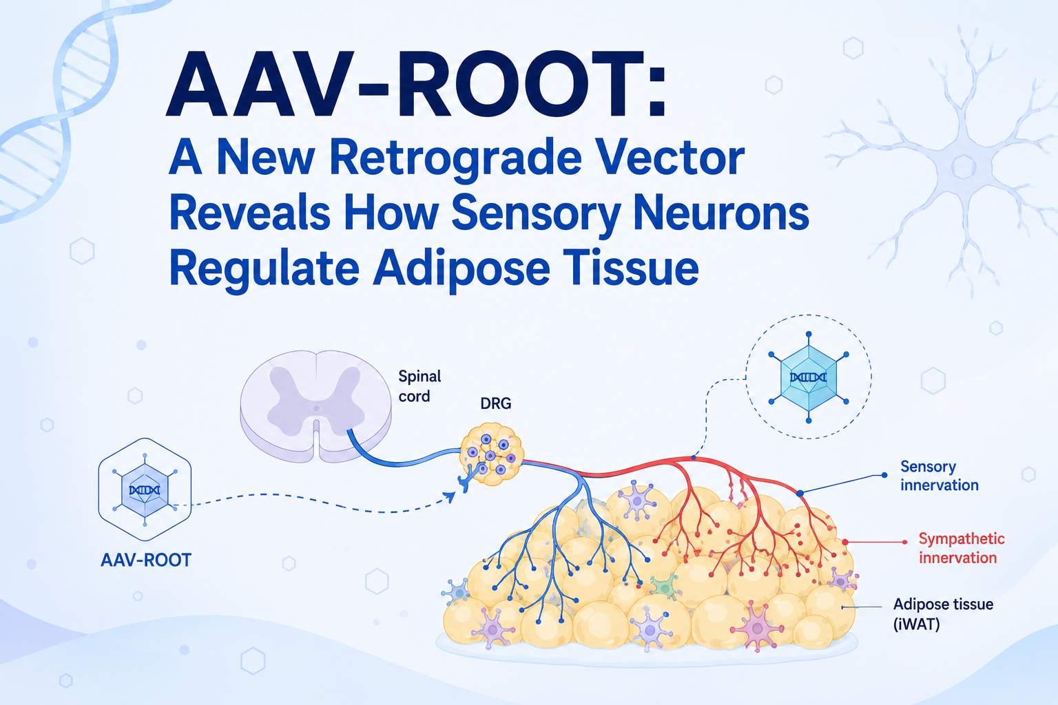
In 2022, a research team led by Li Ye at The Scripps Research Institute addressed this challenge in a study published in Nature. By developing a novel retrograde AAV vector called ROOT (Retrograde vector Optimized for Organ Tracing), they demonstrated that adipose tissue possesses its own dedicated sensory innervation and uncovered an unexpected neural mechanism regulating thermogenesis.
Visualizing Sensory Innervation of Adipose Tissue
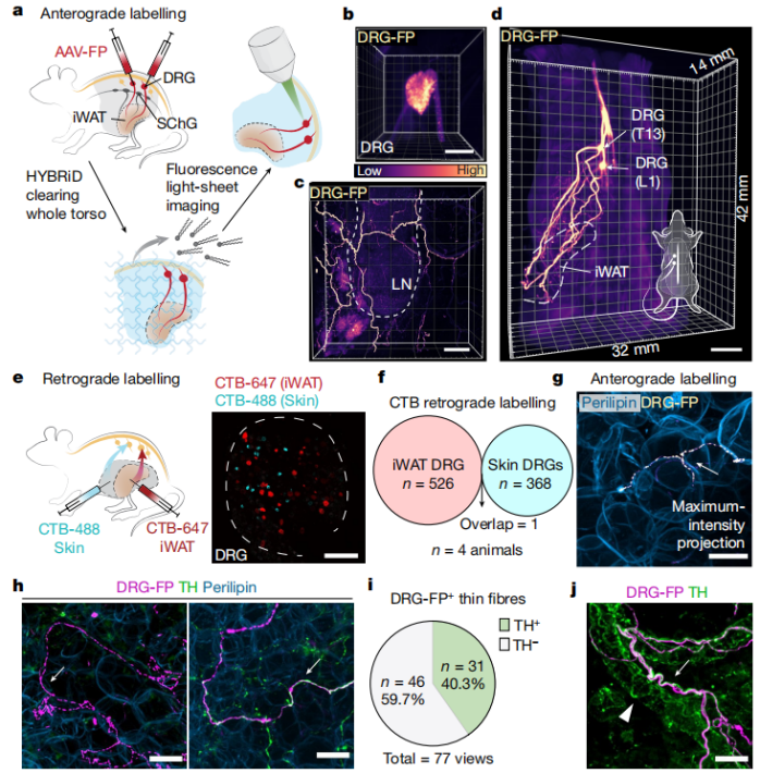
To determine whether dorsal root ganglion (DRG) neurons directly innervate adipose tissue, the researchers first injected an AAV expressing fluorescent proteins into thoracolumbar DRGs. Combined with HYBRiD tissue clearing and light-sheet microscopy, this approach enabled three-dimensional visualization of sensory projections throughout inguinal white adipose tissue (iWAT).
The imaging results clearly revealed abundant sensory axons extending into adipose tissue.
Figure 1: DRG Innervation of Adipose Tissue
To further determine whether these neurons were shared with skin innervation, the team performed dual-color CTB retrograde tracing. Surprisingly, neurons projecting to adipose tissue and those innervating the skin formed two completely separate populations, confirming that adipose tissue is served by its own dedicated sensory neurons.
These findings provide direct anatomical evidence that adipose tissue is not merely regulated by the nervous system—it is itself a sensory organ capable of transmitting information through specialized neural pathways. Rethinking TH as a Marker for Sympathetic Innervation
Another interesting observation emerged during immunostaining.
Traditionally, tyrosine hydroxylase (TH) has been widely used as a marker for sympathetic nerve fibers in adipose tissue. However, the authors found that nearly 40% of sensory nerve terminals were also TH-positive.
This finding suggests that TH labeling alone cannot reliably distinguish sympathetic fibers from sensory fibers in adipose tissue. As a result, previous studies relying exclusively on TH staining may have unintentionally included sensory innervation in their analyses.
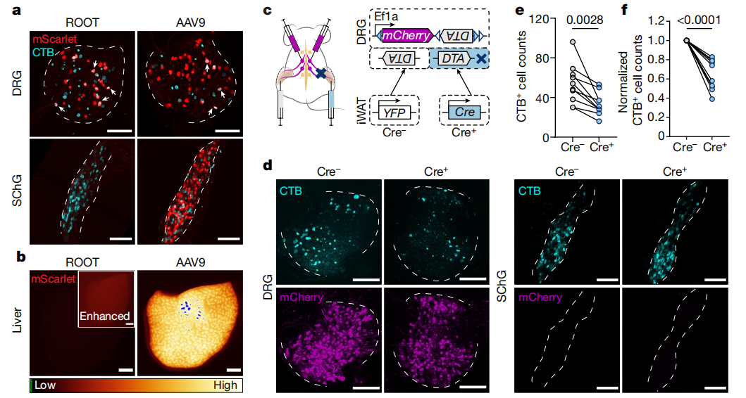
Figure 2: Specific Labeling of Fat-Sensing Neurons
Although this was not the primary focus of the study, it represents an important methodological consideration for future research.
ROOT: A Retrograde AAV Designed for Organ-Specific Neural Tracing
Perhaps the most significant contribution of this work is the development of the ROOT viral vector.
ROOT was generated through directed evolution based on an AAV9 capsid library, aiming to improve retrograde transport from peripheral organs while minimizing off-target labeling.
Compared with conventional retrograde approaches, ROOT efficiently labeled DRG neurons innervating adipose tissue while greatly reducing unwanted transduction in sympathetic chain ganglia, contralateral DRGs, and the liver.
This level of specificity enabled researchers to selectively manipulate adipose sensory neurons, something that had previously been extremely difficult using existing viral tools.
Selectively Removing Sensory Neurons Revealed Their Physiological Function
With ROOT in hand, the researchers next asked an important question:
What role do adipose sensory neurons actually play?
To answer this, they combined ROOT-Cre with a Cre-dependent diphtheria toxin A (DTA) system to selectively eliminate sensory neurons projecting to adipose tissue.
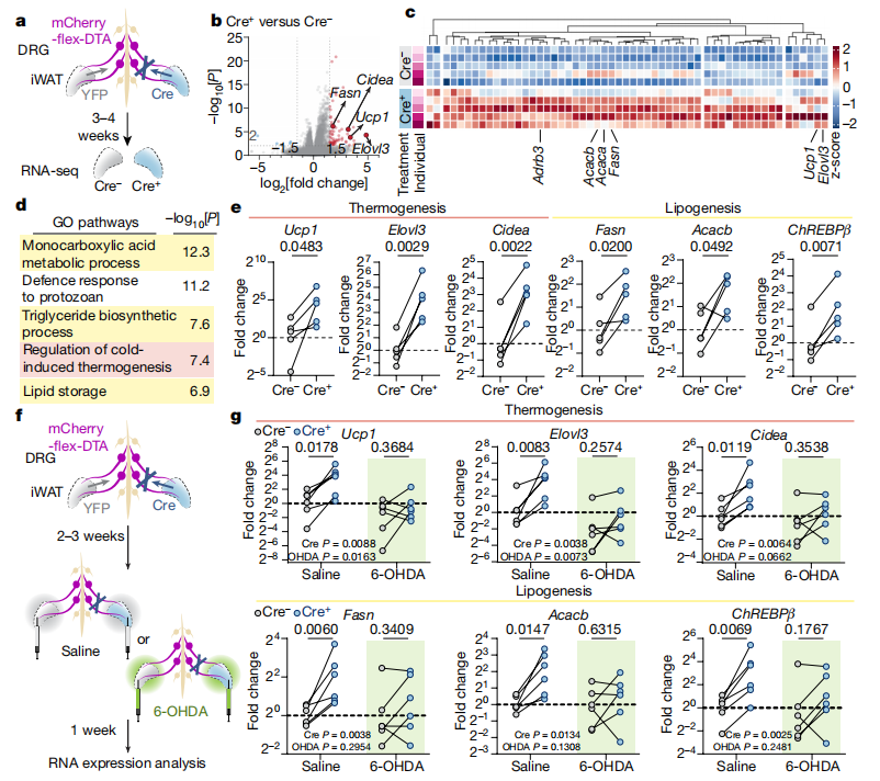
RNA sequencing performed several weeks later revealed a striking transcriptional response.
Genes associated with thermogenesis and lipid metabolism—including Ucp1, Cidea, and Elovl3—were significantly upregulated following sensory neuron ablation.
These molecular changes were accompanied by increased beige adipocyte formation and enhanced thermogenic activity.
However, when sympathetic nerves were chemically ablated using 6-hydroxydopamine (6-OHDA), these effects disappeared entirely.
This finding indicates that sensory neurons regulate adipose tissue indirectly by suppressing sympathetic activity. Once sensory input is removed, sympathetic signaling becomes more active, driving thermogenic gene expression.
Figure 3: Specific Loss of Sensory Neurons Upregulates Thermogenesis-Related Gene Transcription
A New Perspective on Fat–Brain Communication
For many years, communication between adipose tissue and the brain was largely understood as a hormonal process.
This study adds an entirely new dimension to that model.
Rather than serving as passive targets of sympathetic output, adipose tissues actively communicate with the nervous system through dedicated sensory neurons, forming a previously unrecognized neural feedback pathway.
More importantly, the study demonstrates how advances in viral vector engineering can transform neuroscience research.
By enabling highly specific retrograde labeling of peripheral sensory circuits, ROOT provides researchers with a powerful tool for studying organ innervation, sensory feedback, and peripheral neural circuit function.
As organ-specific viral vectors continue to evolve, they are likely to accelerate discoveries not only in metabolic regulation but also across broader areas of peripheral neuroscience and bioelectronic medicine.
Reference
Wang Y, Leung VH, Zhang Y, et al. The role of somatosensory innervation of adipose tissues.Nature. 2022;609:569–574.
Service Type :
Select the service you'd like to purchase.
Order Information(Premade-AAVs)
Please provide us some information about the service you'd like to order.
Order Information(Custom AAV/Lentivirus)
Please provide us some information about the service you'd like to order.
Order Information(Others)
Please provide us some information about the service you'd like to order.