Experimental Tips | Understand Chemogenetics in 3 Minutes, Precisely Control Neuron "Switches"
Release time:2026-02-10 15:09:29
I. Core Basics
(1) Definition of Chemogenetics
Chemogenetics is an interdisciplinary technique that bridges chemical biology and neuroscience. Its core principle involves designing small molecule compounds that interact specifically with ligand-dependent receptors to activate downstream signaling pathways and regulate cellular activity. The DREADDs (Designer Receptors Exclusively Activated by Designer Drugs) technology is the most widely used chemogenetics technique currently.
(2) DREADDs
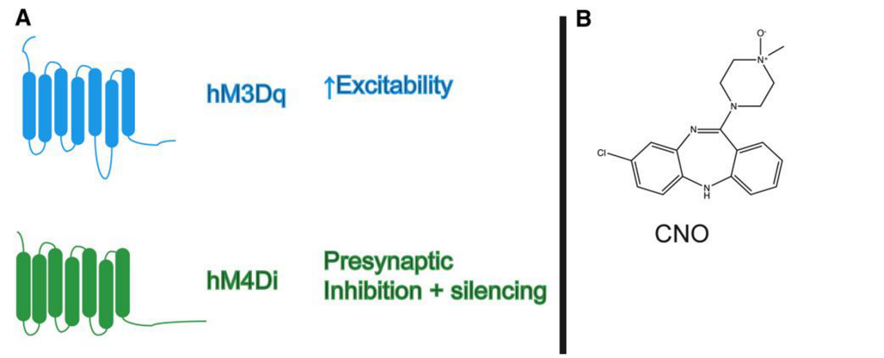
1. Types of DREADD Receptors:
These are genetically engineered G-protein-coupled receptors (GPCRs) with the core characteristic of having very low sensitivity to endogenous ligands (avoiding interference from the body’s own signals) and high affinity for the exogenous ligand CNO. The most commonly used DREADDs can be divided into two categories:
🔹Activating Gq-DREADD — hM3Dq: This is a modified human muscarinic acetylcholine receptor M3 (hM3D), which responds only to CNO and not acetylcholine. Upon binding with CNO, the Gq protein-coupled signaling pathway is activated, triggering phospholipase Cβ (PLCβ) to break down phosphatidylinositol bisphosphate (PIP2). This results in the closure of the KCNQ outward potassium ion channel, leading to membrane depolarization and triggering an action potential.
🔹Inhibitory Gi-DREADD — hM4Di: This is a modified human muscarinic acetylcholine receptor M4 (hM4D), which responds only to CNO and is not regulated by acetylcholine. Upon binding with CNO, the Gi protein-coupled inwardly rectifying potassium ion channels (GIRK) are activated, causing membrane hyperpolarization and inhibiting action potential firing in neurons.
Figure 1: Common DREADDs and their ligand CNO
2. Delivery Vectors:
By using AAV vectors, in combination with specific serotypes and cell-specific promoters, precise and reversible control of specific neuronal activity can be achieved.
3. Core Ligand:
CNO (clozapine N-oxide) is a metabolic byproduct of clozapine, and it binds to artificially expressed receptors in neurons (such as hM3Dq, hM4Di) to precisely regulate neuronal activity. It has the following advantages: 🔹High Selectivity: It specifically activates the modified DREADD receptors and has no effect on natural receptors, completely avoiding non-specific signal interference. 🔹Strong Penetration: It is highly lipophilic and can easily cross the blood-brain barrier, supporting administration through oral or intraperitoneal injection to reach the central nervous system. 🔹Reversibility: It has a fast metabolism rate in vivo. After a single dose, the regulatory effect lasts for about 6 hours, and the effect is reversible after discontinuation of the drug. It also has no significant toxic or side effects.
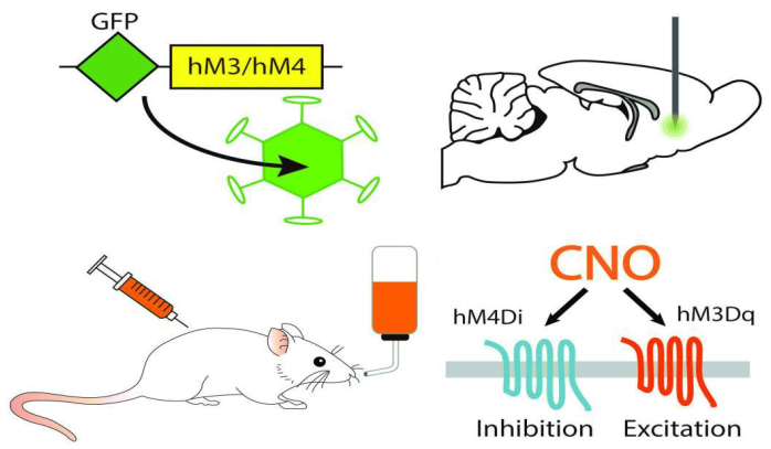
II. DREADDs Experimental Workflow
1. Receptor Selection: Choose the appropriate DREADD receptor based on the experimental goals. Use hM3Dq for activation and hM4Di for inhibition. 2. Virus Vector Construction and Delivery: Clone the DREADDs gene into an AAV vector, combined with a specific AAV serotype and cell-specific promoter, to achieve targeted infection and expression of DREADDs in the in vivo target cells. You can choose to co-express a fluorescent reporter protein (such as mCherry) for tracking and localization purposes. 3. CNO Administration: After sufficient viral expression, regulate neuronal activity by administering CNO through systemic or localized delivery. Set up control groups to exclude non-specific interference. 4. Verification of Manipulation Effectiveness:
🔹Expression Verification: Use tissue sectioning and fluorescence imaging to confirm the location of DREADD expression in the target brain regions.
🔹Functional Verification: Perform electrode recordings to monitor changes in neuronal membrane potential.
5. Phenotype and Functional Analysis: Conduct behavioral and physiological experiments during the CNO drug effect window, comparing results with control groups to validate the functional regulation of target neurons.
Figure 2: Experimental Steps in Chemogenetics
III. CNO Administration Methods and Ligand Expansion
(1) Comparison of CNO Administration Methods
Table 1: Different CNO Administration Methods
Category
Intraperitoneal Injection (IP)
Intracerebral Stereotactic Microinjection
Chronic Long-Term Administration
Route of Administration
Administered via the peritoneum to enter the systemic blood circulation, affecting all central brain regions and peripheral tissues expressing DREADD.
Guided by stereotaxic apparatus, microinjected through a pre-positioned catheter into specific brain regions for precise local drug delivery and targeted control.
Administered through drinking water, food intake, or subcutaneous slow-release pumps for sustained, long-term drug exposure.
Application Scenario
Acute intervention experiments, such as acute behavioral testing or short-term neuronal function regulation; requires simultaneous regulation of multiple expression sites.
Precise regulation of specific brain regions, exploration of neural circuit mechanisms.
Simple and quick operation, no complex surgery required; fast onset, precisely matches acute experimental time windows.
Highly targeted, efficient regulation at low concentrations; supports multi-region intervention, suitable for complex neural circuit studies.
Non-invasive operation, minimal body stress, suitable for continuous intervention from days to weeks.
Disadvantages
Invasive procedure, may cause stress responses in the body, potentially interfering with experimental results.
Complex operation, requires surgical implantation of a catheter; risks of local tissue damage and postoperative infection; pre-experiment needed to determine drug diffusion range.
Drug concentration affected by animal water/food intake, difficult to control precisely, significant individual variability; cannot achieve acute "on/off" regulation.
(2)Other DREADD Ligands
DREADD ligands are a class of pharmacologically inert compounds. In addition to CNO, several optimized ligands have been developed. The key information is as follows:
Mouse: 0.1-0.5 ng / 51-171 ng; Rat: 86-171 ng; 5-30 min onset
Rat: ~10 ng; 15-30 min onset
Rat: ~15 ng; 15-30 min onset
--
Core Advantages
The first widely used DREADD ligand; flexible administration methods; water-soluble formulations are easily accessible
Good water solubility, no co-solvent required; faster onset than CNO; lower non-specific activity
A metabolite of CNO, optimal efficacy; extremely high DREADD specificity; no issues with reverse metabolic conversion; much lower required dose than CNO and C21, minimal off-target effects
Extremely high targeting specificity; fastest onset; can be used with CNO or other muscarinic ligands for bidirectional control
Limitations/Precautions
About 2% of CNO is metabolized into clozapine, causing off-target effects (interaction with serotonin and histamine receptors)
Doses of 0.5-1 mg/kg may activate dopaminergic neurons in the substantia nigra, posing a risk of off-target effects; short duration of action
Limited research accumulation; rat intraperitoneal injection doses not clearly defined
Limited solubility; short duration of action; less research available, optimal dose needs to be determined
Applicable Experimental Scenarios
Basic acute/chronic experiments; behavioral research requiring long-term regulation (oral administration)
Acute experiments requiring rapid DREADD activation; experimental systems sensitive to co-solvents
Behavioral experiments requiring rapid, short-term regulation; multi-region/multi-receptor bidirectional control experiments
IV. Experimental Cases
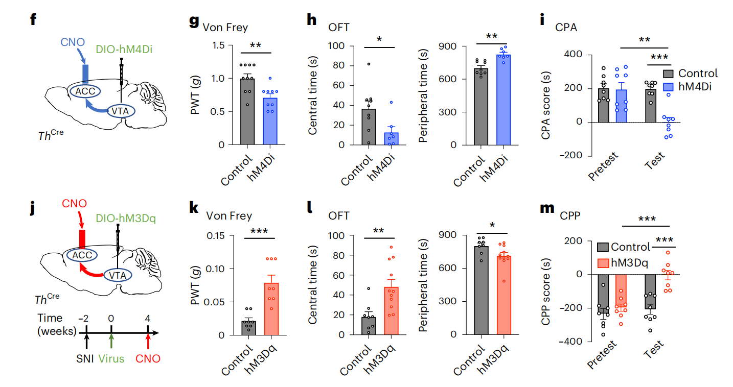
(1) CNO Cannula Administration
1. Short-Term Stimulation
Experimental Animals: ThCre mice virus
Vector: rAAV-EF1α-DIO-hM3D(Gq)-EGFP or rAAV-EF1α-DIO-hM4D(Gi)-EGFP
Injection Method: ACC/VTA, stereotaxic injection into the brain, 1.0×10¹² vg/ml, 300 nl
Administration Protocol: After 3 weeks of viral expression, a guide cannula was implanted and allowed to recover for 1 week; CNO (3 μmol/L) was infused at a rate of 100 nL/min for 0.5 μL, with a 2-minute needle pause; behavioral testing was conducted 5 minutes after infusion.
Experimental Results: To determine whether disactivation of ventral tegmental area dopaminergic neurons (VTADA) projecting to the anterior cingulate cortex (ACC) would induce anxiety-like behavior and comorbid pain, the study injected Cre-dependent hM4Di virus (inhibiting VTADA neurons) into the VTA of ThCre mice. Three weeks after expression, a guide cannula was implanted into the ACC. The results showed that the inhibition group exhibited anxiety-like behaviors in the open field test (OFT), elevated plus maze test (EPM), and light-dark box test (DLB). They also showed reduced mechanical/thermal pain thresholds and aversive behavior in the conditioned place aversion (CPA) test. In mice with spared nerve injury (SNI) 6 weeks post-surgery (chronic neuropathic pain model), activation of this pathway completely reversed hypersensitivity to pain, anxiety, and aversion behaviors, with no significant changes in the sham surgery group. In summary, the VTADA-ACC pathway regulates anxiety-like behavior and hyperalgesia, with VTADA neurons projecting forward to ACCGlu neurons to transmit emotional feedback.
Figure 3: VTADA-ACC Pathway Regulates Anxiety-Like Behavior and Hyperalgesia
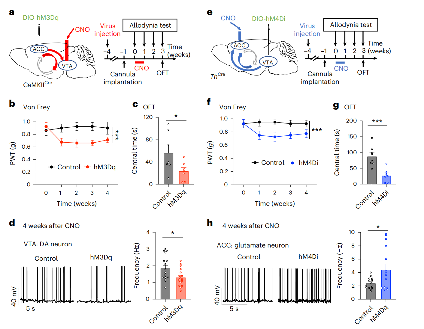
Viral Vectors: rAAV-EF1α-DIO-hM3D(Gq)-EGFP or rAAV-EF1α-DIO-hM4D(Gi)-EGFP
Injection Method: ACC/VTA, stereotaxic injection into the brain, 1.0×10¹² vg/ml, 300 nl
Administration Protocol: After 4 weeks of viral expression, a guide cannula was implanted and allowed to recover for 1 week. CNO was administered once daily for a week to simulate long-term noxious stimulation.
Experimental Results: To further validate the core role of a key positive feedback loop between ACC glutamatergic neurons and VTA dopaminergic neurons (ACC⁺ᴳˡᵘ–VTA⁺ᴳᴬᴮᴬ–VTA⁺ᴰᴬ–ACC⁺ᴳˡᵘ) in the pathogenesis of chronic pain comorbid with emotional disorders, Cre-dependent hM3Dq virus was injected into the ACC of mice with 1-week CNO intervention. The results showed that this intervention decreased the mechanical pain threshold in mice, induced anxiety-like behavior, and these behavioral changes persisted for over 3 weeks. The firing frequency of VTADA neurons was also reduced for more than 4 weeks. In reverse validation, inhibition of the VTADA→ACC pathway similarly induced persistent pain hypersensitivity and anxiety behaviors for ≥3 weeks, accompanied by long-term potentiation of ACCGlu neuronal firing. In summary, both interventions enhanced the ACC-VTA positive feedback loop activity, mediating the long-term maintenance of hypersensitivity and anxiety, confirming the key role of this loop in the progression of chronic pain.
Figure 4: ACC–VTA–ACC Feedback Loop Mediates the Persistent Presence of Neuropathic Pain
(2) Intraperitoneal Injection Administration
1. Short-Term Administration
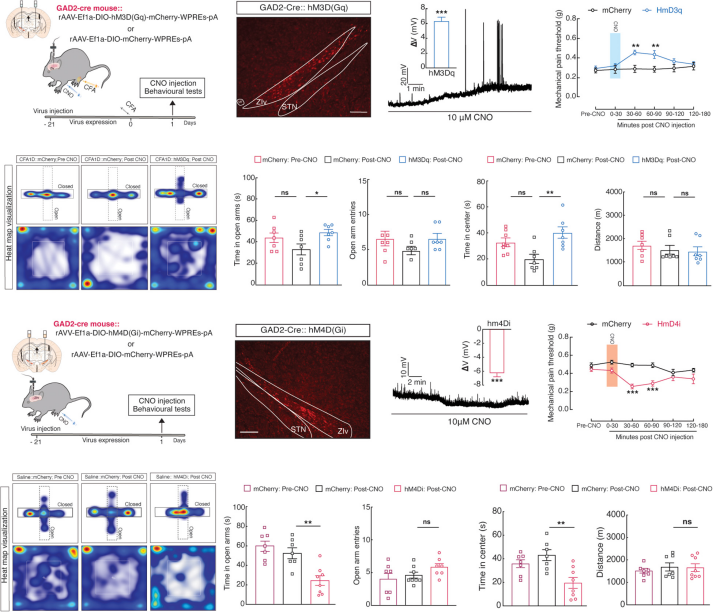
Experimental Animals: GAD2-Cre mice Viral Vectors: rAAV-EF1α-DIO-hM3D(Gq)-mCherry orrAAV-EF1α-DIO-hM4D(Gi)-mCherry Injection Method: ZI region, stereotaxic brain injection, 300 nl, 3 weeks of expression Administration Protocol: 30 minutes before behavioral testing, CNO (1 mg/kg, 0.5 mL) was administered via intraperitoneal injection to activate or inhibit ZIvGABA neuronal activity.
Experimental Results: To investigate the relief effect of activating ZIvGABA neurons on pain-related anxiety behaviors, AAV-DIO-hM3Dq-mCherry was injected into the ZI region of GAD2-Cre mice, followed by intraperitoneal CNO injection to activate ZIvGABA neurons. Electrophysiological validation (current clamp mode) showed that after 10 μM CNO treatment, the neurons depolarized and generated action potentials. In GAD2-Cre mice with AAV-DIO-hM3Dq-mCherry injected into the ZI region, intraperitoneal CNO activation resulted in depolarization and action potential generation. A pain-related anxiety behavior model was established by injecting complete Freund's adjuvant (CFA) into the hindpaw of the mice. Behavioral testing on day 1 after injection showed increased mechanical pain thresholds and reduced anxiety. In normal GAD2-Cre mice injected with AAV-DIO-hM4Di-mCherry, CNO-induced inhibition hyperpolarized neurons, resulting in heightened pain sensitivity and anxiety-like behavior. In summary, the reduced activity of ZIvGABA neurons is a necessary and sufficient condition for acute pain and anxiety induced by CFA.
Figure 5: Regulation of ZIvGABA Activity Modulates Pain Perception and Anxiety-Like Behavior
2. Long-Term Administration
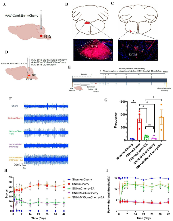
Experimental Animals: SD rats
Viral Vectors: rAAV-EF1α-DIO-hM3D(Gq)-mCherry or rAAV-EF1α-DIO-hM4D(Gi)-mCherry
Administration Protocol: 30 minutes before electroacupuncture (EA) stimulation, CNO (1 mg/kg) was administered via intraperitoneal injection to activate or inhibit neuronal activity in this pathway.
Experimental Results: To clarify the role of the NTS glutamatergic neurons (NTSGlu) to RVLM (rostral ventrolateral medulla) pathway in the regulation of sympathetic nerve activity and spontaneous pain by EA, the anterograde tracing virus rAAV-CaMKIIα-mCherry was injected into the NTS of SD rats, confirming that NTSGlu neurons directly project to the RVLM. Subsequently, chemogenetic approaches were used to inhibit and activate this pathway: inhibition of NTSGlu neurons mimicked the EA effect, significantly reducing the firing frequency of RVLM neurons and spontaneous pain scores; activation of this pathway reversed the effects of EA. It is noteworthy that this pathway had no significant impact on mechanical pain, and its activation could not reverse the improvement of mechanical pain by EA. In summary, electroacupuncture specifically regulates spontaneous pain by inhibiting the NTSGlu-RVLM pathway, suggesting that this pathway is one of the important neural mechanisms underlying EA-induced analgesia.
Figure 6: EA Relieves Spontaneous Pain via the NTSGlu-RVLM Pathway, but Not Mechanical Pain
(3) Oral Administration
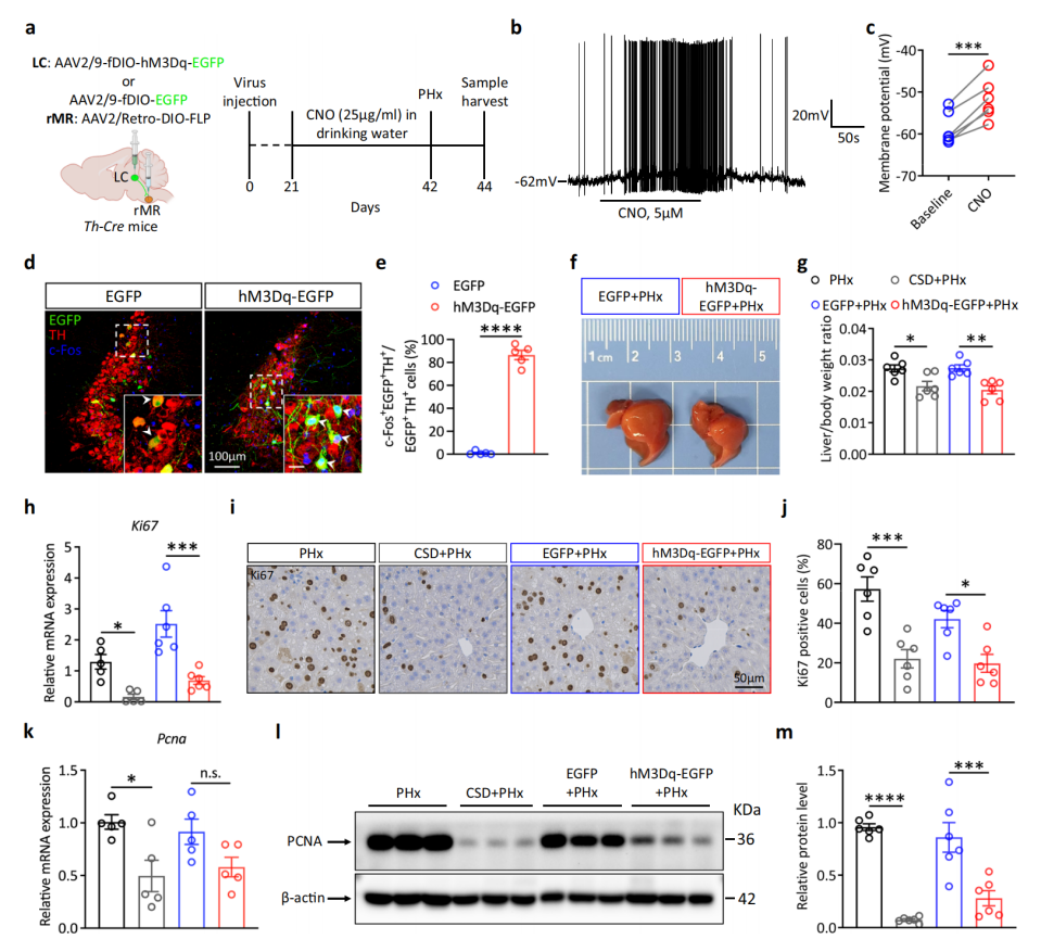
Experimental Animals: Th-Cre mice
Viral Vectors: AAV-EF1α-fDIO-hM3Dq-EGFP
Injection Method: LC, stereotaxic brain injection
Administration Protocol: After 3 weeks of viral injection, CNO was administered through drinking water at a concentration of 25 µg/ml to chronically activate NELC neurons projected by rMR.
Experimental Results: To analyze the regulatory effect of the NELC-5-HTrMR neural circuit on liver regeneration, AAV-fDIO-hM3Dq-EGFP or control virus was injected into the locus coeruleus (LC) of Th-Cre mice, and AAV2/Retro-DIO-FLP was injected into the dorsal raphe nucleus (rMR). CNO was administered through drinking water to chronically activate the NELC neurons projected to rMR. Whole-cell patch-clamp recordings and c-Fos immunostaining confirmed the effective activation of these neurons. The 70% partial hepatectomy (PHx) experiment showed that, compared to the control group, the activation group had a significantly lower liver/body weight ratio and lower gene and protein expression levels of proliferation markers Ki67 and PCNA, indicating that chemogenetic activation of NELC neurons projecting to rMR inhibits liver regeneration, simulating the regulatory effect of chronic stress.
Figure 7: Chemogenetic Activation of NELC Neurons Projecting to rMR Impairs Liver Regeneration
References
[1]Roth BL. DREADDs for Neuroscientists. Neuron. 2016;89(4):683-694. [2] Gao J, Ye T, Chen X, et al. Advances in chemogenetics: a review of DREADDs and its application in psychiatric disorders. Mol Psychiatry. 2026;31(1):480-497. [3]Song Q, Wei A, Xu H, et al. An ACC-VTA-ACC positive-feedback loop mediates the persistence of neuropathic pain and emotional consequences. Nat Neurosci. 2024;27(2):272-285.[4]Farzinpour Z, Liu A, Cao P, Mao Y, Zhang Z, Jin Y. Microglial Engulfment of Spines in the Ventral Zona Incerta Regulates Anxiety-Like Behaviors in a Mouse Model of Acute Pain. Front Cell Neurosci. 2022;16:898346. [5]Chen W, Ma X, Fu YM, Liu CZ, Li HP, Shi GX. Electroacupuncture Regulates Sympathetic Nerve Through the NTSGlu-RVLM Circuit to Relieve Spontaneous Pain in SNI Rats. CNS Neurosci Ther. 2025;31(3):e70327. [6]Zhou Y, Lin X, Jiao Y, et al. A brain-to-liver signal mediates the inhibition of liver regeneration under chronic stress in mice. Nat Commun. 2024;15(1):10361.
With extensive hands-on experience in neuroscience research,
Brain Case Biotech provides a wide range of viral vectors
to support cutting-edge scientific studies. For inquiries or collaboration,
please contact bd@ebraincase.com
Service Type :
Select the service you'd like to purchase.
Order Information(Premade-AAVs)
Please provide us some information about the service you'd like to order.
Order Information(Custom AAV/Lentivirus)
Please provide us some information about the service you'd like to order.
Order Information(Others)
Please provide us some information about the service you'd like to order.