E-mail:BD@ebraincase.com
Tel:+8618971215294
English 中文版
Virus Vector - Viral Vector Production - BrainCaseVirus Vector - Viral Vector Production - BrainCase
  • Home
  • Virus product library
    CRISPRRNAiHSV-helperRV-helperNeurophilic virusCalcium SensorsOptogenetics activationOptogenetics inhibitionChemical geneticsSparse labelingFluorescent proteinBiosensorsRecombinaseApoptosis & AutophagyDisease ModelNeurotoxicityOther
  • Products & Service

    Product Center

    Virus

    VSV-circuit research-vaccine and gene therapy research-BrainCase
    Retrovirus-RCAS-TVA-BrainCase
    Lentivirus Vector - Lentivirus Production - BrainCase
    Rabies Virus Vector - RBV Vector - BrainCase
    Herpes simplex virus-Oncolytic and anterograde tracing-Brain Case
    PRV-retrograd multisynaptic-Peripheral-Btain Case
    AAV-gene therapy vectors-BrainCase

    Animal Model

    Neurological Disease Models-BrainCase
    Tumor animal models-anti-tumor-BrainCase
    Digestive System Disease Animal Model-Brain Case
    Cardiovascular System Disease Animal Models-Brain Case

    Plasmid Construction

    Library Construction
    Plasmid design and construction

    Popular Applications

    Gene Regulation

    Gene Overexpression-Brain Case
    RNA interference(RNAi)-siRNA-Brain Case
    Gene Editing - CRSIPR cloning - BrainCase

    Neural Circuit Function Research

    Optogenetics - BrainCase
    Chemical genetics-DREADDs-Brain Case
    Calcium signal recording-Gels- Brain Case
    GRAB Neurotransmitter Fluorescent Sensor- Brain Case
    Functional Magnetic Resonance Imaging Technology- Brain Case

    Research on the structure of neural circuits

    Direct Input and Output-viral vectors- Brain Case
    Anterograde Mono-synaptic Tracing -HSV- Brain Case
    Antrograde Muti-synaptic Tracing-HSV & VSV-Brain Case
    Retrograde Mono-synaptic Tracing-Rabies Virus-Brain Case
    Retrograde Muti-synaptic Tracing-PRV-Brain Case

    Featured Services

    Nervous System Disease Drug Effect

    Alzheimer's disease-AD-Brain Case
    Depression-mental disorders-Brain Case
    Parkinson's disease--PD-Brain Case
    Epilepsy-an ancient neurological disorder-Brain Case

    AAV Serotype Screening

    AAV Serotypes screening-gene therapy-Brain Case

    Tumorigenicity Test

    Tumorigenicity Testing-Evaluation of tumor models-Brain Case

    Efficacy of Oncolytic Virus

    Oncolytic virus-for cancer therapy-Brain Case
    Herpes Virus Vector-anti-tumor- BrainCase
    Vesicular stomatitis virus-killing tumor cells-Brain Case
  • News
    Corporate News New Product Launch Media Activity Investor News
  • Support
    Literature interpretation Customer article Video Zone FAQs
  • About Us
    Virus product library Products & Service News Support About Us Contact
  • Contact
    Contact Us Join us
  • 中文
    English 中文版
  • Home
  • Support
  • Literature interpretation
  • Support
  • Literature interpretation
  • Customer article
  • Video Zone
  • FAQs

Nature Deep Dive: Microglia - Unveiling of Mechanisms and New Therapeutic Strategies of Neurodegenerative Diseases

Release time:2024-11-04 08:54:39

In the research of neurodegenerative diseases, microglia, as resident immune cells of the central nervous system (CNS), have gradually become a focal point for scientists' in-depth exploration. These cells play an indispensable role in various physiological processes, including synaptic pruning, injury repair, homeostasis maintenance, phagocytosis, and supporting communication between other glial cells and other cell types. Notably, microglia are closely linked to the occurrence and development of various neurodegenerative diseases.

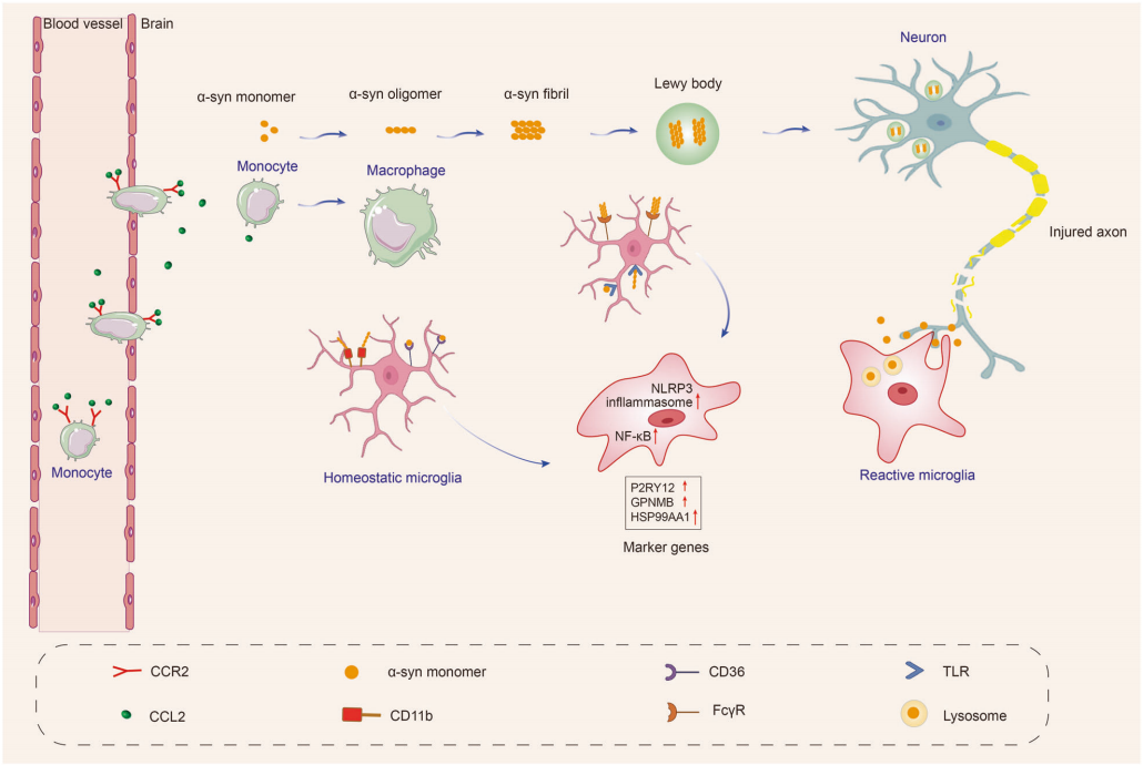
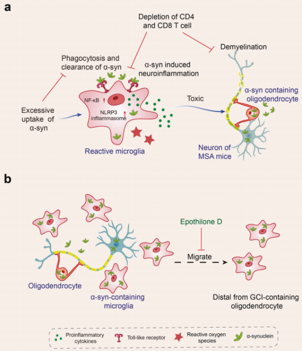
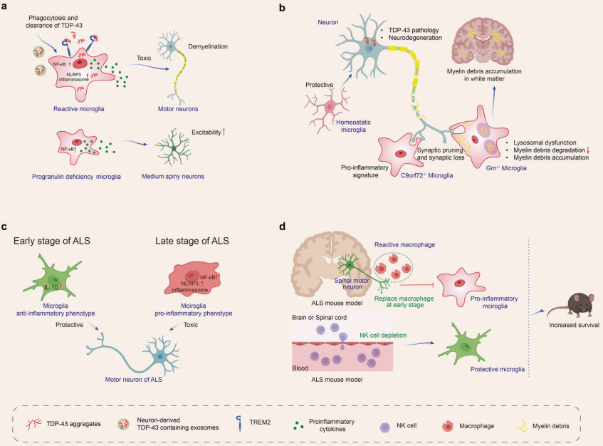
When the central nervous system is damaged or affected by disease, microglia exhibit a complex response, often referred to as "activation." In early studies of microglia, scientists primarily detected the activation state of these cells through morphological observations, specifically by noting their transformation from a branched phenotype in healthy brains to an amoeboid appearance in diseased brains. However, as research has progressed, scientists have found that the activation process of microglia is far more diverse and dynamic than previously anticipated, as reflected in both omics characteristics and functional outcomes. This indicates that microglia present different response patterns in various diseases. Currently, research has observed microglial activation in multiple neurodegenerative diseases, including Alzheimer's disease (AD), Parkinson's disease (PD), multiple system atrophy (MSA), amyotrophic lateral sclerosis (ALS), frontotemporal dementia (FTD), progressive supranuclear palsy (PSP), dementia with Lewy bodies (DLB), and Huntington's disease (HD).


I. Dysfunction of Microglia in Neurodegenerative Diseases

I. Dysfunction of Microglia in Neurodegenerative Diseases

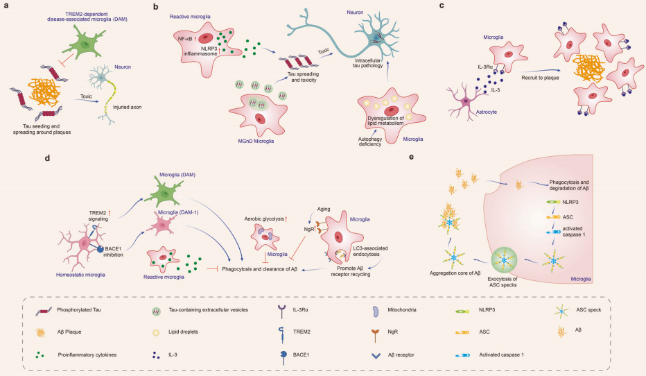
AD is one of the most well-known neurodegenerative diseases, characterized primarily by the accumulation of beta-amyloid (Aβ) plaques and the abnormal aggregation of tau protein, leading to neurofibrillary tangles within neurons. Imaging studies have found that the activation of microglia in AD is associated with tau and amyloid proteins. These activated microglia have been observed to accumulate near Aβ plaques in various regions of the brains of AD mice and in humans post-mortem. In AD mouse models, a subset of microglia, referred to as disease-associated microglia (DAM), was first identified, which participates in the clearance of Aβ.

Research indicates that microglia play a double-edged role in the progression of AD. On one hand, they can phagocytize and clear Aβ and tau proteins, limiting the spread of these pathological substances and potentially offering protective effects in the early stages of the disease. On the other hand, prolonged stimulation of microglia by pathological deposits may lead to dysfunction, promoting neuroinflammation, accelerating the spread of Aβ and tau, and resulting in neurodegeneration.

A substantial body of evidence shows that microglia affect Aβ and tau proteins in various ways:

(1)Aβ binds to microglial TREM2, which promotes the transformation of microglia to the DAM phenotype and activates TREM2 signaling, enhancing phagocytic function. Conversely, loss of TREM2 function impairs microglial phagocytosis of Aβ and increases amyloid protein expression.
(2)LC3-associated phagocytosis (LANDO) in microglia promotes the recycling of Aβ receptors, increasing surface receptors for Aβ and facilitating its clearance. In contrast, AD mice with LANDO deficiency exhibit neurodegeneration and memory deficits.
(3)With aging, the expression of Nogo receptor (NgR) on microglia increases, weakening their phagocytic activity and Aβ clearance. Conversely, AD mice with NgR deficiency show reduced amyloid burden and improved cognitive function.
(4)Inhibition of BACE-1 in microglia promotes the transition of microglial phenotype from homeostasis to stage I disease-associated microglia (DAM-1), thereby enhancing amyloid clearance and improving cognitive abilities in AD mice.
(5)Microglial interaction with astrocytes, after recognizing Aβ deposits, leads to an increase in the expression of the IL-3-specific receptor IL-3Rα on microglia. Astrocyte-derived IL-3 binds to the upregulated IL-3Rα on microglia, enhancing their migration towards Aβ deposits and facilitating the clearance of Aβ aggregates.
(6) APOE3 lipoprotein induces microglia to migrate towards Aβ more rapidly, promoting Aβ uptake and improving cognition.
 

Figure 1: The impact of microglia on Aβ and tau pathology in Alzheimer's disease. Microglia phagocytize Aβ and tau, limiting the spread of Aβ and tau pathology. However, under pathological conditions, microglia may also accelerate the diffusion of Aβ and tau, leading to neurodegeneration.

2. Parkinson's Disease (PD)

PD is the second most common neurodegenerative disease after AD, characterized by the loss of dopaminergic neurons in the substantia nigra and the accumulation of misfolded alpha-synuclein (α-syn) in Lewy bodies. In PD, the activation of microglia begins early and persists throughout the disease course. In 1988, reactive microglia were first observed in the substantia nigra of post-mortem brain tissue from PD patients. Other markers of microglial activation, such as iNOS and cyclooxygenase, as well as the phagocytosis-related marker CD68, are also upregulated in Parkinson's patients. Additionally, PET scans of PD patients show widespread microglial activation.

Microglia possess pattern recognition receptors (PRRs), such as TLRs, NLRs, and SRs, allowing them to detect and respond to various stimuli. When α-syn accumulates extracellularly and is not adequately cleared, it can activate microglia via PRRs, leading to a pro-inflammatory response initiated by interactions between α-syn and membrane receptors. These receptors activate NF-κB and assemble the NLRP3 inflammasome through various mediators, resulting in the production of inflammatory mediators and free radicals, triggering neuroinflammation.

Moreover, the pathways connecting microglia and neurons are closely related to the progression of PD. The CX3CL1-CX3CR1 signaling pathway is crucial for maintaining a healthy balance of microglial activity, regulating chemotaxis and synaptic plasticity, and reducing microglia-mediated inflammation and neurotoxicity. Dysregulation of the CD200-CD200R pathway is associated with increased microglial activation and degeneration of dopaminergic neurons. Additionally, CB1 receptors are abundant in neurons, while CB2 receptors are primarily expressed in microglia in the brain. Animal models of Parkinson's disease suggest that the activation of microglial CB2 receptors has neuroprotective effects and improves motor symptoms.

Figure 2: α-syn induces microglial response in Parkinson's disease
 

3.Multiple System Atrophy (MSA)

MSA is characterized by autonomic dysfunction, Parkinsonian symptoms, and cerebellar ataxia. Clinically, it is divided into two subtypes: MSA with predominant cerebellar ataxia (MSA-C) and MSA with predominant Parkinsonian symptoms (MSA-P). The main pathological features include atrophy of the olivopontocerebellar region and degeneration of the striatonigral pathways.

Research has found that α-syn-induced microglial activation is dependent on microglial Toll-like receptors (TLRs) 1/2 signaling and TLR4 signaling.
In MSA mouse models, α-syn interacts with TLRs and is subsequently phagocytized by microglia. However, excessive uptake of α-syn by microglia can lead to a significant decline in their phagocytic capacity, triggering an inflammatory response that includes activation of NF-κB and NLRP3 inflammasome signaling, production of reactive oxygen species, and upregulation of pro-inflammatory cytokines. This creates a toxic environment, ultimately inducing neurodegeneration. Additionally, the removal of CD4 and CD8 T cells alleviated inflammation and demyelination in α-syn-induced MSA mice. See Figure 3.

Figure 3: The role of microglia in the pathogenesis of multiple system atrophy (MSA).
 

4. Amyotrophic Lateral Sclerosis (ALS) and Frontotemporal Dementia (FTD)

ALS is characterized by the loss of motor neurons in the cortex, brainstem, and spinal cord, including mutations in the C9orf72, SOD1, TDP-43, and FUS genes. FTD is the second most common form of dementia after AD in individuals under 65, characterized by progressive cognitive, behavioral, and language dysfunction, including mutations in genes such as TDP-43, tau, and FUS.

Research has found that microglia can phagocytize pathological protein aggregates such as TDP-43. The interaction of TDP-43 with microglial TREM2 promotes the phagocytosis and clearance of these aggregates. Conversely, microglial phagocytosis of TDP-43 aggregates leads to microglial activation, and when this activation is excessive, it can cause NLRP3 inflammasome activation and upregulation of pro-inflammatory markers, which have neurotoxic effects on motor neurons. Additionally, defects in progranulin in microglia activate NF-κB signaling and promote the release of pro-inflammatory cytokines, leading to hyperexcitability of medium-sized spiny neurons. See Figure 4.

Under disease conditions, progranulin deficiency can also lead to lysosomal dysfunction in microglia, reducing the clearance of deposited proteins and myelin debris, resulting in the accumulation of myelin fragments in white matter. Furthermore, microglia can exacerbate neuronal loss and behavioral abnormalities by increasing synaptic pruning through interactions with complement.

Figure 4: The role of microglia in the pathogenesis of amyotrophic lateral sclerosis (ALS) and frontotemporal dementia (FTD).
 

5. Others

In exploring the relationship between neurodegenerative diseases and microglia, besides the extensively studied diseases such as AD, PD, MSA, ALS, and FTD, progressive supranuclear palsy (PSP), dementia with Lewy bodies (DLB), and Huntington's disease (HD) also demonstrate the crucial role of microglia in disease progression. In PSP, microglial activation is closely related to the accumulation of hyperphosphorylated tau protein (pTau), a process that not only leads to the disruption of microtubule structures and neuronal dysfunction but also promotes the reactive proliferation of microglia and astrocytes, accelerating the process of neurodegeneration. In DLB, the behavior of microglia is similarly complex and variable, transitioning from an early reactive activation state to a later state of atrophy, with phenotypic changes closely linked to the disease stage, accompanied by abnormal aggregation of α-synuclein and coexisting AD-like pathological features. As for HD, microglial activation is closely connected to the abnormal expansion of the CAG repeat sequence in the HTT gene, leading to the misfolding of huntingtin protein and neuronal loss. Multiple signaling pathways, such as NF-κB, kynurenine, and cannabinoid receptor pathways, are involved in this process. Microglia detect and respond to external stimuli through receptors like TLRs, triggering immune responses that further influence disease progression. In summary, the role of microglia in neurodegenerative diseases is multifaceted and complex, with changes in their behavior profoundly impacting the pathological processes and clinical manifestations of these diseases.

II. Potential Microglial Targeted Interventions and Therapeutic Approaches for Neurodegenerative Diseases

Based on the in-depth understanding of the complex relationship between neurodegenerative diseases and microglia, researchers are actively exploring a range of innovative therapeutic strategies aimed at promoting disease treatment by modulating microglial function. The principles and effects of these strategies have been preliminarily validated in cell experiments, animal models, and clinical trials.

1. Modulating Neuroinflammation

Modulating neuroinflammation is one of the most widely used therapeutic targets, focusing on intervening in the activation state of microglia through drugs or other means to reduce the release of harmful inflammatory factors and thereby alleviate neuronal damage. Research has found that using the NLRP3-specific inhibitor OLT1177 to inhibit the NLRP3 inflammasome can rescue cognitive impairments in AD mouse models. Similarly, the NLRP3 inhibitor MCC950 has been shown to suppress inflammasome activation, α-synuclein aggregation, and dopaminergic degeneration in the substantia nigra of PD mouse models, improving motor function.

2. Inhibiting the Synthesis and Secretion of Microglial Exosomes

Microglia release exosomes containing tau, and microglial activation can facilitate tau propagation. Inhibiting the synthesis and secretion of microglial exosomes can help reduce tau proliferation. Studies have shown that early pharmacological blockade of P2RX7 in tau mice significantly impairs microglial exosome secretion, reducing tau accumulation in the brain and thereby improving working and learning memory.

3. Modifying Microglial Metabolic Pathways

Microglial phagocytosis requires substantial energy. When microglia switch their metabolism from oxidative phosphorylation to aerobic glycolysis, sustained aerobic glycolysis can impair their immune function. Research indicates that modulating microglial metabolism can enhance their phagocytic capacity for Aβ. For instance, treatment with sodium rutin (NaR) shifts microglial metabolism from anaerobic glycolysis to mitochondrial oxidative phosphorylation, providing sufficient ATP for Aβ clearance. Additionally, NaR promotes the clearance of Aβ by increasing the expression levels of phagocytosis-related receptors in microglia.

4. Changing Microglial Phenotype

The brain microenvironment can regulate the phenotypic transformation of microglia, promoting their positive functions. In AD, a ROS-responsive polymer micelle system can accumulate in lesioned areas by mimicking Aβ transport pathways. These micelles normalize the oxidative and inflammatory microenvironment, promoting microglial regeneration. In PD, vitamin D protects dopaminergic neurons from inflammation and oxidative stress by inhibiting microglial activation and promoting M2 polarization, increasing the expression of M2 microglial markers such as CD163, CD204, and CD206.

5. TREM2 Gene Intervention Activation

Activating the expression of key genes such as TREM2 is also considered to enhance the clearance capacity of microglia, promoting their effective clearance of pathological proteins like Aβ and tau. Studies have shown that enhancing TREM2 signaling through TREM2 agonist antibodies such as 4D9, AL002c, or tetravalent TREM2, or using a gene delivery system targeting microglia to upregulate TREM2 levels in the brain, can enhance Aβ clearance and alleviate neuroinflammation.


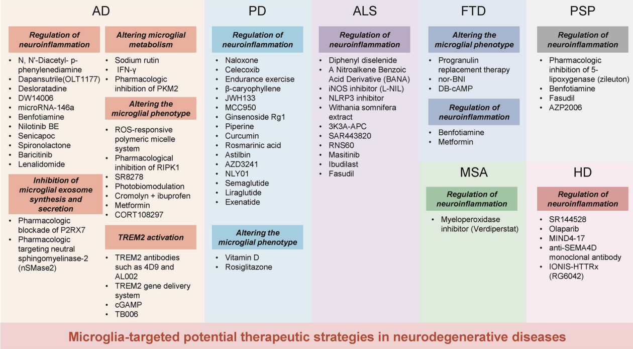
Figure 5: Potential microglial-targeted interventions and therapies for neurodegenerative diseases.

In summary, the role of microglia in neurodegenerative diseases is multifaceted and complex. Through in-depth research and exploration of therapeutic strategies, new insights have been provided for the treatment of neurodegenerative diseases, laying a solid foundation for the future development of more precise and effective therapies.

Brain Case has launched an innovative AAV11 serotype vector, model BC-ZA0182: rAAV-mIBA1-EGFP-WPRE-4×miR-9.T, specifically designed for the efficient and selective transduction of microglia. Furthermore, this vector is flexibly designed to allow for the overexpression or interference of specific genes, or to integrate techniques such as optogenetics, chemogenetics, and calcium signaling recording, thereby providing a powerful genetic manipulation toolset for exploring the functions, mechanisms, and roles of microglia in neurological diseases.


References
Gao C, Jiang J, Tan Y, Chen S. Microglia in neurodegenerative diseases: mechanism and potential therapeutic targets. Signal Transduct Target Ther. 2023 Sep 22;8(1):359. doi: 10.1038/s41392-023-01588-0. PMID: 37735487; PMCID: PMC10514343.


💥Holiday

Special

Offers

Click to fill in the requirements and submit them to us!

Service Type :

Select the service you'd like to purchase.

Order Information(Premade-AAVs)

Please provide us some information about the service you'd like to order.

Detailed requirements:

scroll

Order Information(Custom AAV/Lentivirus)

Please provide us some information about the service you'd like to order.

Gene ID or gene information:

Selection of the reporting gene:

Special Instructions:

scroll

Order Information(Others)

Please provide us some information about the service you'd like to order.

Virus name/Detailed requirements:

scroll

Related products

Nature Review: The Past and Present of AAV Gene Therapy Vectors

Nature Review: The Past and Present of AAV Gene Therapy Vectors

Science: Yulong Li’s Team Develops HaloDA1.0 Far-Red Dopamine Sensor, Enabling Simultaneous Monitoring of Multiple Neurochemical Signals

Science: Yulong Li’s Team Develops HaloDA1.0 Far-Red Dopamine Sensor, Enabling Simultaneous Monitoring of Multiple Neurochemical Signals

Scientific Breakthrough | Nat. Methods | ATLAS: A Tool for Monosynaptic Anterograde Tracing from Defined Starter Neurons

Scientific Breakthrough | Nat. Methods | ATLAS: A Tool for Monosynaptic Anterograde Tracing from Defined Starter Neurons

Literature Interpretation | Nature | How to Detect Glial Phagocytosis of Synapses

Literature Interpretation | Nature | How to Detect Glial Phagocytosis of Synapses

map
{dede:global.cfg_webname/}

Virus product library

CRISPR
RNAi
Neurophilic virus
Optogenetics activation
Biosensors

News

Corporate News
New Product Launch
Media Activity
Investor News

Support

Literature interpretation
Customer article
Video Zone
FAQs
微信

WhatsApp Business Account

Tel: +8618971215294
E-mail: BD@ebraincase.com

Address:-

Address:-

  • Copyright © 2024 Brain Case All Rights Reserved.