In-Depth Interpretation | Tissue Clearing Technologies: A 3D Imaging Tool for Neuroscience
Release time:2025-12-08 16:28:50
In neuroscience research, traditional paraffin or cryosectioning methods have long faced the challenge of fragmentation. These approaches struggle to preserve long-range neuronal projections and the complex connectivity of neural circuits, and mechanical slicing can damage tissue structure or cause information loss.
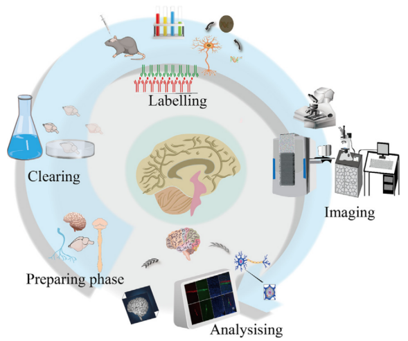
With the breakthrough development of tissue clearing technologies, researchers can now reduce light scattering and homogenize refractive indices through chemical processing, enabling true three-dimensional imaging of intact samples. This innovation has transformed neuroscience, shifting the field from localized observation to system-level analysis.
Figure 1. Schematic of 3D neural imaging enabled by tissue clearing.
I. Technical Principles and Three Main Tissue-Clearing Strategies
The core principle of tissue clearing is to remove substances that cause light scattering or absorption—such as lipids, pigments, and calcium—through chemical treatments. By also matching the refractive indices of the sample and imaging medium, these methods minimize light interference and render tissues optically transparent.
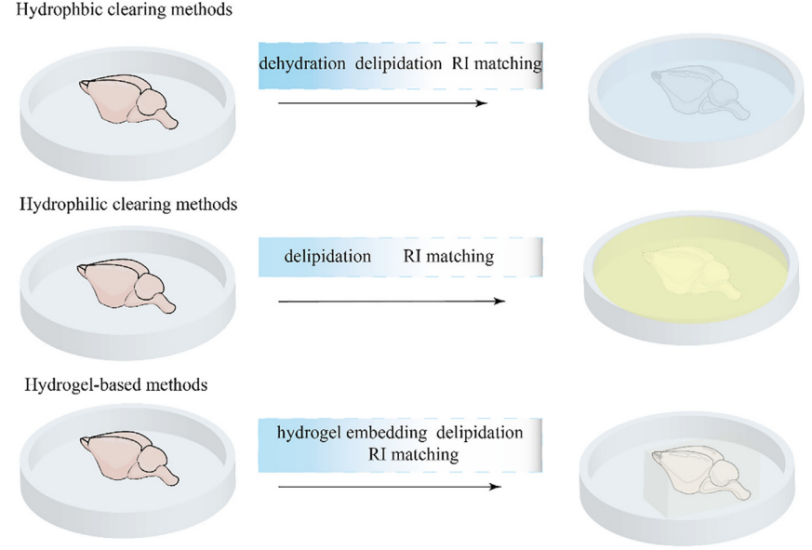
Current mainstream clearing strategies fall into three major categories:
Hydrophobic (organic-solvent) clearing methods These techniques use organic solvents to achieve rapid and efficient clearing of large samples. Representative method: vDISCO.
Hydrophilic (aqueous-based) clearing methods Using water-soluble reagents, these approaches offer excellent biocompatibility and preserve fluorescence well. Representative method: CUBIC.
Hydrogel-based clearing methods By embedding tissues in a hydrogel matrix, these methods fix biomolecules in place, preserving structural and molecular integrity to the greatest extent. Representative method: CLARITY.
Figure 2. Illustration of the three major tissue-clearing strategies.
II. A Panoramic View of Neuroscience Applications: From Basic Research to Clinical Translation
When combined with light-sheet microscopy (for rapid, large-volume imaging), single-cell labeling techniques (such as virus-mediated fluorescent labeling), and AI-driven data analysis tools (including BigStitcher for image stitching and iLastik for cell segmentation), tissue clearing enables efficient interpretation of massive imaging datasets.
This integrated technological workflow has propelled tissue clearing to achieve breakthrough, multidimensional applications across neuroscience—from central nervous system tissues such as the brain and spinal cord to peripheral organs including the liver, kidney, and heart, and from fundamental neural circuit mapping to the investigation of clinical disease mechanisms.
Case 1. Brain Research: Precise Localization of Neuro-Immune Interaction Circuits
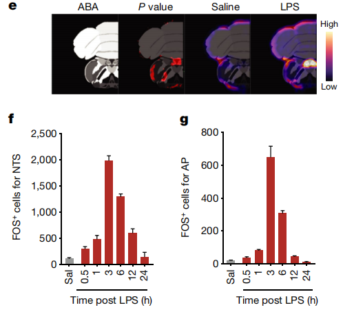
Using the AdipoClear clearing method, the research team performed whole-brain antibody labeling and DBE refractive index matching on fixed mouse brains, followed by light-sheet microscopy for three-dimensional imaging and quantitative analysis of whole-brain FOS signals.
The results showed that lipopolysaccharide (LPS) treatment markedly increased FOS-positive neurons in the nucleus tractus solitarius–area postrema (NTS–AP). Through 3D reconstruction, the dense distribution of activated cells within the dorsal vagal complex of the brainstem was clearly visualized, and neuronal activity changes across multiple brain regions under immune stimulation were dynamically mapped.
This clearing-based imaging strategy enabled the first spatiotemporal atlas of whole-brain neuronal activation under immune stress, precisely identifying the NTS–AP region as a key central node of neuro-immune interaction and providing an essential structural basis for future mechanistic studies.
Figure 3. Whole-brain activity changes during disease-related behaviors.
Case 2. Spinal Cord Research: Anatomical Verification of Injury–Regeneration Circuits
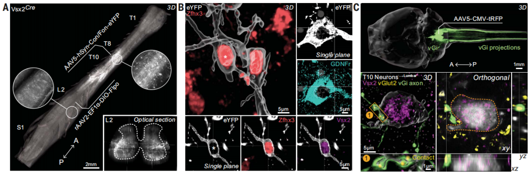
Researchers combined AAV-flex-tdTomato with multi-virus intersectional labeling, followed by decalcification and tissue clearing of intact spinal cords. Light-sheet microscopy enabled continuous longitudinal imaging and 3D visualization of axonal projections from specific neuronal subtypes.
The study revealed, at single-cell resolution, the dense long-range projections from thoracic Vsx2⁺ neurons to the lumbar locomotor center, and verified the striking overlap between regenerated axons after injury and their natural projection patterns.
This clearing–light-sheet imaging platform systematically mapped the intrinsic connectivity of molecularly defined neuronal subtypes and provided direct, comprehensive anatomical evidence supporting the concept that targeted regeneration of lumbar circuits may restore locomotor function.
Figure 4. Neuronal connectivity and projection features.
Case 3. Liver Research: 3D Analysis of Metabolic-Stress–Associated Neuropathy
Using an optimized iDISCO⁺ whole-organ immunolabeling and light-sheet imaging workflow, the research team cleared mouse, non-human primate, and human liver tissues, achieving optical homogeneity via efficient delipidation and refractive index matching.
3D scanning and Imaris reconstruction produced the first complete single-axon–level spatial atlas of intrahepatic sympathetic fibers. Results showed that TH-positive sympathetic axons formed dense, tree-like branches along portal triads, tightly associated with vasculature, while parasympathetic fibers were nearly absent—confirming the sympathetic-dominant innervation pattern of the liver.
Further studies revealed that 20 weeks of high-fat diet reduced sympathetic axon density by over 50%, producing “sympathetic neuropathy.” This pathology was reversible through TNF-α neutralizing antibody treatment or Sarm1 knockout. Additionally, 4 weeks of caloric restriction or leptin replacement fully restored axon density, suggesting new therapeutic targets for metabolic diseases.
Figure 5. 3D neuroanatomy of mouse and primate liver.
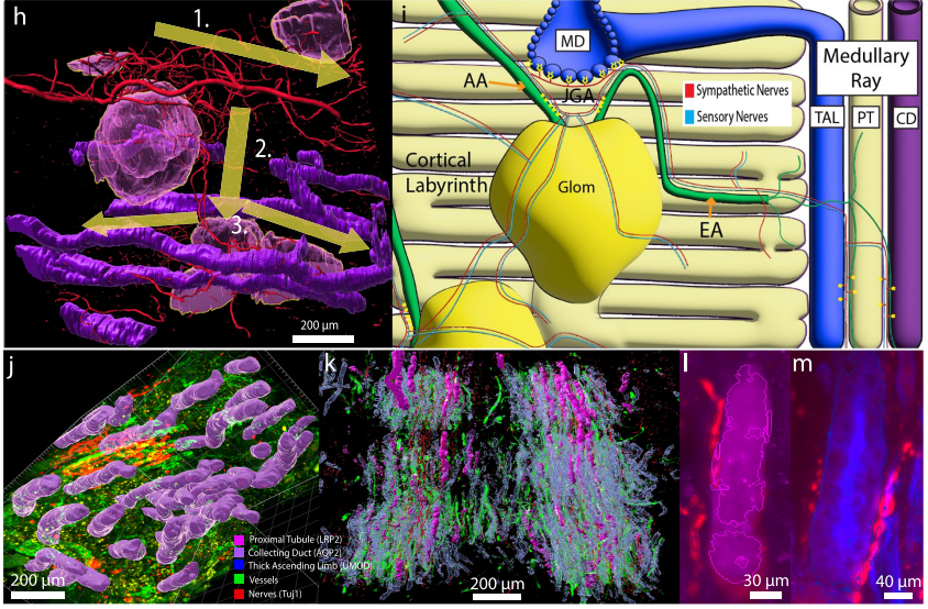
Case 4. Kidney Research: Developmental and Disease-Related Remodeling of Neuro–Nephron Networks
Using CLARITY-SHIELD active clearing combined with light-sheet fluorescence microscopy, the research team performed multi-marker immunostaining and multiscale 3D imaging on 0–75-year human kidney thick sections, rapidly constructing a neurovascular–nephron connectivity atlas spanning the cortex–medulla axis.
Findings showed that glomeruli gain polarity and cluster into “communities” during development, forming reproducible hub-and-spoke neural networks centered on “mother glomeruli,” enabling synchronized inter-nephron communication.
The renal neural network exhibited dynamic changes across lifespan and disease: sparse in newborns, peaking in adulthood, and displaying pathological remodeling in elderly individuals or patients with diabetes or hydronephrosis—characterized by reduced neural connectivity, excessive linkage of sclerotic units, or global neurodegeneration.
Mapping the developmental rules and disease-associated remodeling of human neuro–nephron communities provides a 3D structural basis for understanding renal coordination and developing interventions for chronic kidney disease.
Figure 6. 3D morphometric analyses.
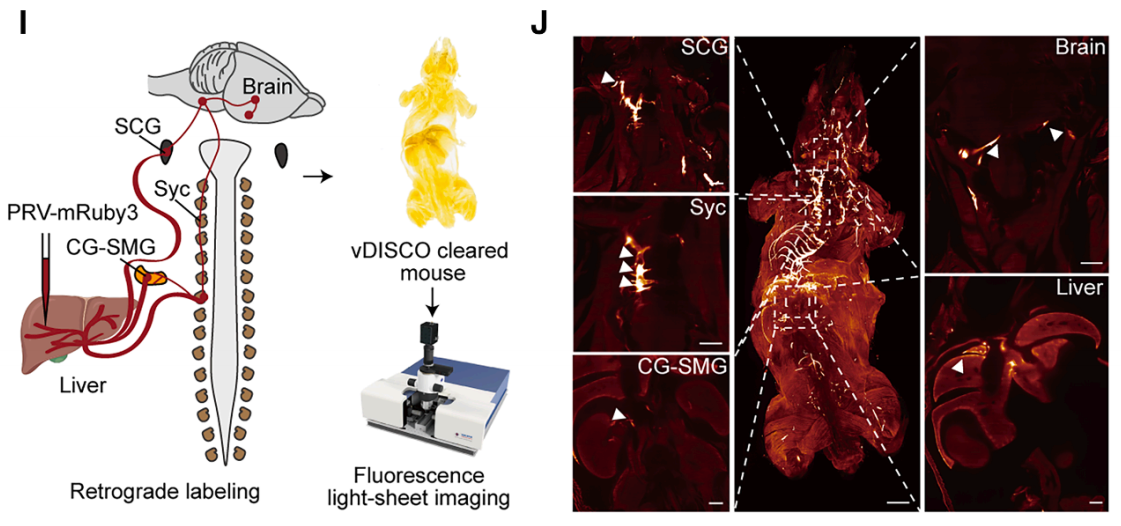
Case 5. Peripheral–Central Integration: Complete Mapping of the Brain–Liver Neural Regulatory Circuit
Using PRV-CAG-mRuby3 viral tracing combined with vDISCO clearing, the research team achieved the first full 3D visualization of the neural circuit connecting the liver to the brain, overcoming the long-standing barrier of cross-organ neural circuit imaging.
The results clearly delineated the projection pathway from the liver to the lateral paragigantocellular nucleus, then to the paraventricular hypothalamic nucleus, and further to the ventromedial hypothalamus, detailing its hierarchical structure and key nodes.
This discovery provides a complete anatomical framework for exploring the neural regulation between the liver and the brain. The circuit may participate in central regulation of hepatic metabolism (e.g., glucose and lipid metabolism) and immune functions, offering new perspectives for understanding metabolic diseases such as diabetes and obesity.
Figure 7. PRV-based tracing and whole-circuit imaging of the brain–liver neural pathway.
III. Technical Summary
Tissue clearing technologies—across the three major approaches of hydrophobic, hydrophilic, and hydrogel-based methods—enable nondestructive 3D imaging of neural tissues from the single-cell level to whole-organ scale. Fundamentally, these techniques reconstruct the optical properties of biological tissues through chemical and physical processes while preserving structural and molecular integrity to the greatest extent, thereby overcoming the inherent fragmentation limitations of traditional sectioning methods.
The deep integration of tissue clearing with light-sheet microscopy, single-cell labeling strategies, and AI-based data analysis has demonstrated powerful value across multiple fields, including neural circuit mapping, mechanisms of neurodegeneration, peripheral nerve–organ interactions, and clinical pathology analysis. Together, these advancements provide core technological support for the transition of neuroscience from localized observation to system-level interpretation.
Brain Case Biotech offers professional tissue clearing services for a wide range of sample types, including brain, liver, and spinal cord, providing researchers with comprehensive, end-to-end technical support. For inquiries or assistance, please contact bd@ebraincase.com
References [1] Ueda HR, Ertürk A, Chung K, et al. Tissue clearing and its applications in neuroscience. Nat Rev Neurosci. 2020;21(2):61-79. doi:10.1038/s41583-019-0250-1 [2] H. Mai, D. Lu. Tissue clearing and its applications in human tissues: A review. VIEW. 2024, 5, 20230046. https://doi.org/10.1002/VIW.20230046. [3] He C, Yuan Y, Gong C, Wang X, Lyu G. Applications of Tissue Clearing in Central and Peripheral Nerves. Neuroscience. 2024;546:104-117. doi:10.1016/j.neuroscience.2024.03.030 [4] Ilanges A, Shiao R, Shaked J, Luo JD, Yu X, Friedman JM. Brainstem ADCYAP1+ neurons control multiple aspects of sickness behaviour. Nature. 2022;609(7928):761-771. doi:10.1038/s41586-022-05161-7 [5] Squair JW, Milano M, de Coucy A, et al. Recovery of walking after paralysis by regenerating characterized neurons to their natural target region. Science. 2023;381(6664):1338-1345. doi:10.1126/science.adi6412 [6] Liu K, Yang L, Wang G, et al. Metabolic stress drives sympathetic neuropathy within the liver. Cell Metab. 2021;33(3):666-675.e4. doi:10.1016/j.cmet.2021.01.012 [7] McLaughlin L, Zhang B, Sharma S, et al. Three dimensional multiscalar neurovascular nephron connectivity map of the human kidney across the lifespan. Nat Commun. 2025;16(1):5161. Published 2025 Jun 3. doi:10.1038/s41467-025-60435-8 [8] Wang J, Sun X, Gong X, et al. Galnt2 neurons in the ventromedial hypothalamus counterregulate hypoglycemia via a brain-liver neurocircuit. Cell Metab. 2025;37(11):2264-2279.e10. doi:10.1016/j.cmet.2025.09.006
Service Type :
Select the service you'd like to purchase.
Order Information(Premade-AAVs)
Please provide us some information about the service you'd like to order.
Order Information(Custom AAV/Lentivirus)
Please provide us some information about the service you'd like to order.
Order Information(Others)
Please provide us some information about the service you'd like to order.