Literature Review | Cell | Meningeal Lymphatics–Microglia Axis Is Closely Linked to Cognitive Function
Release time:2025-07-14 15:33:46
Neurons perform brain functions by integrating excitatory and inhibitory synaptic inputs. The balance between excitation and inhibition (E/I balance) is crucial for neural computation—the process by which the nervous system, particularly the brain, processes and analyzes information through electrical and chemical signaling between neurons to enable perception, learning, memory, decision-making, and more. Disruption of E/I balance is associated with a variety of neuropsychiatric disorders.
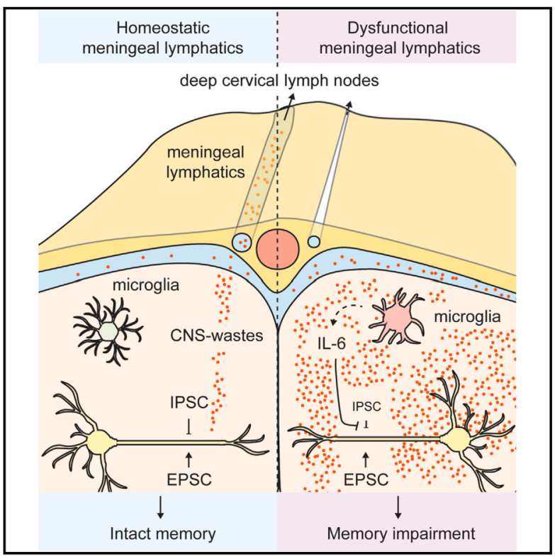
Meningeal lymphatic vessels are responsible for draining cerebrospinal fluid and clearing waste from the central nervous system. Dysfunction of these vessels has been linked to several neurodegenerative diseases, yet the underlying neural mechanisms remain unclear.
On March 21, 2025, Jonathan Kipnis’s team at the Center for Brain Immunology and Glia (BIG), University of Washington, published a study in Cell titled “Meningeal lymphatics-microglia axis regulates synaptic physiology.”The research reveals that the meningeal lymphatics–microglia axis plays a critical role in synaptic physiology and cognitive function. Using surgical and genetic models, the study demonstrates that dysfunction of the meningeal lymphatics disrupts cortical E/I balance and impairs memory. This process is mediated by microglia, with elevated interleukin-6 (IL-6) levels leading to synaptic and behavioral changes. Furthermore, enhancing the function of meningeal lymphatic vessels in aged mice alleviates age-related neurological and cognitive decline, offering a potential therapeutic target for treating age-associated cognitive deterioration.
Memory Impairment Following Surgical Ablation of Meningeal Lymphatic Vessels
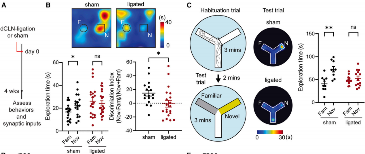
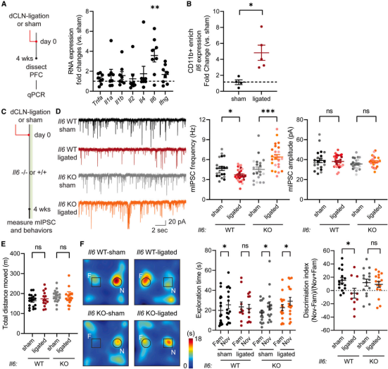
To investigate the role of meningeal lymphatic vessels in neural circuit homeostasis and behavior, researchers performed surgical ligation of the afferent lymphatic vessels that drain cerebrospinal fluid (CSF) to the deep cervical lymph nodes (dCLNs), referred to as “dCLN ligation.” Four weeks later, they assessed the behavioral and electrophysiological characteristics of dCLN-ligated mice compared to sham-operated controls (Figure 1A).
In the novel object recognition test, sham-operated mice spent more time exploring the novel object, while dCLN-ligated mice spent similar amounts of time exploring both objects. This indicates that dCLN-ligated mice were unable to form or retrieve memory of the previously encountered object (Figure 1B).
To validate these findings, researchers used the water Y-maze paradigm (Figure 1C). Sham mice showed a preference for exploring the novel arm, whereas dCLN-ligated mice spent similar time in both arms (Figure 1C). Additional tests confirmed that the memory impairment was not due to changes in motor ability, anxiety, social interaction, or depression-like behavior.
Figure 1. Memory impairment following surgical ablation of meningeal lymphatic vessels
Meningeal Lymphatic Dysfunction Leads to Synaptic E/I Imbalance
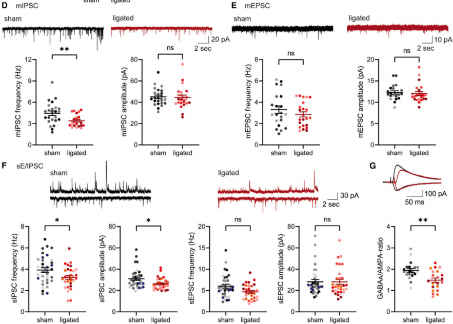
Previous studies have shown that dysfunction of the meningeal lymphatic system can lead to behavioral abnormalities; however, the neural mechanisms linking lymphatic impairment to behavioral changes remain unclear. Since the medial prefrontal cortex (mPFC) is involved in decision-making and cognitive processes, researchers conducted electrophysiological recordings of layer II/III pyramidal neurons in the mPFC. Compared to controls, mice with dCLN ligation exhibited approximately a 20% reduction in the frequency of miniature inhibitory postsynaptic currents (mIPSCs), while the amplitude remained unchanged (Figure 2D). This decrease in mIPSC frequency emerged around 3 weeks after ligation. Similar changes were observed in hippocampal CA1 synapses at 4 weeks post-ligation.
In contrast, excitatory synaptic parameters, including the frequency and amplitude of miniature excitatory postsynaptic currents (mEPSCs), remained unchanged (Figure 2E). Protein analysis showed that the postsynaptic inhibitory marker gephyrin and the presynaptic inhibitory marker VGAT were each reduced by about 20%, while levels of excitatory synaptic molecules such as PSD-95 and vGlut1 were unchanged.
In dCLN-ligated mice, both the frequency and amplitude of spontaneous inhibitory postsynaptic currents (sIPSCs) recorded from individual neurons were reduced (Figure 2F). Additionally, there was a trend toward decreased frequency of spontaneous excitatory postsynaptic currents (sEPSCs). These results suggest that the observed reduction in mIPSC frequency has real physiological consequences that cannot be compensated for by homeostatic mechanisms.
To further validate this, researchers compared inhibitory and paired excitatory synaptic responses in the same neuron under identical stimulation. They electrically stimulated layer I axonal fibers while holding the membrane potential at -70 mV or 0 mV to isolate excitatory or inhibitory responses, respectively. Consistent with previous results, the evoked IPSC/EPSC ratio in neurons from dCLN-ligated mice was approximately 20% lower than in sham-operated controls (Figure 2G).
Microglia Mediate the Synapti / Behavioral Phenotypes Caused by Meningeal Lymphatic Dysfunction
Since meningeal lymphatic vessels are responsible for draining cerebrospinal fluid (CSF), the researchers analyzed the CSF proteome and metabolome of sham-operated and dCLN-ligated mice. The results revealed an increase in certain cellular metabolites. These metabolites may interact with neurons or non-neuronal cells in the brain parenchyma, triggering synaptic and behavioral abnormalities.
Microglia, the resident macrophages of the brain, respond to changes in meningeal lymphatics and monitor neural network activity to optimize circuitry through various mechanisms, including phagocytosis, extracellular matrix remodeling, and modulation of neuronal firing activity.
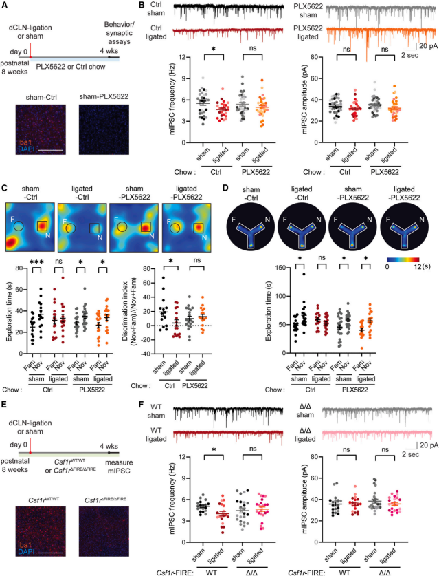
To test whether microglia are necessary for the synaptic and behavioral phenotypes observed in mice with meningeal lymphatic dysfunction, the researchers combined dCLN ligation with microglial depletion. The colony-stimulating factor 1 receptor (CSF1R) signaling pathway is essential for the survival of macrophages, including microglia. Starting from the day of dCLN ligation or sham surgery, mice were fed chow containing the CSF1R antagonist PLX5622. This treatment led to a significant reduction in microglia in the brain (Figure 3A), and the behavioral and synaptic changes observed in dCLN-ligated mice were eliminated (Figures 3B–3D), without affecting general locomotor activity.
To further confirm the role of microglia in mediating the synaptic phenotype following dCLN ligation, researchers ligated the dCLN afferent vessels in Csf1r ΔFIRE/ΔFIRE mice, which specifically lack microglia, while other macrophage populations remain intact (Figure 3E). Consistent with previous findings, Csf1r ΔFIRE/ΔFIRE mice showed no change in mIPSC frequency after dCLN ligation, whereas Csf1r WT/WT littermate controls exhibited a reduction in mIPSC frequency (Figure 3F). No significant changes were observed in mIPSC amplitude in either group (Figure 3F).
On the other hand, dCLN ligation in Rag2 KO mice (which lack T and B cells) still resulted in reduced mIPSC frequency, similar to wild-type (WT) mice.
Figure 3. Microglia mediate the synaptic and behavioral phenotypes in dCLN-ligated mice
dCLN Ligation Induces Morphological, Functional, and Transcriptional Changes in Microglia
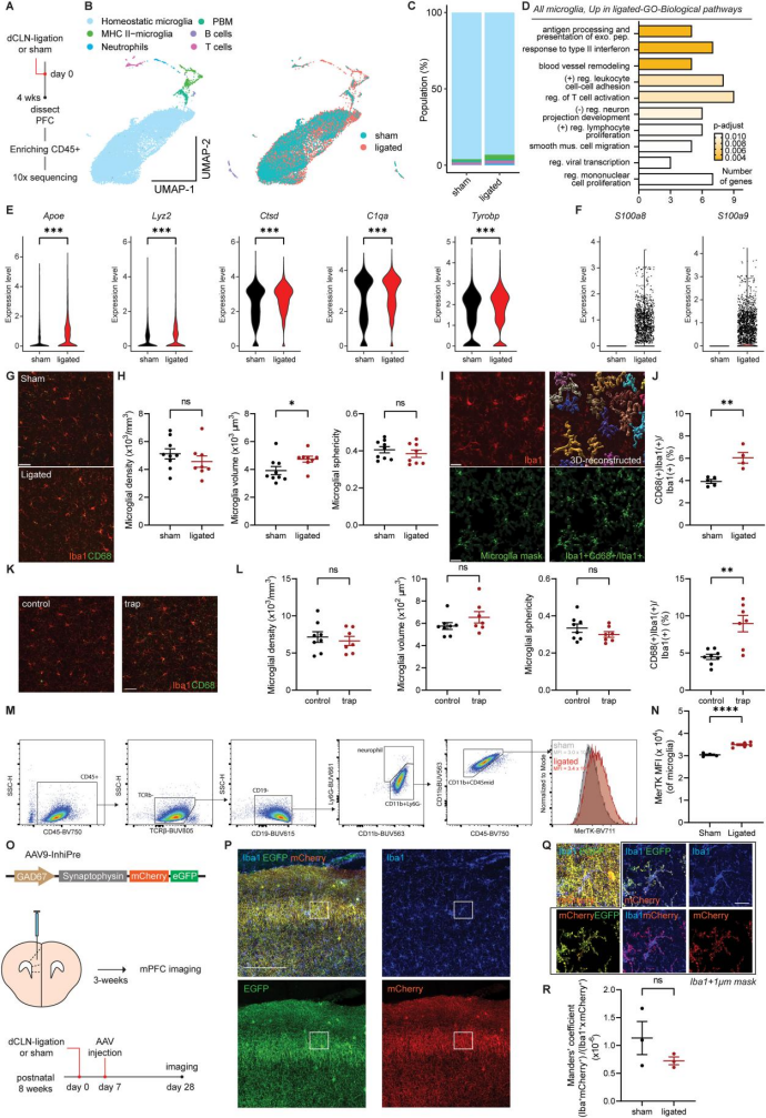
To investigate how dysfunctional meningeal lymphatic vessels affect microglia, researchers isolated CD45⁺ cells from the prefrontal cortex (PFC) of sham-operated and dCLN-ligated mice and performed single-cell sequencing (Figures 4A–4D). The results showed that in dCLN-ligated mice, microglia exhibited upregulated expression of genes associated with phagocytic function and disease-associated microglial signatures, along with increased expression of injury-related molecules (Figure 4F).
Following dCLN ligation, the volume of individual microglial cells increased by approximately 20%, although microglial density and morphological complexity remained unchanged (Figures 4G–4H). Lysosomal volume within microglia increased by about 50% (Figures 4I–4L). Additionally, expression of Mer tyrosine kinase (MerTK), a key receptor involved in phagocytosis, was significantly upregulated in microglia from dCLN-ligated mice (Figures 4M–4N). These findings indicate that microglia respond to meningeal lymphatic dysfunction by altering their transcriptome, morphology, and function.
Microglia are known to engulf synapses during development and in neurodegenerative conditions. To determine whether the observed synaptic phenotype was due to changes in microglial phagocytic activity, the researchers used AAV9-InhiPre (AAV9-GAD67-synaptophysin-mCherry-EGFP) to label inhibitory presynaptic structures. Under physiological pH conditions, both mCherry and EGFP signals are detectable; however, under acidic lysosomal pH, the EGFP signal is quenched, leaving only the mCherry signal.
One week after sham or dCLN ligation surgery, AAV9-InhiPre was injected into the mPFC (Figure 4O). Three weeks post-injection, the PFC was stained with anti-Iba1 antibodies (Figure 4P). The results showed that the volume of synapses engulfed by microglia (mCherry⁺/Iba1⁺) in the mPFC was comparable between sham and dCLN-ligated mice (Figures 4Q–4R).
Figure 4. Alterations in Cortical Microglia in dCLN-Ligated Mice
IL-6 Mediates Synaptic and Behavioral Phenotypes
To investigate the potential mechanisms by which microglia regulate inhibitory synapses, quantitative PCR results showed that Il6 expression in the prefrontal cortex of dCLN-ligated mice increased 3.5-fold (Figure 5A). In the CD11b-positive macrophage population, Il6 expression was also elevated 4 to 6-fold (Figure 5B). Previous studies have demonstrated that acute IL-6 treatment of brain slices can specifically reduce inhibitory synaptic responses, possibly through altered trafficking and/or internalization of γ-aminobutyric acid (GABA) receptors.
To verify whether excess IL-6 mediates the electrophysiological and behavioral phenotypes induced by dCLN ligation, mIPSCs and behavioral tests were performed in Il6 knockout (Il6 KO) mice and age-matched wild-type (WT) mice. These mice were divided into dCLN-ligated and non-ligated groups (Figure 5C).
Compared to sham-operated Il6 KO mice, dCLN-ligated Il6 KO mice exhibited significantly increased mIPSC frequency (Figure 5D). Furthermore, unlike dCLN-ligated Il6 WT littermates, dCLN-ligated Il6 KO mice spent more time exploring the novel object than the familiar one in the novel object recognition test, with no changes in locomotor activity (Figures 5E–5F).
Although the underlying mechanism by which Il6 deletion increases mIPSC frequency after dCLN ligation remains unclear, these results suggest that the IL-6 signaling pathway may be a potential mediator of the dCLN ligation phenotype.
Figure 5. Disappearance of Synaptic and Behavioral Phenotypes Induced by dCLN Ligation in Il6 KO Mice
Classical and Trans IL-6 Signaling Mediate the Effects of Dysfunctional Meningeal Lymphatics on Synapses
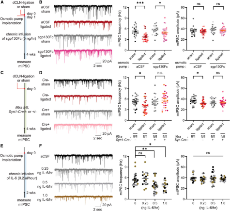
IL-6 is a pro-inflammatory cytokine that signals through two pathways. IL-6 binds to the membrane-bound receptor IL-6Ra, which, together with gp130, initiates downstream signaling (classical IL-6 signaling). Alternatively, IL-6 can bind to soluble IL-6Ra in the extracellular space, forming an IL-6–soluble IL-6Ra complex that activates signaling through membrane-bound gp130 on cells that do not express IL-6Ra (trans IL-6 signaling).
Using sgp130Fc (a soluble gp130 domain fused to an Fc fragment), which binds and neutralizes extracellular IL-6 + soluble IL-6Ra complexes, trans IL-6 signaling can be inhibited. An osmotic pump delivering sgp130Fc or artificial cerebrospinal fluid (aCSF) was implanted into the mPFC of dCLN-ligated mice (Figure 6A). Blocking trans IL-6 signaling partially rescued the reduction in mIPSC frequency (Figure 6B).
Conditional knockout of IL-6Ra in neurons (Il6ra cKO) was generated by crossing Il6ra^fl/fl mice with Syn1-Cre mice, followed by dCLN ligation or sham surgery (Figure 6C). The results showed that the inhibitory synaptic phenotype associated with dCLN ligation was abolished in Il6ra cKO mice (Figure 6D), while excitatory synaptic properties remained comparable between Il6ra cKO and WT mice. This indicates that both classical and trans IL-6 signaling mediate the decrease in mIPSC frequency in the mPFC following dCLN ligation.
Osmotic pumps containing various concentrations of IL-6 (0.25, 0.5, and 1.0 ng IL-6/h) or aCSF were implanted into the mPFC of WT mice, and synaptic phenotypes were assessed after two weeks (Figure 6E). Chronic exposure to 0.5 and 1.0 ng/h IL-6 reduced mIPSC frequency (Figure 6F), while chronic IL-6 treatment had no effect on the frequency or amplitude of mEPSCs. These findings suggest that prolonged IL-6 exposure can alter inhibitory synaptic function.
Figure 6. IL-6 Signaling Pathways Mediate Synaptic and Behavioral Phenotypes in dCLN-Ligated Mice
Enhancing Meningeal Lymphatic Function in Aged Mice Alleviates Age-Related Synaptic and Behavioral Changes
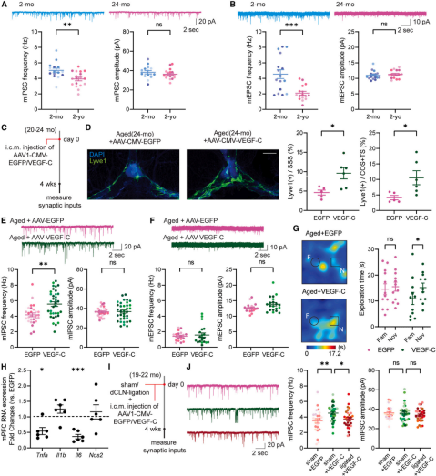
Comparing aged mice (20–24 months old) with young mice (2 months old), researchers found that the frequency of mIPSCs and mEPSCs in layer II/III pyramidal neurons of the medial prefrontal cortex (mPFC) decreased by 22% and 46%, respectively, in aged mice, while their amplitudes remained unaffected (Figures 7A–7B).
Injection of AAV1-CMV-mVEGF-C, which overexpresses vascular endothelial growth factor C (VEGF-C), into the cisterna magna of aged mice enhanced the coverage and function of meningeal lymphatic vessels, increasing the area positive for the lymphatic marker Lyve1 (Figures 7C–7D). Compared to control mice injected with EGFP, VEGF-C treatment restored the reduced mIPSC frequency in the mPFC of aged mice (Figure 7E), while mEPSC frequency and amplitude were unchanged (Figure 7F). Frequencies of spontaneous excitatory and inhibitory postsynaptic currents (sEPSCs and sIPSCs) were both increased. VEGF-C–treated aged mice showed improved performance in the novel object recognition test (Figure 7G), along with reduced levels of Il6 (Figure 7H).
Combining cisterna magna injection of AAV1-CMV-mVEGF-C (or EGFP) with sham or dCLN ligation surgery in aged mice (Figure 7I), the researchers found that VEGF-C treatment increased mIPSC frequency in sham-operated mice (Figure 7J), but this effect was abolished by dCLN ligation (Figure 7J), indicating that the synaptic circuit effects of VEGF-C are mediated by enhanced lymphatic function.
Figure 7. Enhancing Meningeal Lymphatic Function in Aged Mice Mitigates Age-Related Synaptic and Behavioral Changes
Summary
This study is the first to clearly reveal the important regulatory role of the meningeal lymphatics–microglia axis in synaptic physiology and cognitive function, providing a novel perspective for understanding the pathogenesis of neurodegenerative diseases. Although some limitations remain, such as the precise mechanisms underlying the reduction in mIPSC frequency not yet being fully understood, this work has already pointed the way for future research.
The AAVs mentioned in the article can all be packaged by Brain Case. For more information, please contact us at bd@ebraincase.com
Service Type :
Select the service you'd like to purchase.
Order Information(Premade-AAVs)
Please provide us some information about the service you'd like to order.
Order Information(Custom AAV/Lentivirus)
Please provide us some information about the service you'd like to order.
Order Information(Others)
Please provide us some information about the service you'd like to order.