Scientific Breakthrough | Nat. Methods | ATLAS: A Tool for Monosynaptic Anterograde Tracing from Defined Starter Neurons
Release time:2025-06-17 14:52:06
This article introduces a technique called ATLAS (anterograde transsynaptic label based on antibody-like sensors), a method for anterograde transsynaptic tracing. Built on rationally engineered proteins, ATLAS enables genetically defined neurons to be labeled in an anterograde, transsynaptic manner. Using mRNA display, the researchers screened for AMPA.FingR, a protein that binds to the GluA1 receptor, and constructed the ATLAS protein to validate its ability to mediate transsynaptic tracing. Experiments demonstrated that ATLAS achieves strictly anterograde, monosynaptic, and activity-dependent neuronal labeling both in vivo and in vitro. It does not interfere with synaptic transmission and can be used to label diverse neural circuits. The modular design of its components allows for independent replacement or modification, making ATLAS a powerful tool for neural circuit research.
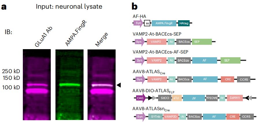
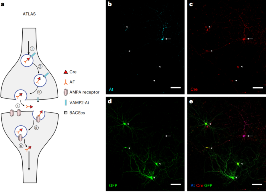
Using mRNA display technology, researchers screened a clone named Fn9.3 from a library that specifically binds to the GluA1 receptor. This clone, termed AMPA.FingR (AF; with FingR referring to intracellular antibody-like fibronectin domains generated via mRNA display), selectively binds to GluA1 receptors and partially localizes to perisynaptic and endocytic regions in neurons. AF can also enter cells via endocytosis. To construct the ATLAS-Cre system, AF was fused with the presynaptic protein VAMP2, and an endogenous BACE1 cleavage site along with an epitope tag (ALFA-tag, abbreviated At) was inserted, resulting in the fusion protein VAMP2-At-BACEcs-AF-Cre. Upon expression, AF-Cre is released into the synaptic cleft, binds to AMPA receptors on the postsynaptic membrane, and enters the nucleus via endocytosis, enabling genetic access to the postsynaptic cell.
Validation of Transsynaptic Tracing In Vitro
ATLAS-Cre was transfected into dissociated neuronal cultures infected with an AAV carrying a floxed-GFP reporter gene (AAV8-DIO-GFP). Some cells were observed expressing both GFP and Cre but not the starter cell marker At, consistent with transneuronal transport of Cre and recombination at the floxed allele. This indicates that Cre had been successfully delivered into the nuclei of postsynaptic cells. These results confirm that ATLAS mediates transsynaptic tracing and enables genetic access to target neurons.
In Vivo Validation of Transsynaptic Tracing
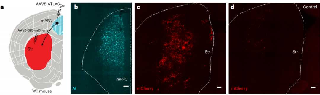
AAV8-ATLAS-Cre was injected into the medial prefrontal cortex (mPFC) of mice, while AAV8-DIO-mCherry was injected into the striatum (Str) to trace unidirectional projections from the mPFC to the Str. One week later, At-positive cells were detected in the mPFC, and a large number of mCherry-positive cells were observed in the Str, indicating that ATLAS successfully mediated transsynaptic transport of Cre, leading to mCherry expression in postsynaptic cells.
Validation in Genetically Defined Neurons
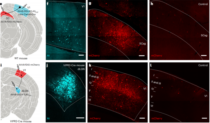
To test cell-type-specific tracing, AAV8-DIO-ATLAS-FLP was co-injected with the lentiviral vector Lenti-CamkII-Cre into the primary visual cortex (V1) of wild-type mice, while AAV8-fDIO-mCherry was injected into the superior colliculus (SC). In the experimental group, At-positive cells were found in V1, and strong mCherry expression was detected in SC, whereas only sparse weakly positive cells were observed in the SC of the control group that received virus injection only in the SC.
In another experiment, AAV8-DIO-ATLAS-FLP was injected into the dorsal lateral geniculate nucleus (dLGN) of VIPR2-Cre mice, and AAV8-fDIO-mCherry was injected into the V1. The results showed At immunostaining specifically in the dLGN and mCherry-positive cells in the V1, demonstrating that ATLAS enables forward transsynaptic tracing from Cre-positive neurons in both wild-type and transgenic mice.
Enhancing Transsynaptic Labeling Efficiency with Exogenous BACE
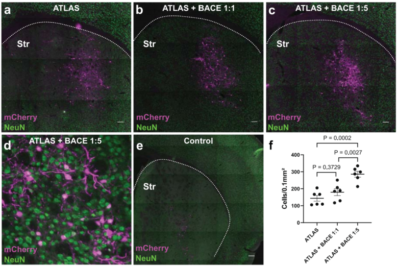
To evaluate how exogenous BACE supplementation affects ATLAS tracing efficiency, AAV8-BACE-HA and AAV8-ATLAS-Cre were co-injected into the mPFC of mice at a 1:1 ratio. This co-injection had no significant effect on the number of mCherry-labeled postsynaptic cells. However, when the two vectors were injected at a 5:1 ratio (BACE:ATLAS-Cre), there was a marked increase in mCherry-positive cells in the Str, with approximately 10% of postsynaptic neurons being labeled. These results suggest that exogenous BACE significantly enhances the labeling density of ATLAS, reaching levels comparable to those achieved by YFV, an anterograde tracing system based on the attenuated yellow fever virus strain YFV-17D.
ATLAS Tracing is Synapse-Specific
Since ATLAS uptake is mediated by the GluA1 receptor—which is absent at inhibitory synapses—any transneuronal labeling observed from inhibitory neurons would likely be nonsynaptic. To test this, AAV8-DIO-ATLAS-FLP and AAV8-fDIO-mCherry were injected into the primary visual cortex (V1) of SST-Cre mice, which express Cre in inhibitory neurons. Two weeks later, nearly all mCherry-labeled cells were co-labeled with the At marker, indicating that ATLAS labeling occurred specifically via synaptic transmission onto postsynaptic neurons.
ATLAS Tracing Does Not Disrupt Synaptic Transmission
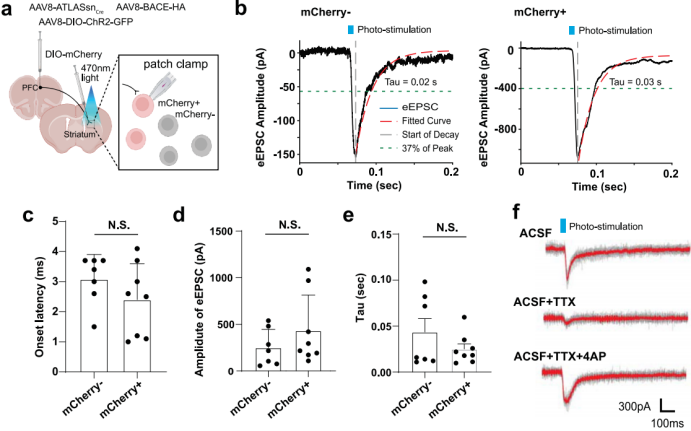
To test whether ATLAS interferes with synaptic transmission, AAV8-ATLASsn-Cre, AAV8-BACE-HA, and AAV8-DIO-ChR2-GFP were co-injected into the mPFC of wild-type mice, while AAV8-DIO-mCherry was injected into the Str. Four weeks later, brain slices were prepared for electrophysiological recordings. When recording from mCherry-labeled postsynaptic neurons, the amplitude of ChR2-evoked excitatory postsynaptic currents (EPSCs) showed no significant increase compared to unlabeled cells. This indicates that ATLAS does not block synaptic transmission and does not induce detectable toxicity.
Electrophysiological Validation of ATLAS Specificity
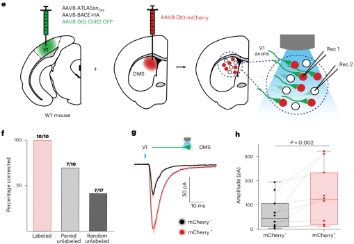
To assess whether ATLAS specifically labels functionally connected neurons, optogenetically evoked currents were recorded in the V1-Str circuit. AAV8-ATLASsn-Cre and AAV8-BACE-HA were co-injected into V1, while AAV8-DIO-mCherry or AAV8-DIO-tdTomato was injected into the dorsomedial striatum (DMS). Two weeks later, AAV8-DIO-ChR2-GFP was injected into the same V1 region to activate ATLAS-labeled presynaptic inputs while minimizing expression competition between ATLAS and ChR2. The results showed that ATLAS-labeled cells were more likely to receive synaptic input, and neurons with stronger synaptic connections were more likely to be labeled, confirming the specificity of ATLAS tracing.
ATLAS Tracing is Strictly Anterograde and Monosynaptic
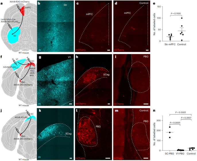
To test whether ATLAS tracing is exclusively anterograde, AAV8-DIO-ATLAS-FLP and Lenti-hsyn-Cre were injected into the Str, while AAV8-fDIO-mCherry was injected into the mPFC. Robust At labeling was observed in the Str, indicating high ATLAS expression, but no mCherry labeling above background was detected in the mPFC. Given that projections from the mPFC to the Str are unidirectional, this confirms that ATLAS does not mediate retrograde labeling.
To assess whether ATLAS tracing is monosynaptic, the V1–superior colliculus (SC)–parabigeminal nucleus (PBG) pathway was examined. AAV8-ATLAS-Cre was injected into V1, and AAV8-DIO-mCherry was injected into SC and PBG. mCherry expression was detected in SC but not in PBG, indicating that ATLAS tracing is restricted to single synapses. Furthermore, when AAV8-ATLAS-Cre was injected into SC and AAV8-DIO-mCherry into PBG, mCherry labeling was observed in PBG, confirming ATLAS can trace SC–PBG projections. These findings demonstrate that ATLAS mediates strictly anterograde and monosynaptic transsynaptic labeling.
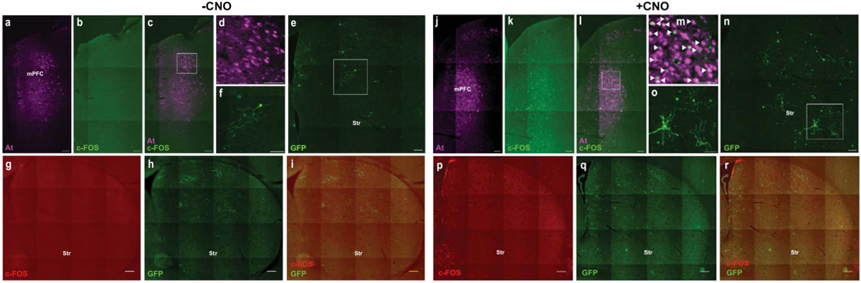
Activity Dependence of ATLAS Tracing
Since ATLAS functions by releasing the AF-recombinase fusion protein from synaptic vesicles, its transsynaptic labeling is likely activity-dependent. To test this in vivo, AAV8-ATLASsn-Cre and AAV8-hHM3Dq-mCherry were injected into the mPFC, and AAV8-DIO-GFP was injected into the Str. Compared to mice without CNO treatment, those treated with CNO showed a significant increase in both the number of GFP-labeled cells and total fluorescence intensity in the Str. These results indicate that ATLAS-mediated transsynaptic labeling is indeed activity-dependent.
Optimization and Application of ATLAS
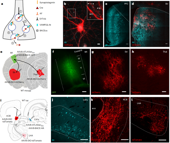
Although VAMP2 is widely used as a presynaptic marker, its overexpression can lead to toxicity. Indeed, expression of ATLAS in the mPFC for 2 or 4 weeks resulted in observable toxicity. To address this, an optimized version of ATLAS was developed by replacing the cytoplasmic domain of VAMP2 with a nanobody against Synaptotagmin-1 (SYTnb), yielding SYTnb-VAMP2Δ-At-BACEcs-AF-Cre (ATLASsn-Cre). When expressed in the mPFC for 2 or 4 weeks, ATLASsn-Cre showed no signs of toxicity. In vitro experiments further supported its functionality: cortical neurons in dissociated culture were transfected with ATLASsn-Cre and infected with AAV8-DIO-mCherry. A large number of cells were labeled with mCherry but not with At, suggesting they were labeled via transsynaptic transfer.
In vivo, AAV8-ATLASsn-Cre and AAV8-BACE-HA were co-injected into the mPFC of mice, while AAV8-DIO-mCherry was injected into the Str. Two weeks later, At expression was detected in the mPFC, and numerous mCherry-labeled cells were found in the Str. The postsynaptic mCherry labeling closely colocalized with At-labeled presynaptic terminals, consistent with transsynaptic labeling mediated by ATLASsn-Cre. Additional tracing of the M1–Str and M1–thalamus circuits further confirmed that ATLASsn-Cre mediates effective transsynaptic labeling.
Labeling Cortico-Subcortical Circuits
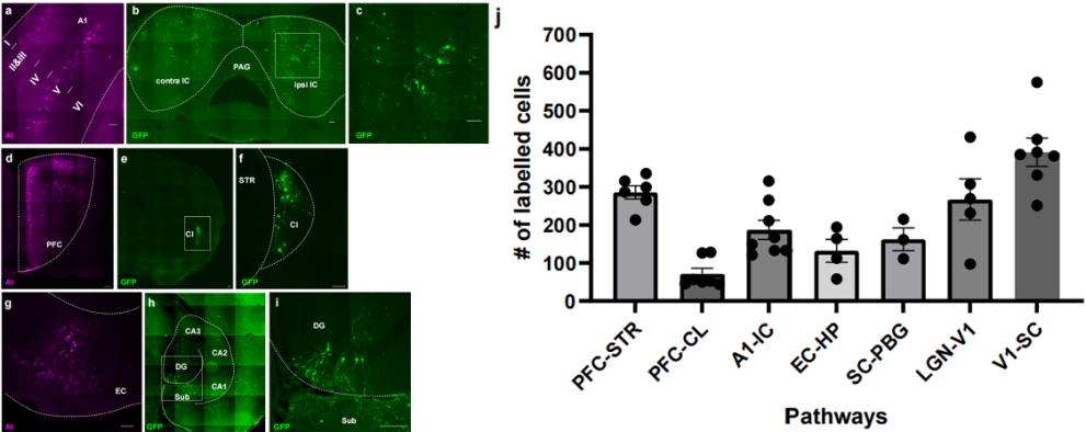
To further validate the versatility of ATLASsn-Cre, three additional neural circuits were traced: (1) auditory cortex (A1) to inferior colliculus, (2) mPFC to claustrum, and (3) entorhinal cortex to hippocampus. In each experiment, AAV8-ATLASsn-Cre and AAV8-BACE-HA were injected into the presynaptic region, and AAV8-DIO-GFP into the corresponding postsynaptic target. After two weeks, At staining was used to identify presynaptic neurons, while GFP indicated postsynaptic labeling. In all cases, both At and GFP labeling were observed, indicating robust transsynaptic tracing. These results demonstrate that ATLASsn-Cre is a broadly applicable tool for mapping diverse neural circuits.
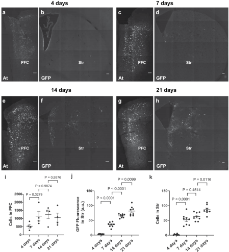
To investigate the temporal dynamics of transsynaptic labeling, AAV8-ATLASsn-Cre and AAV8-BACE-HA were co-expressed in the mPFC, and AAV8-DIO-GFP was expressed in theStr. Mice were analyzed at 4, 7, 14, and 21 days post-injection. Postsynaptic labeled cells began to appear between days 4 and 7, with a significant increase in both the number and intensity of labeling by day 7, and continued growth observed by day 21. In contrast, the number of presynaptic labeled cells stabilized around one week after injection. The number of transsynaptically labeled cells increased markedly during the first 7 days, then the rate of increase slowed, while the total fluorescence intensity continued to grow in a relatively linear fashion. Meanwhile, the number of presynaptic labeled cells remained largely stable after the first week.
Transsynaptic Labeling by ATLAS in Rats
AAV8-ATLASsn-Cre and AAV8-BACE-HA were co-injected into the ventral hippocampal CA1 region of rats, and AAV8-DIO-tdTomato was injected into the nucleus accumbens (ACB). As a result, At staining was observed in CA1, and a large number of tdTomato-positive cells were detected in the ACB. Additionally, tdTomato-labeled axons were found in the lateral hypothalamic area, demonstrating that ATLAS can also mediate transsynaptic labeling in rats.
Conclusion
The ATLAS tracing technique is a rationally designed, anterograde, monosynaptic transsynaptic labeling system with several advantages:
It exclusively performs forward monosynaptic labeling, enabling precise mapping of neuronal connections;
It is non-replicative, non-toxic, and does not interfere with synaptic transmission;
It is activity-dependent, allowing the labeling of behaviorally relevant active neural circuits;
It features a modular design, permitting independent replacement or modification of components to suit different receptors, and is applicable to multiple species.
However, there are also limitations:
It relies on recombinase-based reporter systems, requiring the expression of corresponding genes in postsynaptic cells, which increases experimental complexity;
The overall labeling efficiency is relatively low and time-sensitive, requiring exogenous BACE supplementation to improve performance.
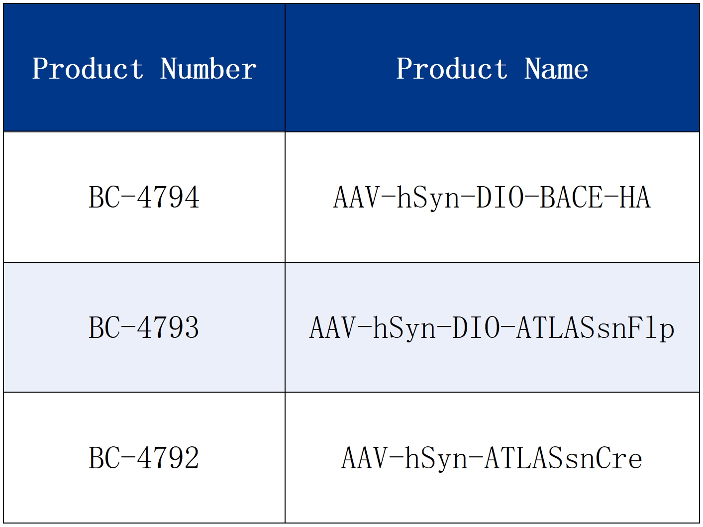
All viruses mentioned in this article are available from Brain Case.
Research on anterograde transsynaptic tracing is limited by the difficulty of tool development, so there are currently few reported methods available for single-neuron-level anterograde tracing. However, each method offers researchers potential solutions to experimental challenges. Braincase Biotech provides a variety of anterograde transsynaptic tools to support your circuit mapping studies. For more details, please visit: https://www.ebraincase.com/support/literature-interpretation/2214.html?1749692246
Service Type :
Select the service you'd like to purchase.
Order Information(Premade-AAVs)
Please provide us some information about the service you'd like to order.
Order Information(Custom AAV/Lentivirus)
Please provide us some information about the service you'd like to order.
Order Information(Others)
Please provide us some information about the service you'd like to order.