Literature Insight | Nature Methods | A “Magic Tool” for Real-Time Observation of Synaptic Structural Dynamics — SynapShot
Release time:2025-05-27 15:11:11
Synapses are the fundamental structural units of the nervous system. Their structural plasticity is crucial for regulating brain function and is closely linked to both physiological and pathological states of the brain. Current methods for studying synaptic organization based on split fluorescent proteins (FPs) face limitations when evaluating synaptic dynamics in vivo, due to the irreversible nature of split FP binding.
On January 8, 2024, the team led by Won Do Heo from the Korea Advanced Institute of Science and Technology (KAIST) published a study in Nature Methods titled “Real-time visualization of structural dynamics of synapses in live cells in vivo.” In this work, the researchers developed a reversible fluorescent protein system—SynapShot—based on a dimerization-dependent fluorescent protein (ddFP) strategy. This system enables real-time detection of complete synaptic structural changes. SynapShot can monitor reversible and bidirectional changes in synaptic structure under physiological stimulation. Its dual-color configuration (green and red) allows simultaneous visualization of two distinct synaptic populations, and it is also compatible with optogenetic technologies.
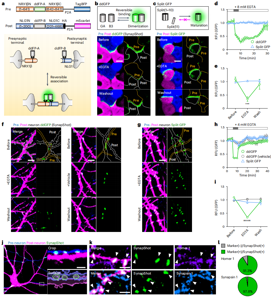
The researchers ingeniously combined ddFPs with engineered synaptic adhesion molecules to construct the SynapShot system. During the design phase, they created various fusion constructs based on previous methods (SynView and mGRASP). In HEK293T cells, they expressed Pre-SynapShot fused to NRX1β and Post-SynapShot fused to NLG1, then co-cultured the two cell types. They found that the combination of NRX1β-GA and NLG1-B3 produced the highest fluorescence intensity at cell contact points (Figure 1a–e).
To verify whether SynapShot accurately marked complete synaptic sites, the team expressed Pre- and Post-SynapShot in cultured hippocampal neurons. Bright green puncta were observed at intersecting points of neurites from different neurons. The ddGFP signal was detected only on dendritic spines adjacent to Pre-SynapShot-expressing neurons and co-expressing Post-SynapShot, indicating high specificity (Figure 1f–i). Immunocytochemistry further confirmed that SynapShot fluorescence co-localized with endogenous excitatory and inhibitory pre- and postsynaptic markers, precisely reporting functional synapse locations (Figure 1j–l).
Monitoring Synaptic Structural Changes with SynapShot
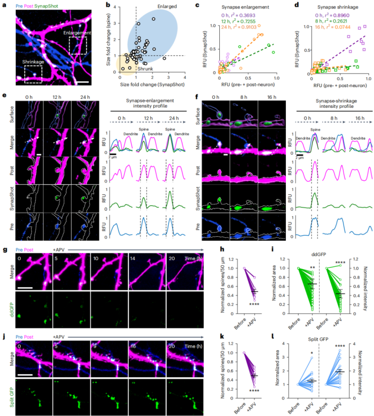
To explore the relationship between synaptic structural dynamics and SynapShot signaling, researchers performed time-lapse imaging over a 24-hour period. They found that SynapShot signal intensity positively correlated with dendritic spine size—enlarged spines showed stronger signals, while shrinking spines showed reduced signals (Figure 2a–f).
To further validate SynapShot's reversibility under physiological stimulation, they treated neurons with an NMDA receptor antagonist (APV) during time-lapse imaging. SynapShot signals significantly decreased and dendritic spines shrank accordingly. In contrast, traditional split-GFP signals remained stable or even increased after APV treatment (Figure 2g–l). This comparison strongly demonstrated that SynapShot reliably reports functionally relevant structural changes in intact synapses.
In a fluorescence recovery after photobleaching (FRAP) experiment, about 50% of SynapShot signal recovered within 30 seconds after bleaching a single synapse. In contrast, SynView—based on irreversible NRX1β-NLG1 interactions—showed no recovery, highlighting SynapShot's unique advantage in tracking synaptic molecular dynamics.
Figure 2. Monitoring complete synaptic structural dynamics with SynapShot.
Dual-Color SynapShot and Compatibility with Optogenetics
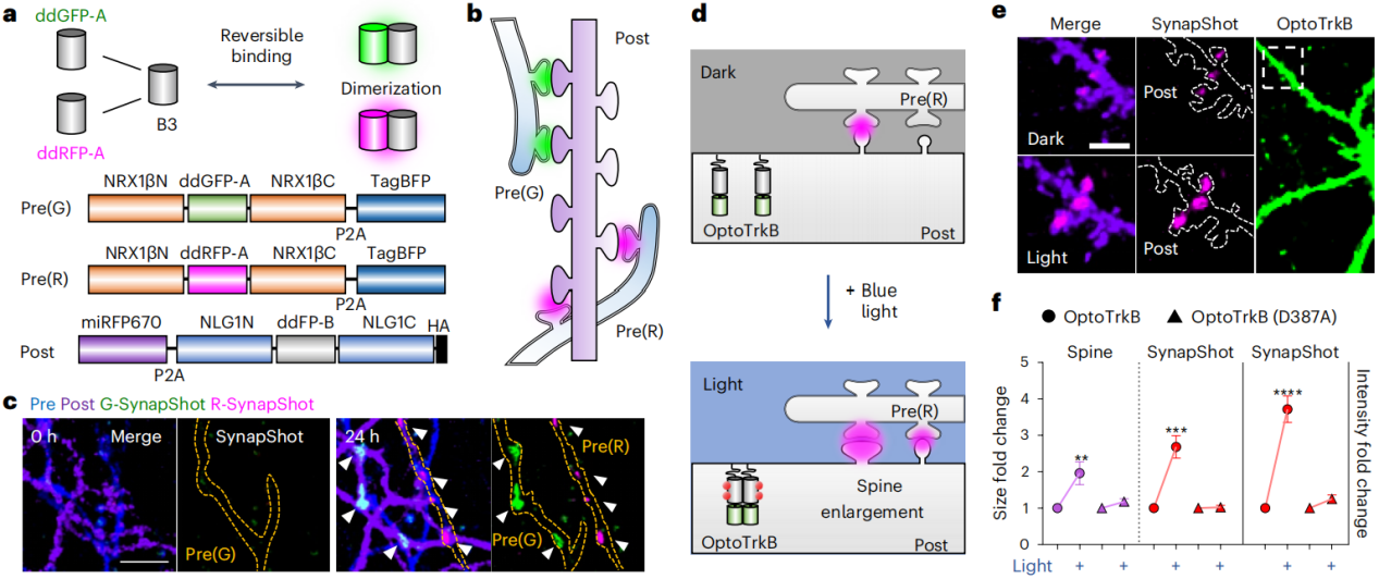
A key feature of ddFPs is that the A copy of ddGFP or red ddFP (ddRFP) can bind the B copy (B3) with similar affinity, producing green or red fluorescence, respectively. Based on this, the researchers developed a red-shifted excitation version of SynapShot, enabling simultaneous visualization of two distinct synaptic populations formed by two presynaptic neurons and one postsynaptic neuron (Figure 3a–b).
By expressing green and red versions of Pre- and Post-SynapShot in different neurons, they detected green and red puncta at different positions on the same dendrite (Figure 3c), making it possible to analyze multiple synaptic inputs on a single postsynaptic neuron.
To test compatibility with optogenetic tools, they used red SynapShot in postsynaptic neurons expressing OptoTrkB. They monitored synaptic changes following endogenous BDNF signaling triggered by light activation. After OptoTrkB activation, dendritic spine size doubled, SynapShot signal intensity increased 2.7-fold, and overall fluorescence intensity increased 3.7-fold. This experiment not only showcased the powerful capabilities of dual-color SynapShot but also emphasized its tremendous potential for precisely regulating and monitoring synaptic dynamics when combined with optogenetic techniques.
Figure 3. Dual-color SynapShot and its compatibility with optogenetics.
Validating SynapShot's Real-Time Synapse Monitoring in Behaving Mice
Researchers designed recombinant adeno-associated viruses (AAVs) encoding Pre-SynapShot (AAV2/5-CAG-GA-Nrx1βc-TagBFP) and Post-SynapShot (AAV2/5-CAG-DIO-B3-NL1C-2A-mScarlet), which were injected into the CA3 region and contralateral CA1 region, respectively. Acute brain slice imaging revealed SynapShot signals in the contralateral CA1, while no signals were detected in the ipsilateral CA1 (which lacked Post-SynapShot) or in CA1 regions expressing only Post-SynapShot. This demonstrated that SynapShot can specifically label synaptic connections in vivo.
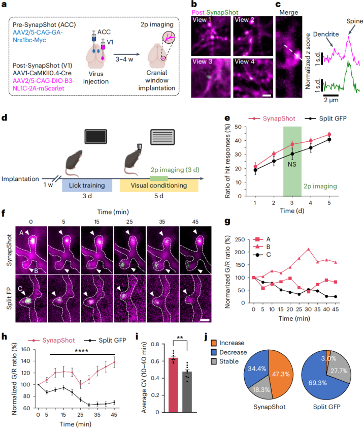
To study synaptic dynamics in live animals, the team expressed Pre-SynapShot via AAV2/5-CAG-GA-Nrx1βc-Myc in anterior cingulate cortex (ACC) neurons, which project axons to the primary visual cortex (V1). Simultaneously, a Cre-dependent Post-SynapShot virus (AAV2/5-CAG-DIO-B3-NL1C-2A-mScarlet) was expressed in V1 neurons (Figure 4a). In vivo two-photon imaging showed sparse SynapShot expression in V1, and ddGFP signals were co-localized with dendrites of neurons from both regions, indicating that axons from ACC neurons formed sparse yet specific synapses with V1 neurons (Figure 4b). Line scan measurements along dendritic shafts and spine heads confirmed that SynapShot signals were restricted to the spine heads (Figure 4c).
The researchers also compared ddGFP signals from SynapShot with split-GFP signals from mGRASP. Mice underwent a 5-day visual discrimination training task (Figure 4d, e), during which licking behavior associated with water rewards increased over time. No behavioral difference was observed between mice expressing SynapShot or mGRASP (Figure 4f). To monitor dynamic changes in SynapShot fluorescence during learning, dendritic imaging of V1 neurons was performed on day 3 of the task. Some spines showed dynamic changes in the ratio of ddGFP (G) to dendritic red fluorescence (R)—the G/R ratio—throughout the task, indicating synaptic strength changes associated with learning could be tracked in vivo. In contrast, most split-GFP signals did not show similar dynamic changes and often diminished at individual synapse points (Figure 4g, h). When analyzing the coefficient of variation of mean fluorescence, SynapShot-labeled synapses exhibited more significant signal changes than those labeled by the split-GFP system. These results suggest that SynapShot detects subtle synaptic changes with greater precision than the mGRASP system (Figure 4i, j).
Figure 4. SynapShot signal in ACC–V1 connections during a visual discrimination task.
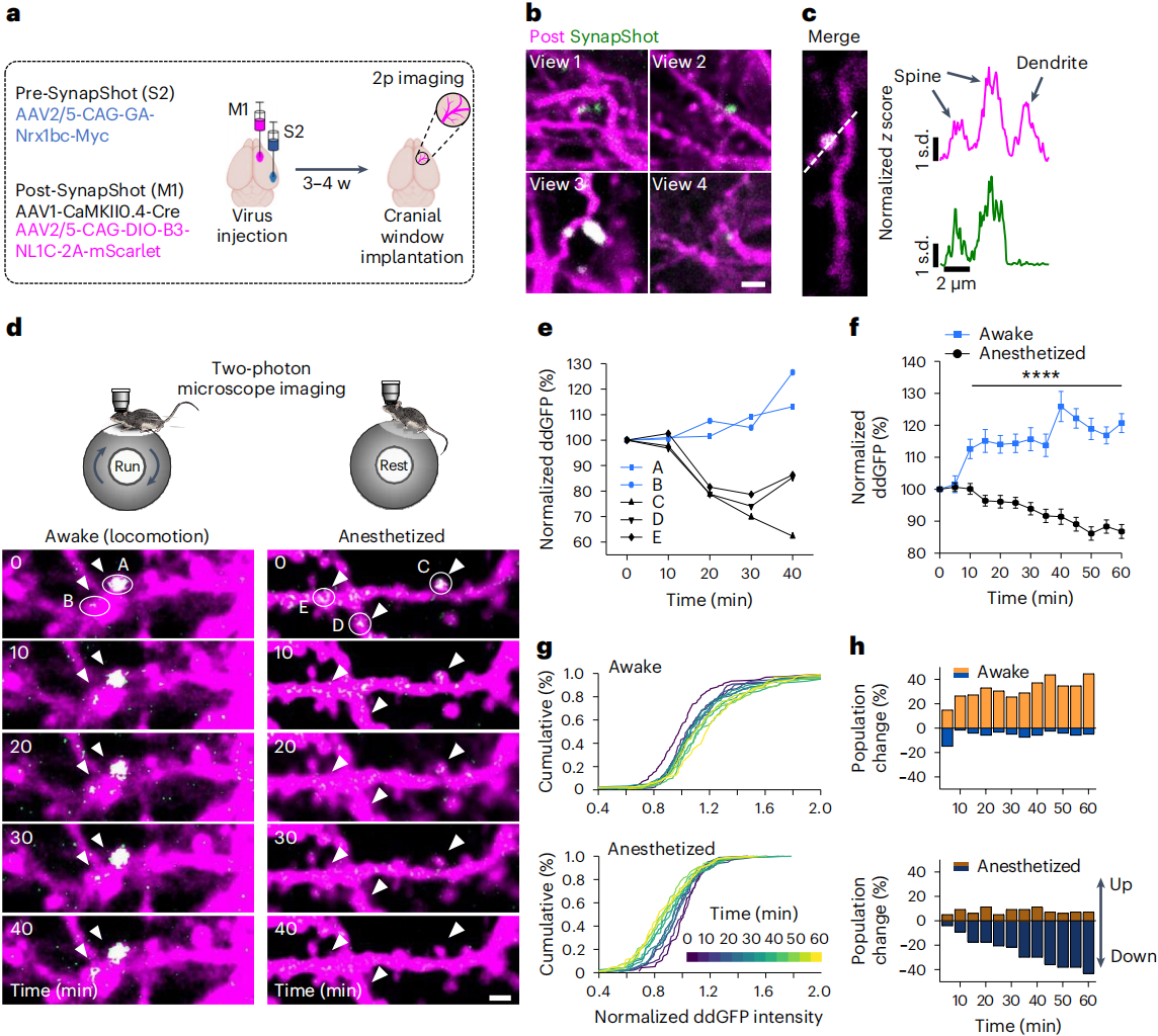
To determine whether these changes reflect learning-related neuronal functional activity, researchers evaluated SynapShot signals in the primary motor cortex (M1) of awake mice through a cranial window,targeting axonal projections from the secondary somatosensory cortex (S2) (Fig. 5a–c).Results showed that during mouse movement, the intensity of individual fluorescence spots in ddGFP signals on the same dendritic spines of M1 neurons gradually increased over time, with fluctuations in the signal;in contrast, in anesthetized mice, the signal gradually decreased (Fig. 5d).Overall, SynapShot signal intensity decreased over time under anesthesia (Fig. 5e, f).A significant difference in average signal intensity was observed between awake and anesthetized states over a 1-hour period (Fig. 5g).At the global level, signal intensity also differed significantly between these two states (Fig. 5h).Taken together, these results demonstrate the utility of SynapShot in monitoring the dynamics of individual synapses in freely moving animals.Furthermore, under the same experimental conditions, mice expressing mGRASP did not show dynamic changes in fluorescence.
Figure 5. Visualizing synaptic structural plasticity in the somatosensory-motor circuit of mice
SynapShot technology has brought a revolutionary advancement to neuroscience research — it acts like a “super microscope,” enabling us to clearly and dynamically observe synaptic changes in real time.From its ingeniously designed concept to its excellent performance in cellular and animal experiments, SynapShot demonstrates tremendous scientific value.However, the technology also has limitations — the ddFP signal is significantly reduced in fixatives such as paraformaldehyde, making it more suitable for real-time visualization of synaptic dynamics in live cells and intact brains.