Literature Interpretation | Nature | How to Detect Glial Phagocytosis of Synapses
Release time:2025-06-05 16:10:54
In the adult hippocampus, synapses are continuously formed and eliminated, but the mechanisms and functions of synapse elimination remain unclear. It is known that astrocytes can phagocytose synapses during development, and researchers have hypothesized that they may play a similar role in the adult brain, contributing to circuit homeostasis. On December 23, 2020, the lab of Won-Suk Chung at the Korea Advanced Institute of Science and Technology (KAIST) published an article in Nature titled "Astrocytes phagocytose adult hippocampal synapses for circuit homeostasis." The authors discovered that in the hippocampal region of adult mice, astrocytes phagocytose excitatory neuronal synapses, and this phenomenon correlates with neuronal activity. The MEGF10 protein in astrocytes plays a key role in this process. Moreover, the researchers developed a molecular indicator to detect glial-mediated synapse elimination and to quantify the types of synaptic connections being removed.
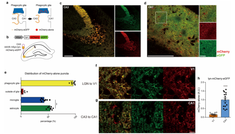
A dual-reporter system based on mCherry-eGFP was employed to monitor glial phagocytosis. This system has been previously used to detect acidification during autophagy. Under neutral pH conditions, both mCherry and eGFP fluoresce normally; however, in acidic environments such as lysosomes, only mCherry retains its fluorescence. To visualize glial phagocytosis, an adeno-associated virus serotype 9 (AAV9) vector was used to deliver a membrane-bound (lyn-tagged) mCherry-eGFP construct driven by the hSyn promoter (Figure 1a,b).
After AAV injection and expression in the CA3 region of the adult mouse hippocampus, most CA3 neurons and their projecting fibers exhibited both mCherry and eGFP signals (Figure 1c,d). However, in the CA1 region—innervated by CA3 neurons—numerous mCherry-only puncta were observed, indicating localization within acidic organelles (Figure 1d). Furthermore, most of these mCherry-only puncta were found within astrocytes or microglia (Figure 1e).
Notably, when the same virus was injected into the lateral geniculate nucleus (LGN) to label thalamic projections to the primary visual cortex, no mCherry-only puncta were observed (Figure 1f-h), suggesting region-specific phagocytic activity.
Figure 1. Development of an mCherry-eGFP reporter system for monitoring glial synaptic phagocytosis.
2. Astrocytic Phagocytosis of Synapses
How can we detect astrocytic phagocytosis of synapses? This can be achieved by designing four specific reporter constructs:
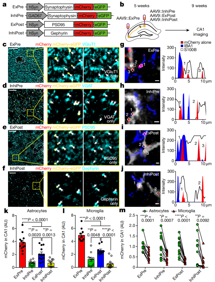
The ExPre reporter, driven by the hSyn promoter, expresses synaptophysin (SYP)-mCherry-eGFP to label excitatory presynaptic structures.
The InhiPre reporter encodes the same fusion protein under the control of the Gad67 (also known as Gad1) promoter, targeting inhibitory presynaptic structures.
The ExPost reporter uses the hSyn promoter to express PSD95-mCherry-eGFP, marking excitatory postsynaptic structures.
The InhiPost reporter also uses hSyn to drive gephyrin (GPHN)-mCherry-eGFP, labeling inhibitory postsynaptic structures (Figure 2a).
When AAV9 vectors carrying these reporters were injected into the CA3 or CA1 regions of the mouse hippocampus (Figure 2b), the fusion proteins specifically colocalized with either excitatory or inhibitory synapses in CA1 (Figure 2c–f). Consistent with data from the lyn-mCherry-eGFP system, numerous mCherry-only puncta—indicative of engulfment into acidic organelles—were found at both excitatory and inhibitory synapses in CA1 (Figure 2g–j). Notably, astrocytes and microglia engulfed significantly more excitatory than inhibitory synapses.
Figure 2. Astrocytes primarily engulf CA1 synapses in the adult hippocampus.
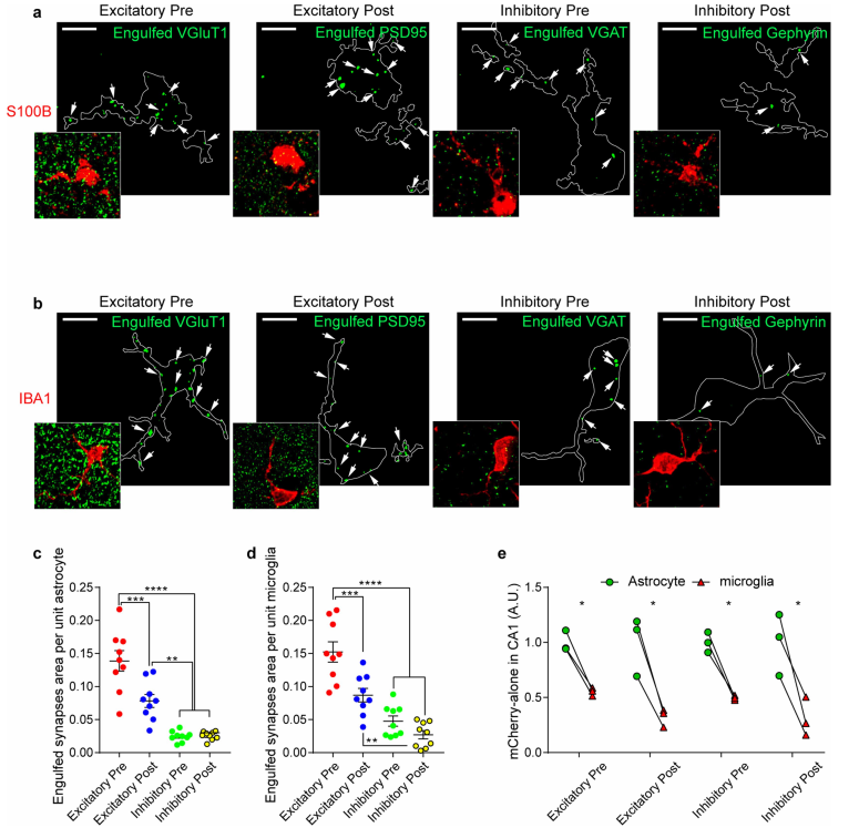
Immunohistochemistry further revealed that glial cells contained more VGLUT1⁺ or PSD95⁺ excitatory puncta than VGAT⁺ or Gephyrin⁺ inhibitory puncta (Figure 3a–d), supporting the conclusion that excitatory synapses are the main targets of glial phagocytosis in the adult CA1. Additionally, astrocytes showed a greater number of engulfed excitatory or inhibitory synaptic mCherry-only puncta than microglia (Figure 3e). These findings suggest that while microglia are traditionally viewed as the brain's primary phagocytes, astrocytes play a dominant role in synapse elimination during normal synaptic remodeling in the adult CA1.
Figure 3. Astrocytes and microglia engulf more excitatory synapses.
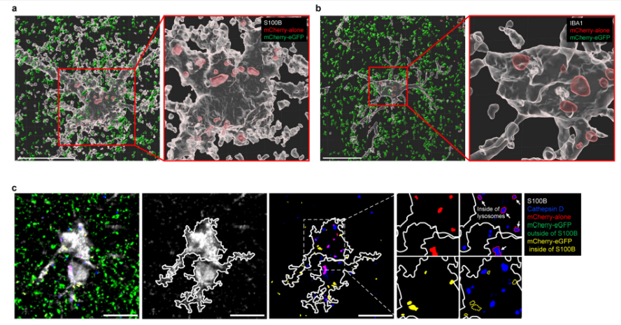
Detailed analysis of ExPre-derived puncta within astrocytes showed that 93% were mCherry-only, while only 4.6% showed both mCherry and eGFP signals (Figure 4a, c). In contrast, both signals were comparably present in early endosomes, indicating that eGFP is lost during transport from endosomes to lysosomes. Some mCherry-only puncta also colocalized with synaptic markers, representing recently engulfed synaptic materials.
Figure 4. Subcellular localization of mCherry-eGFP and mCherry-only puncta in glial cells.
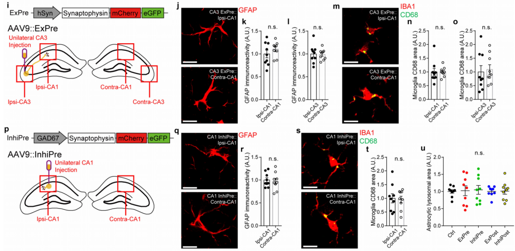
Electrophysiological recordings confirmed that AAV-mediated expression of fluorescent reporters did not induce abnormal synaptic properties (sEPSCs and sIPSCs), nor did it affect excitability or membrane characteristics. Furthermore, no signs of reactive gliosis were observed in hippocampal tissues following AAV injection (Figure 5i–u). Finally, astrocytic phagocytosis of excitatory synapses in the hippocampus remained consistent across ages from 3 to 9 months.
In summary, these results demonstrate ongoing endogenous synaptic remodeling in the adult hippocampus, with astrocytes actively contributing by continuously engulfing synapses.
Figure 5. AAV-based synaptic phagocytosis reporters do not alter synaptic properties or induce reactive gliosis.
3. Quantification of Glial Phagocytosis
To quantify glial phagocytosis, brain slice images were acquired using confocal microscopy and analyzed with ImageJ software and the Diana plugin to count synaptic puncta. Synapses were categorized and counted by type—excitatory synapses (VGLUT1⁺ or PSD95⁺) and inhibitory synapses (VGAT⁺ or Gephyrin⁺)—to assess changes in synapse numbers under different conditions.
Synaptic structures were further examined using scanning electron microscopy (SEM) and transmission electron microscopy (TEM). 3D reconstruction allowed precise localization of astrocytic phagocytic sites and enabled detailed analysis of structural parameters such as synaptic density, total vesicle number, and docked vesicle number. These morphological data provided insights into how astrocytic phagocytosis affects synaptic architecture.
To isolate mCherry-alone puncta, the eGFP signal was subtracted from the mCherry signal. Any mCherry puncta that overlapped even minimally with eGFP were excluded from further quantification and image processing. Single-plane confocal images were then analyzed for individual mCherry-alone, green, and merged red-green signals. Colocalization analysis was performed using the Diana plugin.
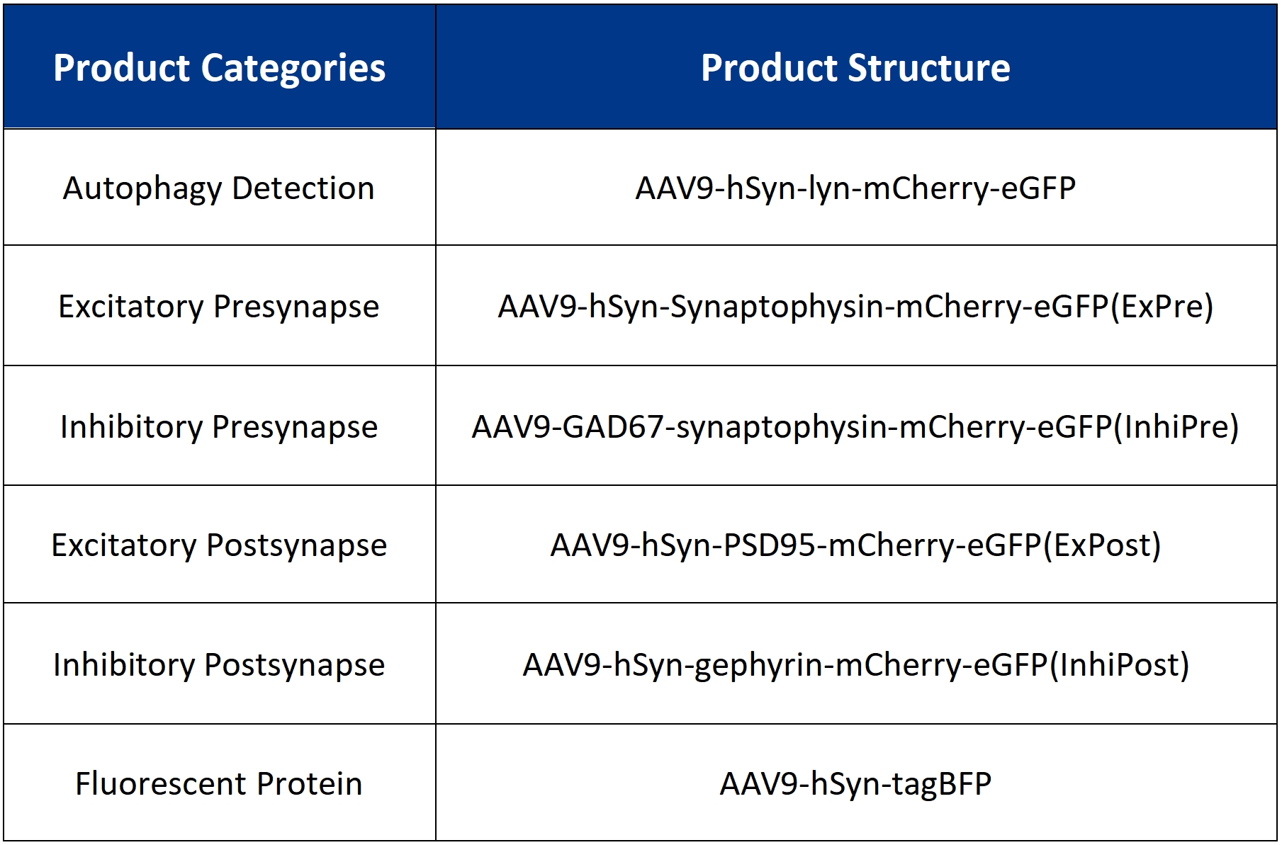
Viral vectors used in this study:
Contact Us
BrainCase offers packaging services for the viruses listed above and provides personalized customization services. For more information, please contact bd@ebraincase.com
Service Type :
Select the service you'd like to purchase.
Order Information(Premade-AAVs)
Please provide us some information about the service you'd like to order.
Order Information(Custom AAV/Lentivirus)
Please provide us some information about the service you'd like to order.
Order Information(Others)
Please provide us some information about the service you'd like to order.