CRISPR-Cas9 Technology: Precise, Efficient, and Reliable Custom Gene Editing Services
Release time:2025-02-07 16:13:13
Gene editing, which involves the precise and targeted modification of an organism's genetic material, is one of the most significant advancements in the field of molecular biology. It has profound applications, from revealing fundamental biological processes to advancing medicine, agriculture, and biotechnology. The programmability of CRISPR-Cas nucleases allows them to create site-specific double-strand breaks in DNA, enabling rapid genome editing technology. CRISPR-Cas9 is a type II system within the CRISPR framework, and the typical Cas9 protein (SpCas9) from Streptococcus pyogenes was the first Cas nuclease used for genome editing. To cleave exogenous genes, only one cleavage protein (Cas9) is needed. Due to its relative simplicity in design and operation, it has been widely applied in gene editing, facilitating gene knockouts, knock-ins, and transcriptional activation.
Ⅰ. Principle of CRISPR-Cas9 Technology
1. Composition of the CRISPR-Cas9 System
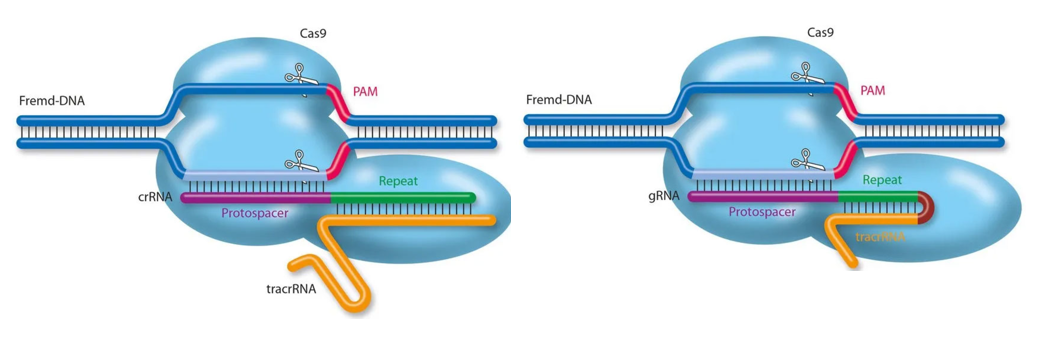
Figure 1: The natural CRISPR-Cas9 system (left) and the modified CRISPR-Cas9 system (right) (image source: internet).
The CRISPR-Cas9 system is an adaptive immune defense mechanism originating from bacteria and archaea, used to protect against the invasion of foreign viruses and plasmids. The natural CRISPR-Cas9 system consists of three components: SpCas9 (referred to as Cas9), crRNA, and tracrRNA. The crRNA and tracrRNA form a guide RNA (gRNA) through local base pairing, which, when bound to the Cas9 protein, directs the protein to recognize and cleave the target DNA sequence (Figure 1, left). To simplify experimental design and enhance the stability of the gRNA, researchers have fused crRNA and tracrRNA into a single RNA molecule [1], referred to as sgRNA (single-guide RNA) (Figure 1, right).
2.Working Mechanism of CRISPR-Cas9
The working mechanism of CRISPR-Cas9 gene editing technology can be divided into the following steps: (1) Target Gene Double-Strand Break: A single-strand RNA guide molecule (sgRNA) that is complementary to the target DNA sequence is designed and introduced into the target cell along with the Cas9 protein complex. Under the specific guidance of the sgRNA, the Cas9 protein reaches a specific location in the genome, where it cleaves the double-stranded DNA (dsDNA), creating a double-strand break (DSB). (2) DNA Break Repair: When the cell repairs the DNA break caused by the cleavage, mutations can be introduced, enabling gene knockout, insertion, or replacement. This process can be achieved through two main DNA repair mechanisms: Non-Homologous End Joining (NHEJ) and Homologous Directed Repair (HDR).
The modified CRISPR-Cas9 system has become the preferred tool for researchers in gene editing. For the CRISPR-Cas9 protein to successfully recognize the target sequence, two conditions must be met: · The 5' end of the sgRNA (approximately 20 nt for SpCas9, or 21 nt for SaCas9) must base-pair precisely with the target DNA sequence; ·The 3' end of the target DNA must contain an appropriate PAM sequence. For SpCas9, the PAM sequence is typically NGG.After CRISPR-Cas9 cuts the target DNA, it generates a DSB, which serves as the foundation for all gene editing based on nucleases.
Ⅱ. CRISPR-Cas9 Gene Editing Applications
CRISPR-Cas9 gene editing technology has two main applications:
Gene Knockout (KO): By inducing a DSB in the target gene using CRISPR-Cas9, the cell repairs the break through the NHEJ repair mechanism. During this repair process, insertions or deletions (indels) may occur, leading to a loss of function of the target gene. This is a commonly used method for gene knockout. Knock in (KI)or Replacement : Using the HDR mechanism, specific mutations or exogenous sequences are introduced at a precise location in the target gene. This requires providing a homologous DNA template containing the desired changes. The cell uses this template to repair the DSB via HDR, achieving precise editing at the targeted site. Additionally, CRISPR-Cas9 gene editing technology can also be used for transcriptional regulation of genes: CRISPR interference (CRISPRi) and CRISPR activation (CRISPRa). CRISPRi involves using a catalytically inactive Cas9 (dCas9) fused with a transcriptional repressor (such as KRAB). The sgRNA guides dCas9 to the promoter region of a specific gene, thereby inhibiting its transcriptional activity. In contrast, CRISPRa involves fusing dCas9 with a transcriptional activator (such as VP64), and the sgRNA guides it to the target gene's promoter region to enhance its transcriptional activity.
Ⅲ Customer Article: Technical Case Sharing
Case 1: Gene Knockout
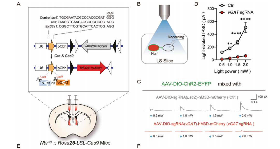
On August 26, 2022, the team of Zhu Yingjie from the Shenzhen Institute of Advanced Technology, Chinese Academy of Sciences, published an article titled "A circuit from lateral septum neurotensin neurons to tuberal nucleus controls hedonic feeding" in Molecular Psychiatry (IF 15.9) [2]. The article mainly explores the role of neurotensin (Nts)-positive neurons in the lateral septum (LS) in regulating hedonic feeding. The study found that by using tetanus toxin (TeNT)-mediated synaptic inactivation or optogenetic methods to silence Nts-positive neurons, the intake of palatable food could be specifically promoted. Conversely, activating Nts-positive neurons through chemogenetics suppressed the intake of general food. In addition, the study used CRISPR-Cas9 gene editing technology to reduce the release of GABA transporter vGAT and Nts from Nts neurons in the LS. The results suggest that GABA release may inhibit hedonic feeding, while strong activation of Nts neurons can suppress overall food intake. This research revealed that the inhibitory circuit from the Nts neurons in the LS projecting to the tuberal nucleus (TU) in the hypothalamus plays a key role in regulating hedonic feeding. It provides insights for developing new therapies aimed at hedonic feeding, which can be used to treat obesity and related metabolic diseases.
Figure 2: Experimental Design for vGAT and Nts Knockdown Mediated by CRISPR-Cas9 Technology
Case 2: Gene Replacement (Point Mutation)
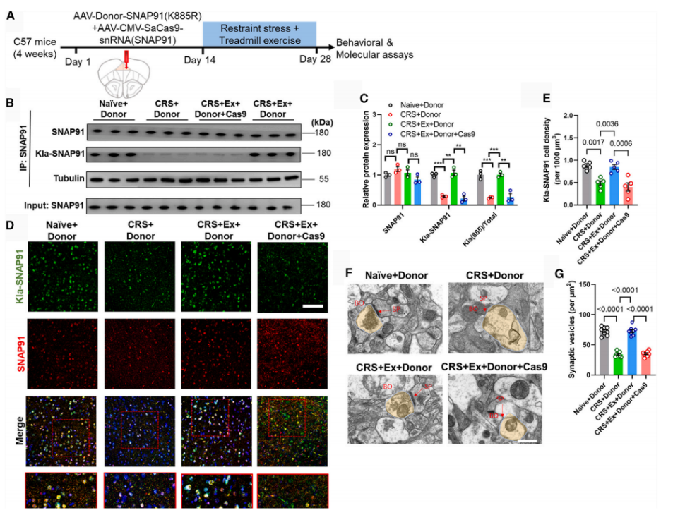
On August 19, 2024, the Zhang Li research team from the Guangdong-Hong Kong-Macau Institute of Central Nervous System Regeneration, Jinan University, published an article titled "Physical exercise mediates cortical synaptic protein lactylation to improve stress resilience" in Cell Metabolism [3]. The article primarily investigates how physical exercise enhances stress resilience by promoting lactylation modification of cortical synaptic proteins. The study found that chronic stress damages the lactylation of cortical proteins, while treadmill training can restore this lactylation pattern. Based on proteomics quantitative analysis of lactylation modifications and customized antibodies, it was found that the lactylation level of lysine at position 885 of SNAP91 was significantly increased after exercise. Further, the study used CRISPR-Cas9 technology to introduce a point mutation (K885R) at the 885th position of the SNAP91 protein to inhibit its lactylation activity and expressed this mutated protein in the medial prefrontal cortex (mPFC) of mice. The results showed that the absence of SNAP91 lactylation impaired the structure and function of synapses (Figure 3). In vivo two-photon imaging revealed that the mPFC neuronal network activity was significantly downregulated in the mice expressing the mutated protein, accompanied by a suppression of the exercise-induced anti-anxiety effect. This suggests that exercise modulates SNAP91 protein lactylation to maintain cortical neuronal activity and exert anti-anxiety effects. The lactylation of SNAP91 is critical for the exercise-mediated anti-anxiety effect. This finding unveils the non-metabolic role of lactate in regulating brain function and provides new insights into how the brain adapts to energy demands during exercise.
Figure 3: Impact of the SNAP91 Point Mutation Model Mediated by CRISPR-Cas9 Technology on Synaptic Structure and Function
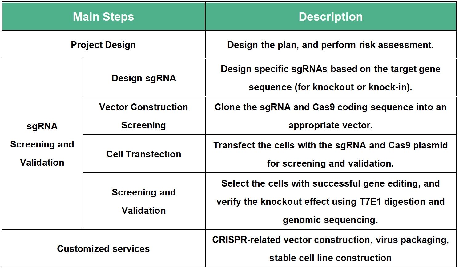
Ⅳ CRISPR-Cas9 Technology Service Process
References
[1]Jinek M, Chylinski K, Fonfara I, Hauer M, Doudna JA, Charpentier E. A programmable dual-RNA-guided DNA endonuclease in adaptive bacterial immunity. Science. 2012 Aug 17;337(6096):816-21.
[2]Chen Z, Chen G, Zhong J, Jiang S, Lai S, Xu H, Deng X, Li F, Lu S, Zhou K, Li C, Liu Z, Zhang X, Zhu Y. A circuit from lateral septum neurotensin neurons to tuberal nucleus controls hedonic feeding. Mol Psychiatry. 2022 Dec;27(12):4843-4860.
[3]Yan L, Wang Y, Hu H, Yang D, Wang W, Luo Z, Wang Y, Yang F, So KF, Zhang L. Physical exercise mediates cortical synaptic protein lactylation to improve stress resilience. Cell Metab. 2024 Sep 3;36(9):2104-2117.
Contact Us
Brain Case can provide customers with a full range of vector construction, virus packaging and stable cell line construction services. If you are interested in customized services such as gene knockout and knock-in based on CRISPR-Cas9 technology, please contact bd@ebraincase.comfor details or to place an order.
Service Type :
Select the service you'd like to purchase.
Order Information(Premade-AAVs)
Please provide us some information about the service you'd like to order.
Order Information(Custom AAV/Lentivirus)
Please provide us some information about the service you'd like to order.
Order Information(Others)
Please provide us some information about the service you'd like to order.