E-mail:BD@ebraincase.com
Tel:+8618971215294
English 中文版
Virus Vector - Viral Vector Production - BrainCaseVirus Vector - Viral Vector Production - BrainCase
  • Home
  • Virus product library
    CRISPRRNAiHSV-helperRV-helperNeurophilic virusCalcium SensorsOptogenetics activationOptogenetics inhibitionChemical geneticsSparse labelingFluorescent proteinBiosensorsRecombinaseApoptosis & AutophagyDisease ModelNeurotoxicityOther
  • Products & Service

    Product Center

    Virus

    VSV-vaccine and gene therapy research
    Retrovirus-RCAS-TVA
    Lentivirus Vector-Lentivirus Production
    Rabies Virus Vector-RBV Vector
    Herpes simplex virus-Oncolytic and anterograde tracing
    PRV-retrograd multisynaptic-Peripheral
    AAV-gene therapy vectors-neuroscience

    Animal Model

    Neurological Disease Models
    Tumor animal models-anti-tumor
    Digestive System Disease Animal Model
    Cardiovascular System Disease Animal Models

    Plasmid Construction

    Library Construction
    Plasmid design and construction

    Popular Applications

    Gene Regulation

    Gene Overexpression-Brain Case
    RNA interference(RNAi)-siRNA-Brain Case
    Gene Editing - CRSIPR cloning - BrainCase

    Neural Circuit Function Research

    Optogenetics - BrainCase
    Chemical genetics-DREADDs-Brain Case
    Calcium signal recording-Gels- Brain Case
    GRAB Neurotransmitter Fluorescent Sensor- Brain Case
    Functional Magnetic Resonance Imaging Technology- Brain Case

    Research on the structure of neural circuits

    Direct Input and Output-viral vectors- Brain Case
    Anterograde Mono-synaptic Tracing -HSV- Brain Case
    Antrograde Muti-synaptic Tracing-HSV & VSV-Brain Case
    Retrograde Mono-synaptic Tracing-Rabies Virus-Brain Case
    Retrograde Muti-synaptic Tracing-PRV-Brain Case

    Featured Services

    Nervous System Disease Drug Effect

    Alzheimer's disease-AD
    Depression-mental disorders
    Parkinson's disease--PD
    Epilepsy-an ancient neurological disorder

    AAV Serotype Screening

    AAV Serotypes screening-gene therapy

    Collaboration Products

    Kiryl Piatkevich' s lab
    Zhifei Fu' s lab
    Yulong Li‘ s lab

    Efficacy of Oncolytic Virus

    Oncolytic virus-for cancer therapy-Brain Case
    Herpes Virus Vector-anti-tumor- BrainCase
    Vesicular stomatitis virus-killing tumor cells-Brain Case
  • News
    Corporate News New Product Launch Media Activity Investor News
  • Support
    Literature interpretation Customer article Video Zone FAQs
  • About Us
    Virus product library Products & Service News Support About Us Contact
  • Contact
    Contact Us Join us
  • 中文
    English 中文版
  • Home
  • Support
  • Literature interpretation
  • Support
  • Literature interpretation
  • Customer article
  • Video Zone
  • FAQs

Neuron Activity-Dependent Tool Series: scFLARE2

Release time:2025-01-14 11:07:48
The brain contains a large number of neurons with functional heterogeneity. In response to stimuli, specific neuron groups assemble into imprints, forming functional encoding units at both the cellular and circuit levels. To analyze these functional encoding units, IEG (immediate early gene) expression is widely used as an indirect representation of neuronal activation. However, the transition from neuronal activation to IEG expression involves a series of cellular electrochemical activities, resulting in insufficient temporal resolution and sensitivity to represent the rapid dynamics of the nervous system in real-time.

In 2017, Wang et al. developed the FLARE tool, a novel light- and calcium-dependent neuronal activity labeling system, capable of genetically tagging specifically activated neurons within the acute experimental time window [1]. Compared to systems such as TRAP2 and RAM that rely on drugs as inducers, the scFLARE2 system can rapidly respond to neuronal activity with a time window of 1 to 30 minutes.


1. Principle

The FLARE tool is based on the modular gene expression structure of the Tango protein interaction system, calcium detection by GECIs (genetically encoded calcium indicators), and the light-responsive properties of the light-oxygen-voltage (LOV) domain. It achieves high spatiotemporal resolution for light-controlled, calcium-dependent labeling of active neuronal clusters and target gene expression.

I
n this system, calmodulin (CaM) and the CaM-binding peptides M13/M2, components of GECIs, replace the GPCR and β-arrestin elements of the Tango system. The transcription factor tTA is linked to one component of the system through a cleavage sequence recognized by tobacco etch virus protease (TEV Protease, TEVp) and anchored to the cell membrane. The other component is fused with TEVp.

When intracellular Ca²⁺ concentration increases, Ca²⁺ binds to CaM, inducing a conformational change that recruits M13/M2, bringing the two separate structural modules into close proximity. At this point, exposure to blue light triggers a conformational change in the LOV domain, exposing the hidden TEVp cleavage site. The subsequent TEVp-mediated cleavage releases tTA from the membrane, allowing it to enter the nucleus and activate the expression of target genes. This process enables light-controlled, calcium-dependent activity labeling.


2. Example Application

Case 1:

On December 15, 2023, the research team led by Sheena A. Josselyn at the University of Toronto published a study titled “Examining memory linking and generalization using scFLARE2, a temporally precise neuronal activity tagging system” in the journal Cell Reports [2].

In this study, researchers injected AAV-scFLARE2 and AAV-TRE-mCherry viruses into the lateral amygdala (LA) and allowed expression for four weeks. They paired auditory conditioned stimuli (CS) with footshock unconditioned stimuli (US) during training to induce fear responses in mice in response to the tone. During the training, mice were exposed to blue light (BL) of varying durations (3 or 10 minutes) and frequencies (0.25 or 20 Hz). Memory tests were conducted 24 hours later, and cFos immunostaining was used to identify neurons active during memory retrieva
l.

The results demonstrated that even applying BL for as short as 3 minutes during training, scFLARE2 effectively labeled active neurons in the LA. This finding highlights the shorter time window of scFLARE2 compared to IEG-based labeling systems.

Figure 1: Using scFLARE2 to label active lateral amygdala neurons in a single threat memory experiment


The researchers further used scFLARE2 and TRE-eNpHR3.0-mCherry to label active neurons during auditory threat training by applying blue light (BL). During the test phase, they applied red light (RL) to inhibit scFLARE2-labeled neurons expressing the RL-sensitive inhibitory opsin eNpHR3.0. Neurons active during the test phase were identified using cFos immunohistochemistry.

The results showed that RL inhibition of scFLARE2-labeled neurons reduced cFos expression and disrupted memory retrieval during the test phase.

Figure 2: Functional validation of scFLARE2's effective labeling in a single threat memory experiment using optogenetics


Case 2

On November 15, 2024, the research team led by C. K. Kim from the Department of Neuroscience at the University of California published an article titled “Isolation of psychedelic-responsive neurons underlying anxiolytic behavioral states” in the journal Science [3].

Using GRIN lens-based calcium imaging, the researchers observed that the injection of the psychedelic compound 2,5-dimethoxy-4-iodoamphetamine (DOI) immediately increased the activity of approximately 54% of neurons in the medial prefrontal cortex (mPFC). To further investigate the transcriptomic characteristics of DOI-activated neuronal populations, the team injected AAV-scFLARE2 and AAV-TRE-GFP into the mPFC of mice and implanted optical fiber cannulas for blue light delivery to label DOI-activated mPFC neurons.

Mice were treated with DOI or saline, followed by 15 minutes of blue light stimulation to drive scFLARE2 labeling of activated neurons. The mice were sacrificed 24 hours later for histological analysis. Results showed that mice injected with DOI expressed more GFP+ cells compared to saline-treated control mice, with approximately 40% of scFLARE2-expressing cells activated in DOI-treated mice.

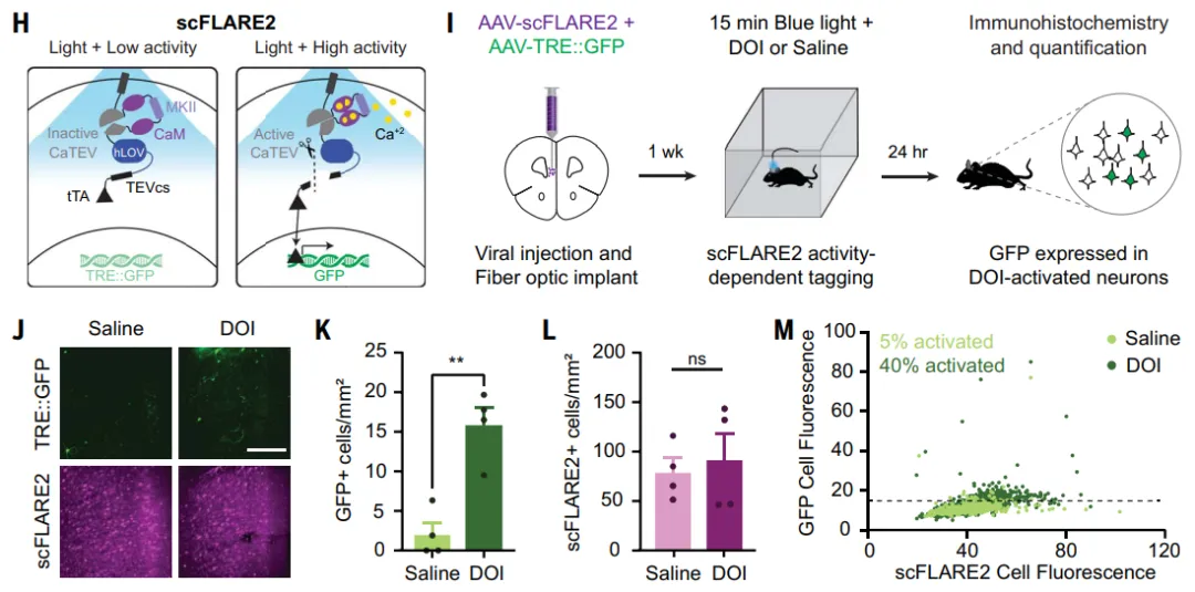
Figure 3: Labeling DOI-activated neurons using scFLARE2


To investigate whether reactivation of DOI-labeled neurons has anxiolytic effects, the researchers used scFLARE2 to label DOI-activated neurons and injected the excitatory optogenetic virus AAV-TRE-bReaChES into the mPFC region. The results showed that, compared to the saline group, DOI-treated mice exhibited reduced burying behavior 30 minutes after treatment, though this effect did not persist into the next day. However, on the second day, optogenetic activation of DOI-labeled neurons significantly reduced burying behavior, and the time spent by mice in the open arms of the EPM (elevated plus maze) was significantly increased. These results indicate that light stimulation of DOI-activated neurons in the mPFC 24 hours after drug administration is sufficient to reproduce the acute anxiolytic effect of the drug.

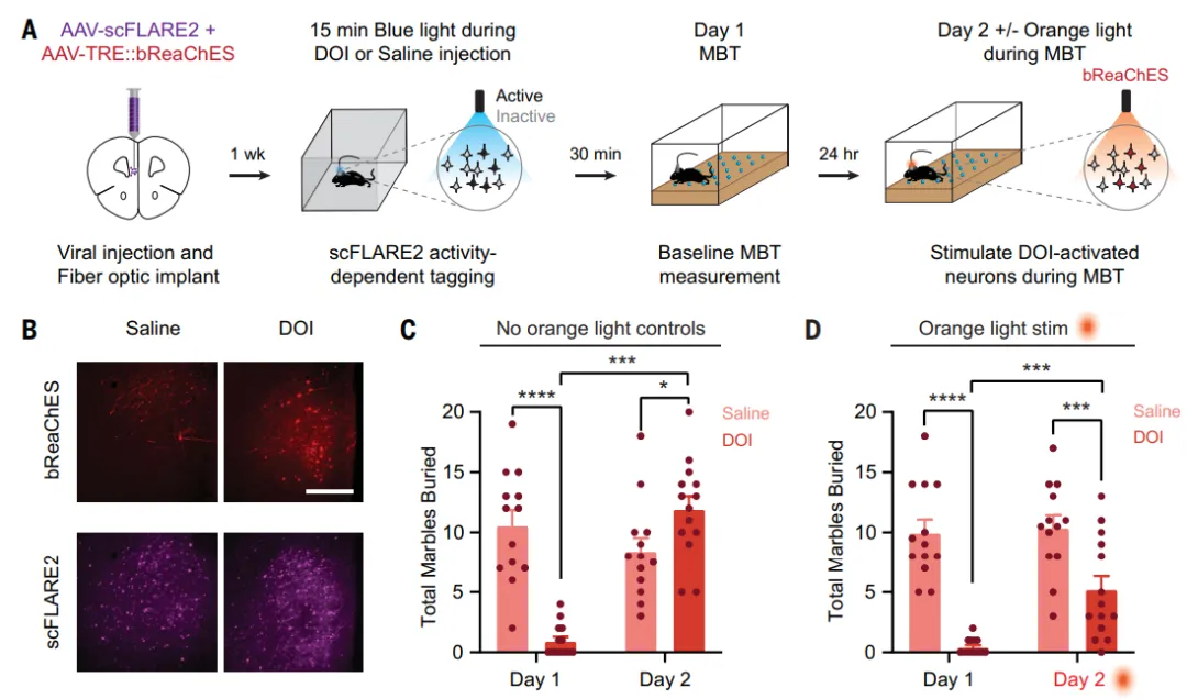
Figure 4: Reactivation of DOI-labeled neurons exerts anxiolytic effects without inducing a psychedelic state.

 
References:
[1] Wang W, Wildes CP, Pattarabanjird T, Sanchez MI, Glober GF, Matthews GA, Tye KM, Ting AY. A light- and calcium-gated transcription factor for imaging and manipulating activated neurons. Nat Biotechnol. 2017 Sep;35(9):864-871. doi: 10.1038/nbt.3909. Epub 2017 Jun 26. PMID: 28650461; PMCID: PMC5595644.
[2] Jung, J. H., Wang, Y., Rashid, A. J., Zhang, T., Frankland, P. W., & Josselyn, S. A. (2023). Examining memory linking and generalization using scFLARE2, a temporally precise neuronal activity tagging system. Cell reports, 42(12), 113592. https://doi.org/10.1016/j.celrep.2023.113592
[3] Muir J, Lin S, Aarrestad IK, Daniels HR, Ma J, Tian L, Olson DE, Kim CK. Isolation of psychedelic-responsive neurons underlying anxiolytic behavioral states. Science. 2024 Nov 15;386(6723):802-810. doi: 10.1126/science.adl0666. Epub 2024 Nov 14. PMID: 39541450.


Brain Case can provide customized virus packaging services. If you have any needs, please contact bd@ebraincase.com


Service Type :

Select the service you'd like to purchase.

Order Information(Premade-AAVs)

Please provide us some information about the service you'd like to order.

How did you hear about BrainCase Biotech or our products?

Detailed requirements:

scroll

Order Information(Custom AAV/Lentivirus)

Please provide us some information about the service you'd like to order.

Gene ID or gene information:

Selection of the reporting gene:

Special Instructions:

scroll

Order Information(Others)

Please provide us some information about the service you'd like to order.

Virus name/Detailed requirements:

scroll

Related products

Literature Interpretation | Nature | OptoXRs: Light-controlled tools for cellular signaling and precise regulation mammalian behavior

Literature Interpretation | Nature | OptoXRs: Light-controlled tools for cellular signaling and precise regulation mammalian behavior

Immunity|Miao Jing’s team at the Beijing Institute for Brain Research develops a fluorescent sensor for chemokine CXCL10 to map spatiotemporal immune signaling

Immunity|Miao Jing’s team at the Beijing Institute for Brain Research develops a fluorescent sensor for chemokine CXCL10 to map spatiotemporal immune signaling

Breakthrough | WESTLAKE UNIVERSITY TEAM DEVELOPS ADVANCED RED GENETICALLY ENCODED CALCIUM SENSOR “SOMAFRCAMPI” WITH SENSITIVITY MATCHING TO GCAMP FOR NEURONAL IMAGING IN VIVO

Breakthrough | WESTLAKE UNIVERSITY TEAM DEVELOPS ADVANCED RED GENETICALLY ENCODED CALCIUM SENSOR “SOMAFRCAMPI” WITH SENSITIVITY MATCHING TO GCAMP FOR NEURONAL IMAGING IN VIVO

Literature Insight | Combining Optogenetics, Calcium Imaging, and Retrograde Viral Tracing in Organoids: Decoding Cortico-Striatal Circuit Connectivity

Literature Insight | Combining Optogenetics, Calcium Imaging, and Retrograde Viral Tracing in Organoids: Decoding Cortico-Striatal Circuit Connectivity

map
{dede:global.cfg_webname/}

Virus product library

CRISPR
RNAi
Neurophilic virus
Optogenetics activation
Biosensors

News

Corporate News
New Product Launch
Media Activity
Investor News

Support

Literature interpretation
Customer article
Video Zone
FAQs
微信

WhatsApp Business Account

Tel: +8618971215294
E-mail: BD@ebraincase.com

Address:-

Address:-

  • Copyright © 2024 Brain Case All Rights Reserved.