Client Article | Molecular Psychiatry | Zhejiang University Team Reveals That Gut Microbiota Contribute to the Pathology of Bipolar Depression by Regulating Synaptic Plasticity and Dopamine Transmission in the VTA–mPFC Pathway
Release time:2026-01-08 16:49:21
Bipolar disorder (BD) is a severe psychiatric condition characterized by recurrent episodes of mania/hypomania and depression, with a global prevalence of approximately 2%. Its clinical presentation is highly complex, and the rate of misdiagnosis remains high. Previous studies have shown that alterations in synaptic plasticity and neurotransmitter dysregulation in the medial prefrontal cortex (mPFC) are involved in the pathogenesis of BD; however, the underlying mechanisms have not yet been fully elucidated. In recent years, the gut microbiota has been recognized as a key component of gut–brain axis regulation. Dysbiosis of the gut microbiota is closely associated with psychiatric disorders, suggesting that microbial communities may influence BD-related behavioral phenotypes by modulating neural plasticity and neurotransmitter systems.
Recently, Shaohua Hu, Professor at the Mental Health Center of the First Affiliated Hospital, Zhejiang University School of Medicine, and Jianbo Lai, Distinguished Research Fellow, together with teams from the School of Brain Science and Brain Medicine at Zhejiang University, the State Key Laboratory of Extreme Optics and Instrumentation, and the Liangzhu Laboratory led by Wei Gong and Scott, published a research article entitled “Gut microbiota modulates synaptic plasticity, connectivity, and dopamine transmission in the VTA–mPFC pathway in bipolar depression” in Molecular Psychiatry. This study demonstrates that the gut microbiota of patients with BD weakens the structural connectivity and functional responsiveness of the dopaminergic VTA–mPFC pathway, impairs synaptic plasticity in the mPFC, and consequently induces depression-like behaviors. These findings provide new experimental evidence for understanding the gut–brain axis mechanisms underlying bipolar disorder.
Fecal Microbiota Transplantation From Patients With BD Induces Bipolar Depression–Like Behaviors in Mice
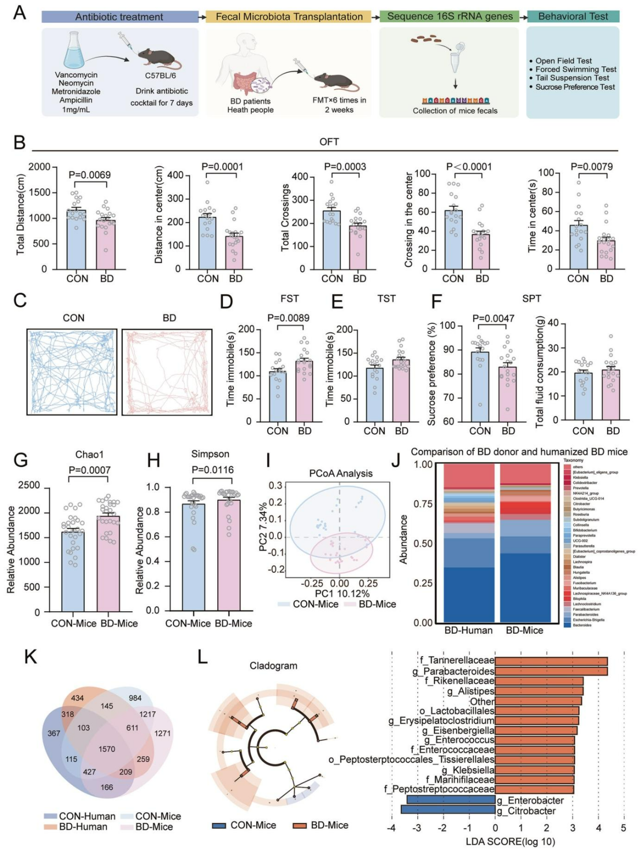
To determine whether the gut microbiota of patients with BD during the depressive phase can induce bipolar depression–like behaviors in mice, researchers performed fecal microbiota transplantation (FMT) via oral gavage. Fecal samples were collected within 24 hours after transplantation, followed by a series of behavioral tests (Figure 1A). Combined antibiotic treatment caused a slight decrease in body weight in mice, with no significant difference between the BD and control (CON) groups. Behavioral assessments showed that, compared with the CON group, BD-FMT mice exhibited significantly reduced total travel distance, distance traveled in the center area, total crossings, center crossings, and time spent in the center in the open-field test (Figure 1B, C). In the forced swim test, immobility time was significantly increased, indicating despair-like behavior (Figure 1D), whereas no significant increase in immobility time was observed in the tail suspension test (Figure 1E). In the sucrose preference test, total fluid intake did not differ between groups, but sucrose preference was significantly reduced, suggesting anhedonia (Figure 1F). Taken together, transplantation of fecal microbiota from patients with BD induced core features of bipolar depression in recipient mice. Further experiments demonstrated that this bipolar depression–like mouse model responded positively to lithium treatment (a mood stabilizer) but not to fluoxetine (a classical antidepressant). This drug sensitivity profile is consistent with clinical features of bipolar depression, thereby validating the face validity of the model.
FMT Successfully Reshapes the Gut Microbiota of Recipient Mice
The study first eliminated endogenous gut microbiota in recipient mice through antibiotic (ABX) treatment, followed by FMT from either patients with BD or healthy donors. 16S rRNA sequencing revealed significant differences in gut microbial composition between the two groups. In α-diversity analyses, the BD group showed higher Chao1 and Simpson indices (Figure 1G–H), while no differences were observed in the Shannon index or species richness. Principal coordinates analysis (PCoA) demonstrated clear separation of microbial communities between groups (Figure 1I). LEfSe analysis identified enrichment of six inflammation-associated bacterial genera in the BD group, along with a relative reduction in several beneficial genera, such as Citrobacter and Enterobacter (Figure 1L). Further analysis showed a high degree of overlap between the microbial profiles of BD-FMT mice and those of human BD patients, with 259 shared bacterial species (Figure 1J, K). Collectively, these findings indicate that FMT effectively established a mouse model harboring gut microbiota characteristics of patients with BD.
Figure 1. Colonization of Gut Microbiota From Patients With BD Alters Mouse Behavior and Achieves Humanization of the Microbiome
Transcriptomic and Structural Alterations in the Medial Prefrontal Cortex (mPFC) Reveal Synaptic Mechanisms Underlying Bipolar Depression–Like Behaviors
To investigate how the microbiota–gut–brain axis regulates bipolar depression–like behaviors, the study focused on the mPFC, a key brain region involved in emotional regulation. RNA sequencing revealed 202 upregulated and 64 downregulated genes in the mPFC of BD mice, with clear separation between the two groups in sample clustering analyses (Figure 2A–C). Gene Ontology (GO) enrichment analysis showed that downregulated genes were mainly associated with dendritic spine plasticity, whereas upregulated genes were primarily involved in immune responses (Figure 2D–E). Gene set enrichment analysis (GSEA) further identified “postsynaptic translation” as the most significantly downregulated biological process, a pathway critical for long-term synaptic plasticity (Figure 2F–G). Consistently, qPCR experiments confirmed reduced expression of genes involved in postsynaptic translation, including Rpl, Rps, Eef, and Uba. These findings indicate that the transplanted microbiota impairs long-term synaptic plasticity in the mPFC.
At the structural level, SIN-pal-GFP virus was injected into the mPFC to sparsely label neurons, enabling analysis of dendritic spine density and morphology in pyramidal neurons located in layers II/III and V of the mPFC, which primarily process long-range inputs (Figure 2H). The results showed a significant reduction in total dendritic spine density in mPFC pyramidal neurons of BD mice (Figure 2I–J). Although the densities of all spine subtypes (thin, stubby, and mushroom spines) showed non-significant decreases, the reduction in mushroom spines was particularly pronounced (Figure 2J), suggesting impaired synaptogenic capacity with subtype-specific vulnerability. Collectively, these results demonstrate that gut microbiota from patients with BD induces bipolar depression–like behaviors by disrupting postsynaptic translation and long-term synaptic plasticity in the mPFC and reducing dendritic spine density.
Figure 2. Transcriptomic and Dendritic Spine Alterations in the mPFC as Potential Mechanisms of Bipolar Depression–Like Behaviors
Reduced Monosynaptic Connectivity Plasticity of mPFC Neurons
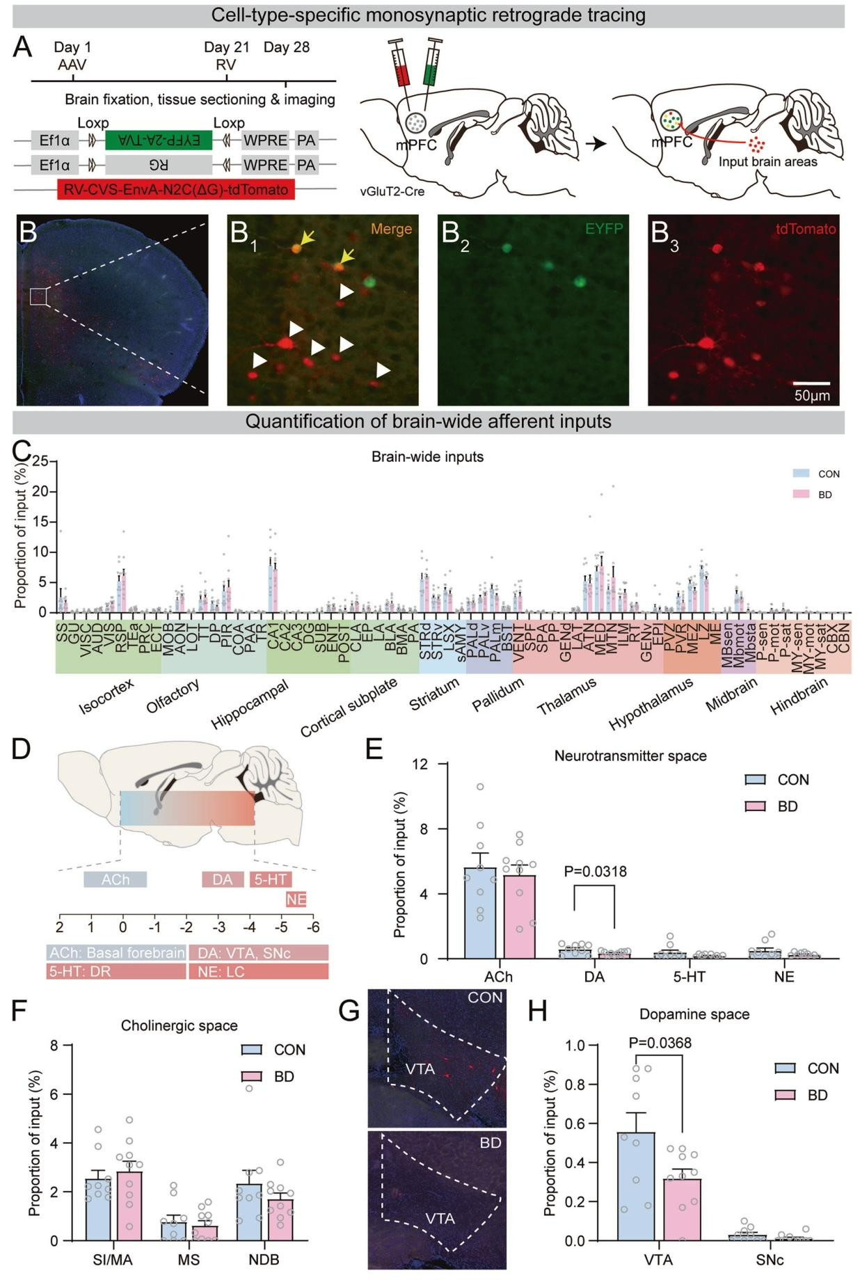
To determine the effects of FMT on synaptic connectivity of glutamatergic neurons in the mPFC of BD mice, monosynaptic whole-brain input mapping was performed using rabies virus–mediated retrograde trans-synaptic tracing (Figure 3A). More than 80% of starter cells were located in mPFC layers II/III and V, with no difference in starter cell density between groups (Figure 3B). Whole-brain imaging revealed monosynaptic inputs to mPFC glutamatergic neurons from multiple brain regions (Figure 3C).
Focusing on neurotransmitter systems closely associated with BD pathology, four major systems with well-defined nuclei were analyzed: the basal forebrain (acetylcholine), midbrain VTA/SNc (dopamine), dorsal raphe nucleus (serotonin), and locus coeruleus (norepinephrine), along with their projections to mPFC glutamatergic neurons. The results showed that only the dopaminergic system exhibited a significant reduction in viral labeling in the BD group compared with controls (Figure 3E). Further region-specific analysis identified the VTA–mPFC pathway as the key circuit with weakened connectivity, while inputs from other major brain regions showed no significant differences between groups (Figure 3G–H). These findings suggest that gut microbiota transplantation selectively impairs the dopaminergic VTA–mPFC pathway, which may represent a critical neural circuit basis for bipolar depression–like behaviors.
Figure 3. Reduced Dopaminergic Connectivity to mPFC Neurons
Monosynaptic Connectivity of mPFC Neurons Projecting to the Basolateral Amygdala (BLA)
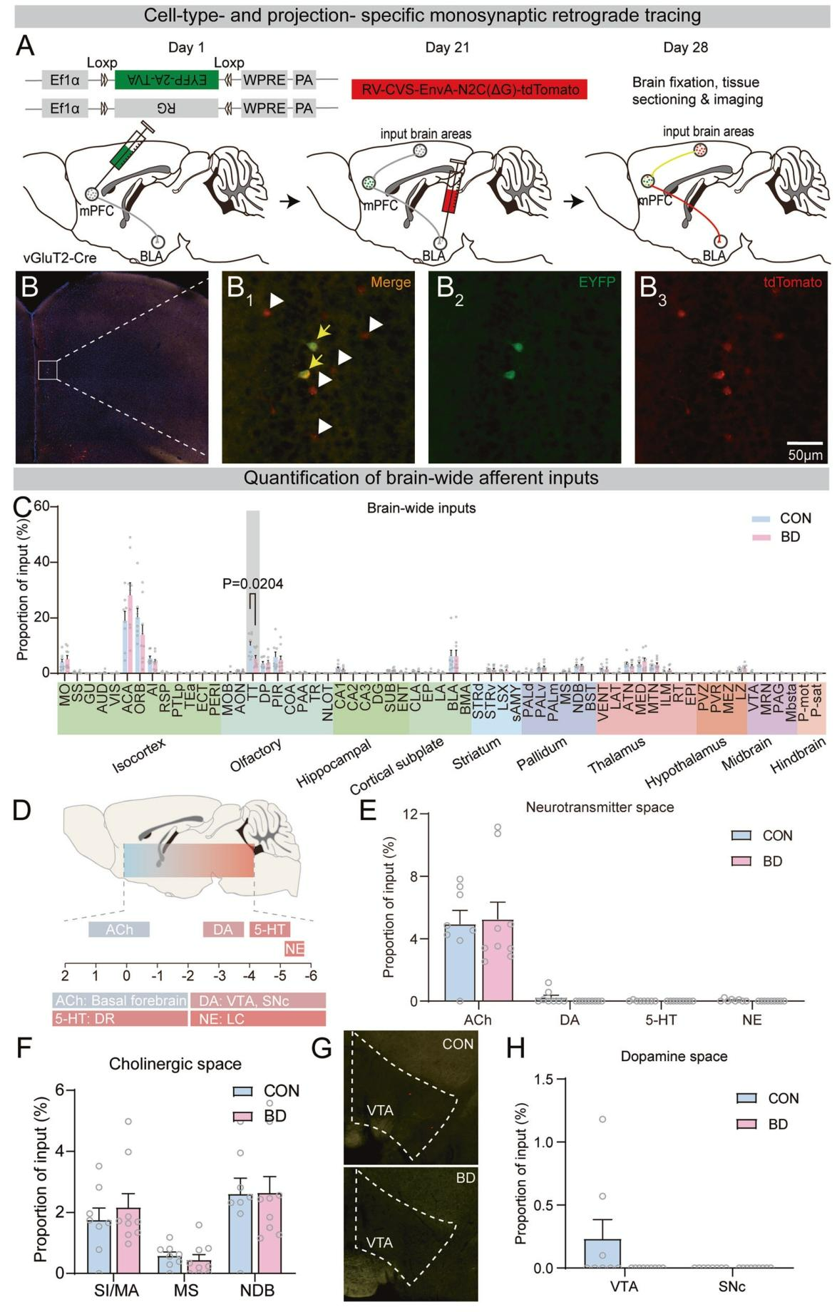
Given that the BLA is highly relevant to bipolar disorder and receives dense projections from the mPFC, the study next examined the effects of gut microbiota transplantation on synaptic inputs to BLA-projecting mPFC glutamatergic neurons using a trans-synaptic viral tracing strategy. An AAV helper virus cocktail (AAV-EF1α-DIO-EGFP-T2A-TVA and AAV-EF1α-DIO-G) was injected into the mPFC, followed three weeks later by targeted injection of rabies virus (RV-CVS-EnvA-N2C(ΔG)-tdTomato) into the BLA. After one week of expression, whole-brain monosynaptic inputs were imaged (Figure 4A).
Validation showed that starter cells were concentrated in the prelimbic–infralimbic complex of the mPFC, with approximately 70% located in layers II/III, and no difference in starter cell density between groups (Figure 4B). Whole-brain imaging revealed significantly reduced or absent inputs from the tegmental tract (TT) and the VTA in BD mice (Figure 4C). Further analyses of inputs from neurotransmitter systems (including acetylcholine and dopamine) as well as regions such as the thalamus and hippocampus showed no significant differences between groups (Figure 4D–H). Together, these results indicate region-specific remodeling of monosynaptic inputs to BLA-projecting mPFC neurons in BD mice, characterized primarily by loss of TT and VTA inputs.
Figure 4. Monosynaptic Connectivity of mPFC Neurons Projecting to the BLA
Structural and Functional Dopamine Deficiency in VTA Dopaminergic Neurons Projecting to the mPFC
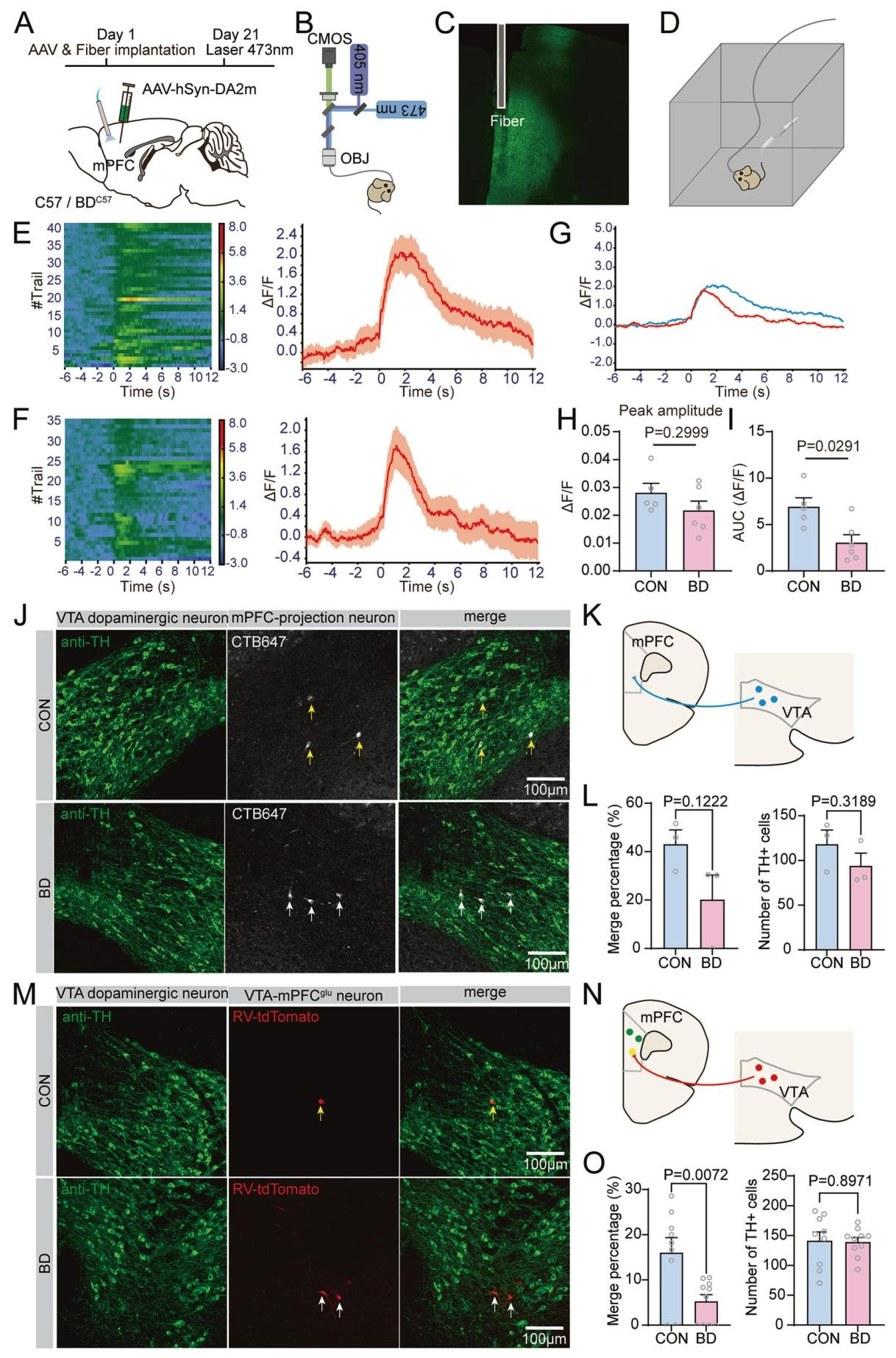
To determine the functional significance of structural defects in the mPFC–VTA connection in BD mice, the study considered evidence linking altered prefrontal–VTA connectivity in BD patients to reward processing and anhedonia, as well as the role of the VTA as a major dopaminergic center. An AAV expressing the dopamine indicator AAV-DA2m was injected into the mPFC (Figure 5A). One month later, fiber photometry combined with tail-pinch stimulation was used to monitor real-time dopamine responses in mPFC neurons (Figure 5B–D). Compared with controls, BD mice showed significantly reduced dopamine release amplitude and overall dopamine responses in the mPFC upon stimulation (Figure 5F–I), indicating functional dopamine deficiency in mPFC neurons.
Having established structural and functional dopamine deficits in the mPFC of BD mice, the study next examined changes in projections from VTA dopaminergic neurons to mPFC glutamatergic neurons. Retrograde tracing with CTB injected into the mPFC, combined with immunostaining to identify dopaminergic neurons, revealed a decreasing trend in the number of VTA dopaminergic neurons projecting to the mPFC in BD mice (Figure 5J–L). Furthermore, combining rabies virus–mediated monosynaptic tracing with tyrosine hydroxylase (TH) immunostaining demonstrated a significant reduction in the number of VTA dopaminergic neurons forming synaptic connections with mPFC glutamatergic neurons in BD mice (Figure 5M–O). These findings provide direct anatomical evidence that BD mice exhibit selective synaptic projection deficits from VTA dopaminergic neurons to mPFC glutamatergic neurons, supporting the observed structural and functional dopamine abnormalities.
Figure 5. Structural and Functional Dopamine Deficiency in VTA Dopaminergic Neurons Projecting to the mPFC
Summary
In this study, transplantation of gut microbiota from patients with BD into mice successfully established a mouse model exhibiting depression-like behaviors. Mechanistically, the gut microbiota derived from patients with BD selectively disrupts the structural connectivity and functional transmission of the dopaminergic VTA–mPFC pathway via the gut–brain axis, leading to reduced synaptic plasticity in the mPFC and, consequently, the emergence of depression-like behaviors. These findings provide a novel microbiota–neural circuit perspective for understanding the pathophysiology of bipolar disorder and suggest that the gut microbiota may represent a potential therapeutic target for this disease.