Customer Article | Nature Neuroscience | Team Led by Yingjie Zhu at SIAT Reveals a Key "Gas-Brake" Mechanism in the Brain That Regulates Addiction
Release time:2025-09-19 14:31:55
Substance use disorder (a medical term referring to a range of problems affecting physiological, psychological, and social functioning caused by repeated use of psychoactive substances such as alcohol and drugs) is a global public health challenge. Social status plays an important role in addiction vulnerability, but the underlying neural mechanisms remain unclear. The dopaminergic system is closely associated with addiction, and the mesolimbic and mesocortical dopamine pathways may play distinct roles in reward and addictive behaviors.
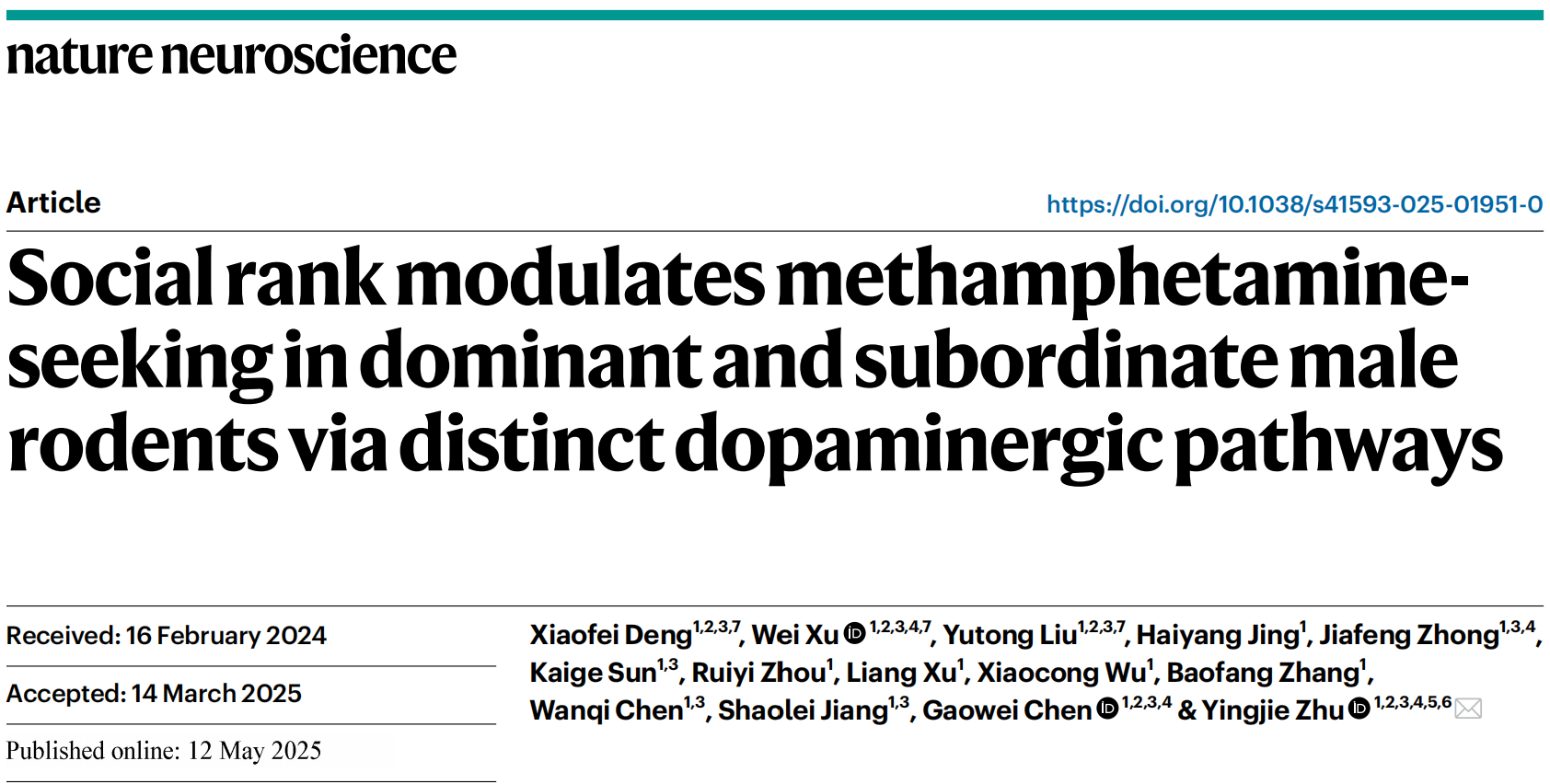
On May 12, 2025, a research team led by Dr. Yingjie Zhu from the Shenzhen Institute of Advanced Technology, Chinese Academy of Sciences, published a study in Nature Neuroscience titled “Social rank modulates methamphetamine-seeking in dominant and subordinate male rodents via distinct dopaminergic pathways.”
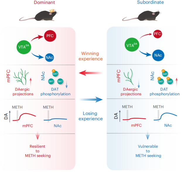
The study reveals that social hierarchy significantly influences methamphetamine (METH)-seeking behaviors in rodents. Dominant male rodents exhibit denser mesocortical dopaminergic projections and show greater resistance to METH-seeking behavior. In contrast, subordinate males demonstrate enhanced dopaminergic function in the mesolimbic pathway, making them more susceptible to METH. Experiences of winning or losing may reshape the dopaminergic system, thereby preventing or promoting METH-seeking behavior, respectively.
Dominant males exhibit resistance to METH-seeking behavior
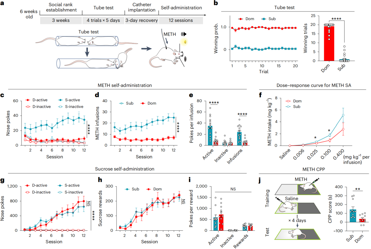
A tube test was used to determine social hierarchy in the rodent model. Male rats in late adolescence (6 weeks old) were pair-housed for 3 weeks (Fig. 1a), followed by 5 days of tube testing to assess stable social hierarchy between paired rats (Fig. 1b). The tube test results were validated by comparing them with other social rank paradigms such as aggression assays and hot spot tests, confirming its effectiveness in identifying social status. After tube testing, rats were single-housed and subjected to 12 days of fixed-ratio 1 (FR1) METH self-administration training.
The results showed that subordinate rats (Sub) progressively exhibited increased METH-seeking and intake behaviors during training, while dominant rats (Dom) showed little to no such tendency (Fig. 1c–e). Specifically, dominant rats had significantly fewer active nose-pokes, inactive nose-pokes, and infusions compared to subordinates, with no increase over time (Fig. 1c–e). This pattern was also observed in mice and under different METH doses in rats (Fig. 1f). In contrast, both dominant and subordinate groups displayed similar behaviors in sucrose consumption tests (Fig. 1g–i).
In the 0.25 mg/kg conditioned place preference (CPP) test for METH, subordinate animals showed a strong preference response, whereas dominant animals responded much less (Fig. 1j). A similar pattern was observed in the 10 mg/kg morphine CPP test. These findings suggest that social hierarchy effectively predicts individual susceptibility to seeking and consuming addictive substances.
Figure 1. Dominant males resist METH-seeking behavior in social rank tests
METH-induced dopamine release differs between dominant and subordinate individuals
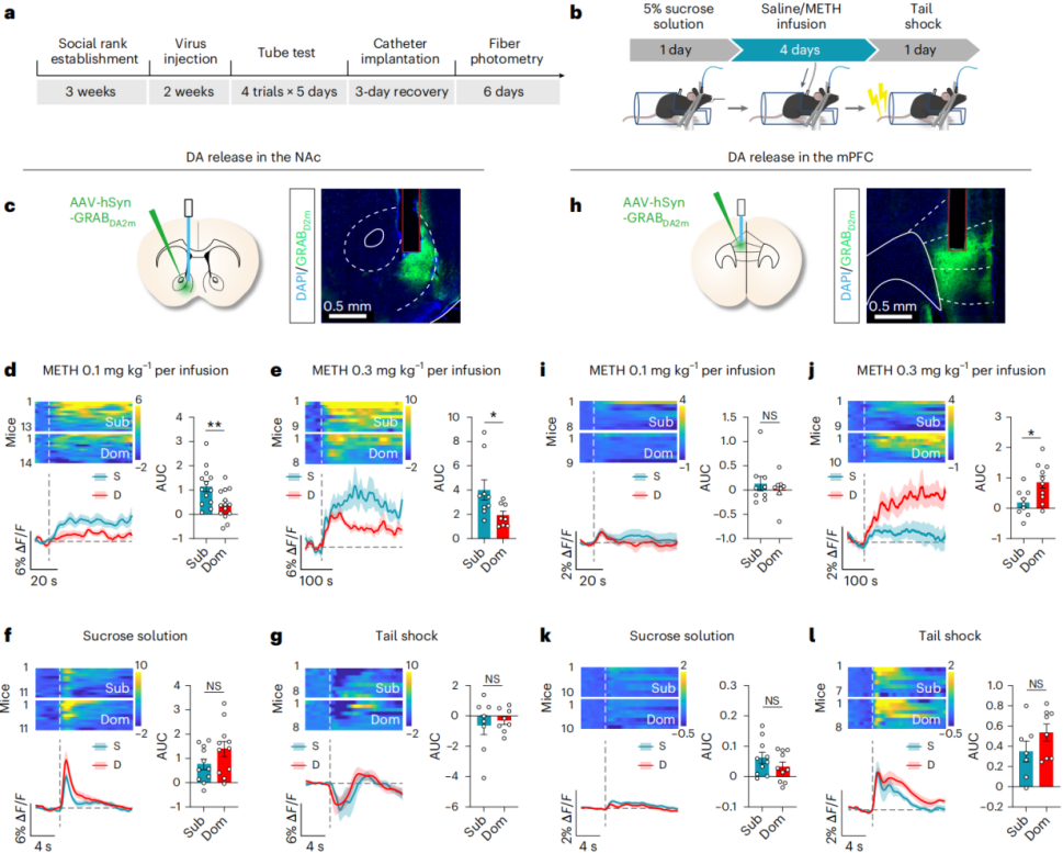
Since METH exerts its reinforcing effects primarily by increasing dopamine (DA) levels in the brain, the researchers hypothesized that social rank may influence METH-seeking behavior by modulating DA dynamics. To test this, they expressed the GRAB DA2m dopamine sensor in the nucleus accumbens (NAc) and medial prefrontal cortex (mPFC) of male mice and used fiber photometry to record real-time DA release (Fig. 2a–c, h).
The results showed that in the NAc, both 0.1 mg/kg and 0.3 mg/kg doses of METH induced significantly greater DA elevations in subordinate mice compared to dominant mice (Fig. 2d–e). In contrast, in the mPFC, only the 0.3 mg/kg dose of METH significantly increased DA levels in dominant mice (Fig. 2j), while the 0.1 mg/kg dose had no significant effect on either group (Fig. 2i). Saline infusion did not induce any changes in DA levels.
Similar patterns were observed in active METH self-administration experiments: dominant mice showed lower DA release in the NAc but higher DA release in the mPFC compared to subordinates. Moreover, neither 5% sucrose licking nor tail shock produced significant differences in DA release between the two groups in either brain region (Fig. 2f–g, k–l). These findings indicate that social hierarchy differentially modulates METH-induced DA elevation in the NAc and mPFC.
Regarding the encoding of natural rewards and aversive stimuli, the study found that sucrose consumption triggered substantial DA release in the NAc, while tail shock suppressed it (Fig. 2f–g). In contrast, tail shock significantly increased DA release in the mPFC, with sucrose having minimal effect (Fig. 2k–l). Furthermore, optogenetic activation of the mesolimbic dopaminergic pathway induced conditioned place preference, whereas activation of the mesocortical pathway did not. These results suggest that the mesolimbic and mesocortical dopamine pathways play distinct roles in reward processing, and likely contribute differently to addiction vulnerability.
Figure 2. METH induces distinct DA release patterns in mesolimbic and mesocortical pathways in dominant vs. subordinate males
Dominant males exhibit reduced mesolimbic dopaminergic function
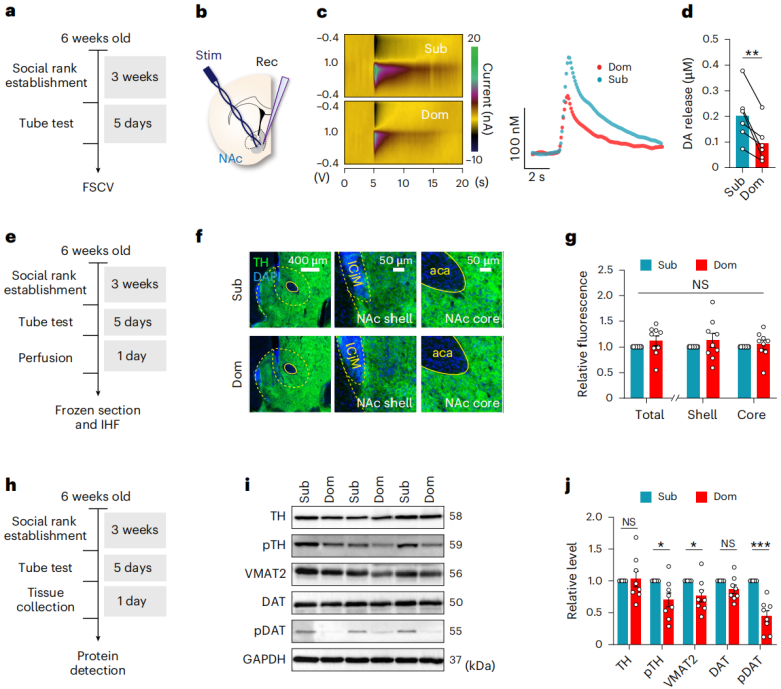
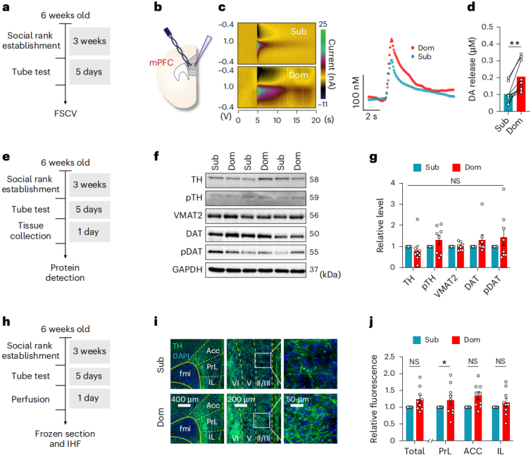
Building on previous findings that dominant and subordinate males exhibit different levels of METH-induced DA release in the nucleus accumbens (NAc) and mPFC, the researchers hypothesized that there may be biochemical and anatomical differences within the mesolimbic-cortical pathways. To investigate the specific features of mesolimbic DA function in dominant male mice, they employed fast-scan cyclic voltammetry (FSCV) to measure electrically evoked DA release in NAc brain slices (Fig. 3a–b).
The results showed that under single-pulse stimulation, subordinate mice released approximately twice as much DA in the NAc as dominant mice (Fig. 3c–d). However, tyrosine hydroxylase (TH) immunostaining revealed no significant differences in the density of dopaminergic terminals in the NAc between the two groups (Fig. 3e–g).
At the protein expression level, both groups had similar total TH expression, but dominant mice exhibited lower levels of phosphorylated TH (pTH) and vesicular monoamine transporter 2 (VMAT2). While the expression of dopamine transporter (DAT) was comparable between groups, dominant mice showed lower levels of phosphorylated DAT (pDAT) (Fig. 3h–j).
In summary, dominant males display reduced mesolimbic dopaminergic function, primarily due to the dephosphorylation of TH and DAT, along with downregulation of VMAT2. In addition, dominant males exhibited reduced phosphorylation of ERK (pERK), which is known to phosphorylate DAT at the Thr53 site. Subsequent experiments will focus on the role of pDAT within the mesolimbic system.
Figure 3. Dominant males show reduced dopaminergic function in the mesolimbic pathway
To further investigate potential differences in the mesocortical dopaminergic pathway between dominant and subordinate groups, fast-scan cyclic voltammetry (FSCV) was used to assess DA release in mPFCbrain slices following electrical stimulation of nerve terminals (Fig. 4a–b). In contrast to the findings in the nucleus accumbens (NAc), dominant males showed significantly greater evoked DA release in the mPFC under single-pulse stimulation (Fig. 4c–d).
However, protein expression levels of TH, phosphorylated TH (pTH), VMAT2, DAT, and pDAT in the mPFC showed no significant differences between the two groups (Fig. 4e–g). Immunostaining results revealed a higher density of TH-positive dopaminergic terminals in the prelimbic (PrL) subregion of the mPFC in dominant males, but no such differences were observed in the infralimbic (IL) or anterior cingulate cortex (ACC) regions (Fig. 4h–j).
Although dopaminergic terminals in the PFC may originate from multiple brain regions, whole-brain imaging using sparse labeling combined with synchronized scanning and volumetric imaging via VISoR (Volumetric Imaging with Synchronized on-the-fly-scan and Readout) technology allowed for the tracing of axons from individually labeled ventral tegmental area (VTA) dopaminergic neurons. The results showed that, while the number of labeled neurons was similar between groups, dominant mice had more dopaminergic neurons projecting to the mPFC, and their axon terminals were denser than those of subordinates. These findings suggest that the enhanced dopaminergic innervation of the mPFC in dominant males originates from denser projections of VTA DA neurons.
Figure 4. Dominant males exhibit denser dopaminergic projections in the mesocortical pathway
Dephosphorylation of DAT in the NAc suppresses METH-seeking behavior
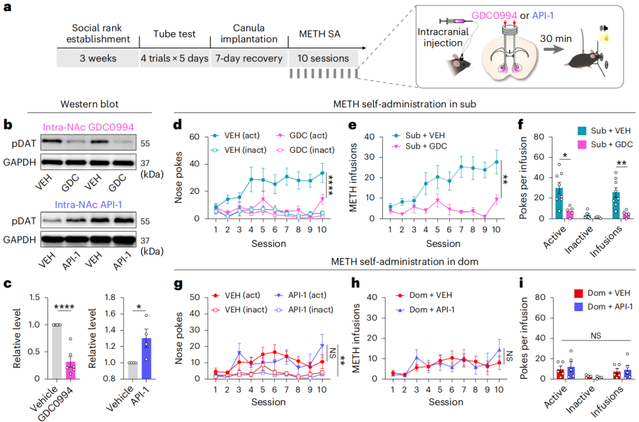
To explore the role of DAT phosphorylation in the reinforcing effects of METH, the researchers intracranially injected GDC0994, an ERK (extracellular signal-regulated kinase) inhibitor, into the nucleus accumbens (NAc) of subordinate male mice (Fig. 5a) to inhibit phosphorylation of DAT at the Thr53 site. The results showed that local injection of GDC0994 into the NAc reduced pDAT levels (Fig. 5b–c) and significantly suppressed METH self-administration behavior in subordinate mice (Fig. 5d–f), as well as reduced their METH conditioned place preference (CPP) scores.
To further determine whether enhancing DAT phosphorylation in the NAc would promote METH-seeking in dominant mice, the researchers injected API-1—an inhibitor of peptidyl-prolyl cis-trans isomerase (PIN1)—into the NAc of dominant male mice, aiming to increase DAT phosphorylation at Thr53. While API-1 did elevate pDAT levels in the NAc (Fig. 5b–c), it did not enhance METH-seeking behavior during self-administration or in the CPP test in dominant mice (Fig. 5g–i).
Neither GDC0994 nor API-1 altered the social rank of the mice. To investigate whether global activation of the mesolimbic pathway alone could drive METH-seeking in dominant mice, AAV-DIO-ChR2 was injected into the VTA of DAT-Cre dominant mice, and optical fibers were implanted in the NAc. Optogenetic activation of the VTA→NAc dopaminergic pathway significantly increased METH-seeking and intake behavior in dominant mice.
Figure 5. Dephosphorylation of DAT in the NAc suppresses METH-seeking in subordinate males
Mesocortical activation enhances winning probability and strengthens METH resistance in subordinates
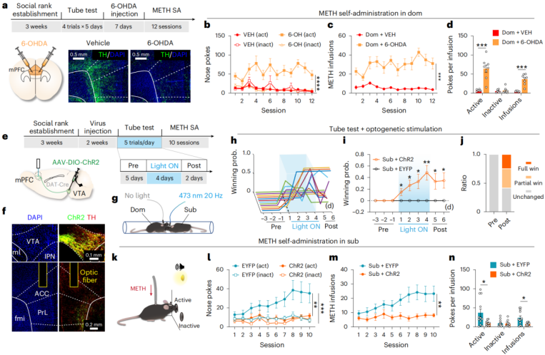
Given that dominant animals exhibit denser mesocortical dopaminergic projections, it was hypothesized that this may underlie their resistance to METH-seeking behavior. To test this, 6-hydroxydopamine (6-OHDA) was injected into the mPFC of dominant male rats to selectively lesion the mesocortical pathway. The results showed a significant reduction in TH-positive dopaminergic terminals (Fig. 6a), which subsequently enhanced METH-seeking behavior in the dominant rats (Fig. 6b–d).
To further determine whether activating the mesocortical pathway in subordinate males could suppress METH-seeking, AAV-DIO-ChR2 was injected into the VTA of DAT-Cre subordinate male mice, and optical fibers were implanted in the mPFC (Fig. 6e–f). The results showed that optogenetic activation during tube testing gradually increased the subordinates’ winning probability, leading to an elevation in social rank (Fig. 6g–j), and significantly reduced METH-seeking behavior (Fig. 6k–n).
Interestingly, subordinate mice that managed to win in the tube test after optogenetic stimulation showed the most pronounced reduction in METH-seeking behavior, while those that remained subordinate exhibited relatively higher METH-seeking. There was a significant negative correlation between METH injection frequency and winning probability.
Taken together, these findings demonstrate that the mesocortical pathway plays a key role in supporting resistance to METH-seeking behavior, and that this resistance is modulated by the individual’s experience of winning.
Figure 6. Mesocortical activation promotes winning and enhances resistance to METH
Winning experience suppresses METH-seeking behavior in subordinates
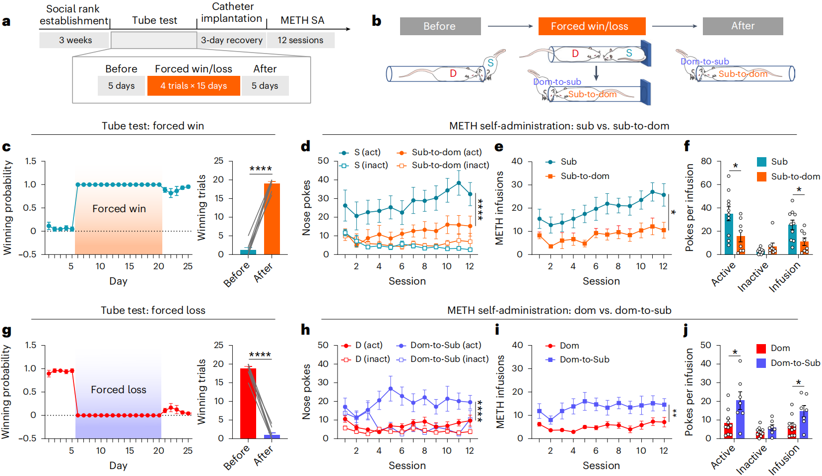
To investigate whether changes in social rank affect METH-seeking behavior, a forced win/loss paradigm was applied to male rats. In this experiment, subordinate rats were forced to win by blocking the opponent’s exit during the tube test (Fig. 7a–b), while dominant rats were subjected to forced defeat (Fig. 7c–g). After three weeks of training (5 days per week), a standard tube test was conducted to evaluate their new social status.
The results showed that some originally subordinate rats maintained consistent victories in the tube test following forced-win training (“Subordinate-to-Dominant” group), while some originally dominant rats exhibited persistent losses after forced-defeat training (“Dominant-to-Subordinate” group). The “Dominant-to-Subordinate” group also showed a notable increase in depression-like behaviors.
Compared to untrained subordinates, the “Subordinate-to-Dominant” group showed significantly reduced METH-seeking behavior (Fig. 7d–f). In contrast, subordinate rats that underwent forced-win training but did not successfully win continued to display high METH-seeking behavior. The “Dominant-to-Subordinate” group exhibited increased METH-seeking behavior (Fig. 7h–j). Even if these rats did not ultimately shift to subordinate status, the experience of forced defeat alone was sufficient to increase their METH-seeking.
In summary, changes in social hierarchy can alter intrinsic patterns of METH-seeking behavior. Winning experiences can suppress METH-seeking in subordinates, while experiences of defeat can enhance METH-seeking in dominant individuals.
Figure 7. Winning experience suppresses METH-seeking behavior in subordinates
Winning experience reshapes the mesolimbic and mesocortical dopaminergic systems
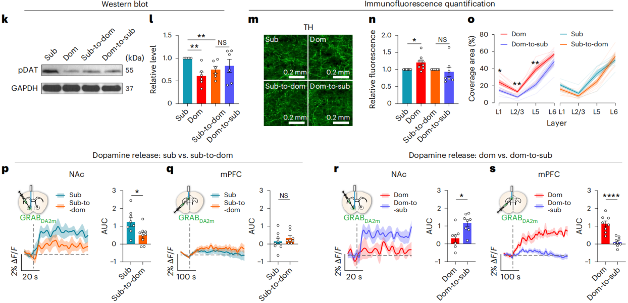
To determine whether changes in social status also affect the mesolimbic and mesocortical dopaminergic pathways, the researchers conducted Western blotting and immunofluorescence analyses. The results showed that forced win/loss training eliminated the original differences in pDAT and pERK expression in the NAc, as well as the differences in dopaminergic terminal distribution in the mPFC between originally dominant and subordinate animals (Fig. 8k–n).
Notably, compared to the original subordinate group, the “Subordinate-to-Dominant” group exhibited a significant reduction in pDAT levels in the NAc (Fig. 8k–l). In the mPFC, the disappearance of differences was mainly due to a reduction in dopamine terminal density in layers I–V of the “Dominant-to-Subordinate” group, rather than an increase in the “Subordinate-to-Dominant” group (Fig. 8o).
To confirm whether these changes impacted dopamine release, the researchers used neurotransmitter sensors to monitor DA dynamics in the NAc and mPFC of male mice. In the “Subordinate-to-Dominant” group, METH-induced DA release in the NAc was significantly reduced, while DA signaling in the mPFC remained unchanged (Fig. 8p–q). Conversely, in the “Dominant-to-Subordinate” group, METH-induced DA release in the NAc was significantly elevated, and DA release in the mPFC was markedly reduced (Fig. 8r–s).
In summary, changes in social hierarchy reshape the mesocortical and mesolimbic dopaminergic systems, thereby mediating the reversal of METH-seeking behavior.
Figure 8. Winning experience reshapes the mesolimbic and mesocortical dopaminergic systems
Females are susceptible to METH regardless of social rank
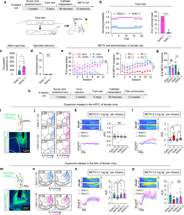
Tube test observations revealed that female rats exhibit strong METH-seeking and intake behaviors regardless of their social rank (Fig. 9a–g), whereas in males, these behaviors are influenced by social hierarchy.
In the mPFC, dominant female rats showed higher METH-induced DArelease than subordinate females, but overall, both female groups exhibited lower DA release than dominant males (Fig. 9h–i). In the NAc, METH-induced DA release did not differ between the two female groups and was higher than that of dominant males; however, among males, subordinates showed higher DA release than dominants (Fig. 9m–p).
Dominant females had a higher density of TH-positive dopaminergic terminals in the ACC and prelimbic (PrL) regions of the mPFC, but the number of terminals in layers 2/3 of the PrL was lower than in dominant males. This may relate to the females’ lower METH-induced DA release and higher METH-seeking behavior. Within female groups, no differences were observed in pDAT levels in the NAc, and these levels were comparable to those of subordinate males but higher than dominant males. This may explain the females’ higher DA release and greater METH-seeking behavior. Overall, female rats display distinct differences from males in both METH-seeking behavior and associated dopaminergic system features.
Figure 9. Females are susceptible to METH regardless of social rank
Summary
Social hierarchy influences METH-seeking behavior through mesocortical and mesolimbic dopaminergic pathways. The mesocortical dopaminergic projections and function in dominant males play a crucial role in resisting METH seeking, while enhanced mesolimbic dopaminergic function in subordinate males increases their susceptibility to addiction. Winning experience can suppress METH-seeking behavior by reshaping the dopaminergic system. Female susceptibility to METH may be independent of social rank, and their underlying neural mechanisms differ from those of males.
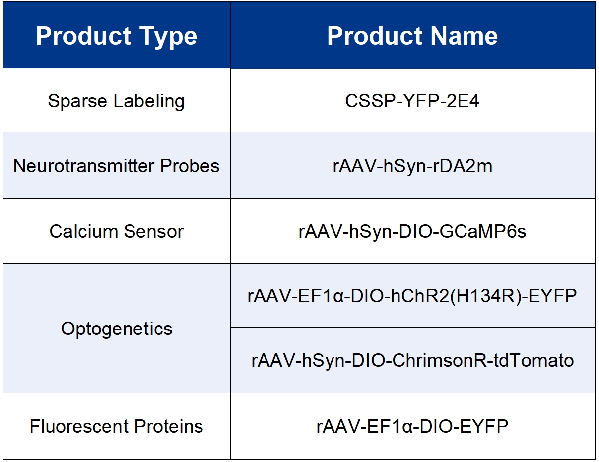
All the viral tools used in this study are available from Brain Case Biotech.