E-mail:BD@ebraincase.com
Tel:+8618971215294
English 中文版
Virus Vector - Viral Vector Production - BrainCaseVirus Vector - Viral Vector Production - BrainCase
  • Home
  • Virus product library
    CRISPRRNAiHSV-helperRV-helperNeurophilic virusCalcium SensorsOptogenetics activationOptogenetics inhibitionChemical geneticsSparse labelingFluorescent proteinBiosensorsRecombinaseApoptosis & AutophagyDisease ModelNeurotoxicityOther
  • Products & Service

    Product Center

    Virus

    VSV-circuit research-vaccine and gene therapy research-BrainCase
    Retrovirus-RCAS-TVA-BrainCase
    Lentivirus Vector - Lentivirus Production - BrainCase
    Rabies Virus Vector - RBV Vector - BrainCase
    Herpes simplex virus-Oncolytic and anterograde tracing-Brain Case
    PRV-retrograd multisynaptic-Peripheral-Btain Case
    AAV-gene therapy vectors-BrainCase

    Animal Model

    Neurological Disease Models-BrainCase
    Tumor animal models-anti-tumor-BrainCase
    Digestive System Disease Animal Model-Brain Case
    Cardiovascular System Disease Animal Models-Brain Case

    Plasmid Construction

    Library Construction
    Plasmid design and construction

    Popular Applications

    Gene Regulation

    Gene Overexpression-Brain Case
    RNA interference(RNAi)-siRNA-Brain Case
    Gene Editing - CRSIPR cloning - BrainCase

    Neural Circuit Function Research

    Optogenetics - BrainCase
    Chemical genetics-DREADDs-Brain Case
    Calcium signal recording-Gels- Brain Case
    GRAB Neurotransmitter Fluorescent Sensor- Brain Case
    Functional Magnetic Resonance Imaging Technology- Brain Case

    Research on the structure of neural circuits

    Direct Input and Output-viral vectors- Brain Case
    Anterograde Mono-synaptic Tracing -HSV- Brain Case
    Antrograde Muti-synaptic Tracing-HSV & VSV-Brain Case
    Retrograde Mono-synaptic Tracing-Rabies Virus-Brain Case
    Retrograde Muti-synaptic Tracing-PRV-Brain Case

    Featured Services

    Nervous System Disease Drug Effect

    Alzheimer's disease-AD-Brain Case
    Depression-mental disorders-Brain Case
    Parkinson's disease--PD-Brain Case
    Epilepsy-an ancient neurological disorder-Brain Case

    AAV Serotype Screening

    AAV Serotypes screening-gene therapy-Brain Case

    Tumorigenicity Test

    Tumorigenicity Testing-Evaluation of tumor models-Brain Case

    Efficacy of Oncolytic Virus

    Oncolytic virus-for cancer therapy-Brain Case
    Herpes Virus Vector-anti-tumor- BrainCase
    Vesicular stomatitis virus-killing tumor cells-Brain Case
  • News
    Corporate News New Product Launch Media Activity Investor News
  • Support
    Literature interpretation Customer article Video Zone FAQs
  • About Us
    Virus product library Products & Service News Support About Us Contact
  • Contact
    Contact Us Join us
  • 中文
    English 中文版
  • Home
  • Support
  • Customer article
  • Support
  • Literature interpretation
  • Customer article
  • Video Zone
  • FAQs

Client Publication | Advanced Science | Discovery of Glial Cell Interaction Mechanism in Depression by the Teams of Jin Yu and Yuqiu Zhang

Release time:2025-07-04 13:54:51
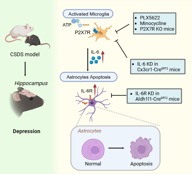
Numerous studies have shown that microglial activation and astrocytic atrophy/loss are key pathological features of depression. During stress, large amounts of extracellular ATP (eATP) are released, which—recognized as damage-associated molecular patterns (DAMPs)—can specifically trigger microglial activation via the P2X7 receptor (P2X7R). However, the specific activation state of microglia under elevated eATP conditions, and how this leads to astrocytic atrophy and loss, remain unclear.

A collaborative study by Professor Jin Yu from the School of Basic Medical Sciences and Professor Yuqiu Zhang from the Institute of Brain Science at Fudan University reveals that chronic stress induces microglial activation in the hippocampus, leading to the release of interleukin-6 (IL-6), which in turn causes astrocytic atrophy and loss. These mechanisms are associated with stress-induced depression-like behaviors. On March 20, 2025, this research was published online as a Research Article in Advanced Science, titled “Microglia-Derived Interleukin-6 Triggers Astrocyte Apoptosis in the Hippocampus and Mediates Depression-Like Behavior.”

https://doi.org/10.1002/advs.202412556
 
In this study, the researchers employed a chronic social defeat stress (CSDS) model to induce anxiety- and depression-like behaviors in mice. Through 3D reconstruction, magnetic bead-based astrocyte isolation, transcriptomic sequencing, and transmission electron microscopy, they observed that CSDS not only induced behavioral phenotypes of anxiety and depression but also led to astrocyte atrophy and apoptosis in the hippocampus.

Microglial activation was also identified as a key pathological feature induced by the CSDS model. Notably, microglial activation in the hippocampus occurred earlier than astrocytic changes following each episode of social defeat stress, suggesting that activated microglia might play a crucial role in driving astrocyte apoptosis. Further investigation revealed significantly elevated IL-6 expression in activated microglia in the CSDS model compared to controls. In vitro experiments subsequently confirmed that IL-6 released by activated microglia could induce astrocyte apoptosis.

Based on these findings, the researchers demonstrated that either genetic deletion of the P2X7R or pharmacological inhibition of microglial activation using minocycline markedly alleviated CSDS-induced microglial activation, IL-6 release, astrocyte apoptosis, and depression-like behaviors. These results highlight the critical role of P2X7R-mediated microglial activation and IL-6 release in promoting astrocyte apoptosis and depressive phenotypes under chronic stress.

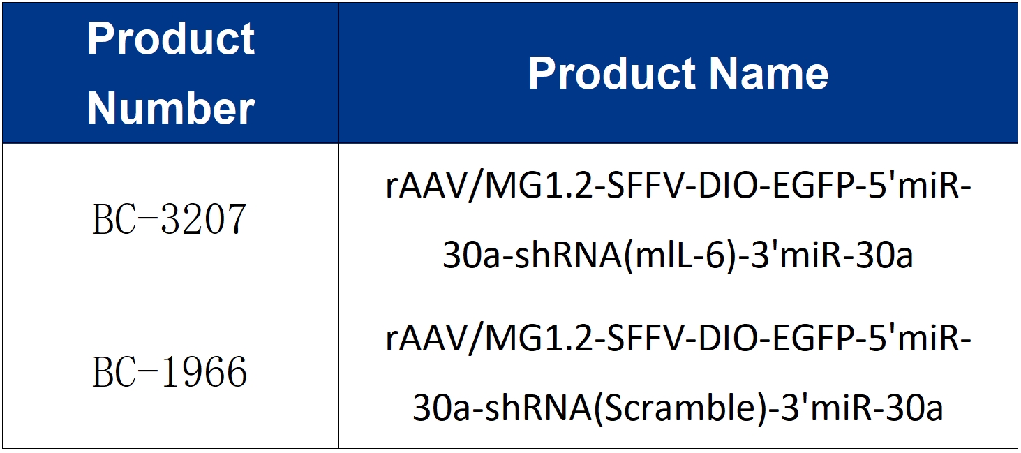
Finally, selective knockdown of IL-6 in microglia or IL-6 receptor (IL-6R) in astrocytes using genetic approaches also effectively mitigated CSDS-induced anxiety- and depression-like behaviors and astrocytic atrophy. These results suggest that the IL-6/IL-6R signaling pathway is a key driver of astrocyte apoptosis and depressive phenotypes in the CSDS model.

In summary, this study reveals that chronic stress-induced upregulation of IL-6 in microglia leads to astrocyte apoptosis, thereby promoting the development of depression-like behaviors in mice. These findings offer potential therapeutic targets for the clinical treatment of depression.

Schematic: Illustration of IL-6 released by microglia in the hippocampus inducing astrocyte apoptosis and mediating anxiety- and depression-like behaviors.


The viral tools used in this study were provided by Brain Case.
For inquiries or orders, please contact bd@ebraincase.com


💥Holiday

Special

Offers

Click to fill in the requirements and submit them to us!

Service Type :

Select the service you'd like to purchase.

Order Information(Premade-AAVs)

Please provide us some information about the service you'd like to order.

Detailed requirements:

scroll

Order Information(Custom AAV/Lentivirus)

Please provide us some information about the service you'd like to order.

Gene ID or gene information:

Selection of the reporting gene:

Special Instructions:

scroll

Order Information(Others)

Please provide us some information about the service you'd like to order.

Virus name/Detailed requirements:

scroll

Related products

Client Publication | Advanced Science | Discovery of Glial Cell Interaction Mechanism in Depression by the Teams of Jin Yu and Yuqiu Zhang

Client Publication | Advanced Science | Discovery of Glial Cell Interaction Mechanism in Depression by the Teams of Jin Yu and Yuqiu Zhang

Client Publication | Neuron | Liping Wang and Xuemei Liu’s Team at SIAT Uncovers Neural Mechanisms Behind Individual Differences in Innate Fear Responses

Client Publication | Neuron | Liping Wang and Xuemei Liu’s Team at SIAT Uncovers Neural Mechanisms Behind Individual Differences in Innate Fear Responses

Customer Publication | Sci. Adv. | Collaboration between Siyu Zhang from Shanghai Jiao Tong University and Min Xu from the Institute of Neuroscience, CAS reveals a whole-brain input-output map of basal forebrain cholinergic neuron subtypes.

Customer Publication | Sci. Adv. | Collaboration between Siyu Zhang from Shanghai Jiao Tong University and Min Xu from the Institute of Neuroscience, CAS reveals a whole-brain input-output map of basal forebrain cholinergic neuron subtypes.

Client Article | Cell Metabolism | Liu Danqian’s Team at the Institute of Neuroscience, CAS, Decodes the Oxidation-Sleep Code in Substantia Nigra Neurons

Client Article | Cell Metabolism | Liu Danqian’s Team at the Institute of Neuroscience, CAS, Decodes the Oxidation-Sleep Code in Substantia Nigra Neurons

map
{dede:global.cfg_webname/}

Virus product library

CRISPR
RNAi
Neurophilic virus
Optogenetics activation
Biosensors

News

Corporate News
New Product Launch
Media Activity
Investor News

Support

Literature interpretation
Customer article
Video Zone
FAQs
微信

WhatsApp Business Account

Tel: +8618971215294
E-mail: BD@ebraincase.com

Address:-

Address:-

  • Copyright © 2024 Brain Case All Rights Reserved.