E-mail:BD@ebraincase.com
Tel:+8618971215294
English 中文版
Virus Vector - Viral Vector Production - BrainCaseVirus Vector - Viral Vector Production - BrainCase
  • Home
  • Virus product library
    CRISPRRNAiHSV-helperRV-helperNeurophilic virusCalcium SensorsOptogenetics activationOptogenetics inhibitionChemical geneticsSparse labelingFluorescent proteinBiosensorsRecombinaseApoptosis & AutophagyDisease ModelNeurotoxicityOther
  • Products & Service

    Product Center

    Virus

    VSV-vaccine and gene therapy research
    Retrovirus-RCAS-TVA
    Lentivirus Vector-Lentivirus Production
    Rabies Virus Vector-RBV Vector
    Herpes simplex virus-Oncolytic and anterograde tracing
    PRV-retrograd multisynaptic-Peripheral
    AAV-gene therapy vectors-neuroscience

    Animal Model

    Neurological Disease Models-BrainCase
    Tumor animal models-anti-tumor-BrainCase
    Digestive System Disease Animal Model-Brain Case
    Cardiovascular System Disease Animal Models-Brain Case

    Plasmid Construction

    Library Construction
    Plasmid design and construction

    Popular Applications

    Gene Regulation

    Gene Overexpression-Brain Case
    RNA interference(RNAi)-siRNA-Brain Case
    Gene Editing - CRSIPR cloning - BrainCase

    Neural Circuit Function Research

    Optogenetics - BrainCase
    Chemical genetics-DREADDs-Brain Case
    Calcium signal recording-Gels- Brain Case
    GRAB Neurotransmitter Fluorescent Sensor- Brain Case
    Functional Magnetic Resonance Imaging Technology- Brain Case

    Research on the structure of neural circuits

    Direct Input and Output-viral vectors- Brain Case
    Anterograde Mono-synaptic Tracing -HSV- Brain Case
    Antrograde Muti-synaptic Tracing-HSV & VSV-Brain Case
    Retrograde Mono-synaptic Tracing-Rabies Virus-Brain Case
    Retrograde Muti-synaptic Tracing-PRV-Brain Case

    Featured Services

    Nervous System Disease Drug Effect

    Alzheimer's disease-AD-Brain Case
    Depression-mental disorders-Brain Case
    Parkinson's disease--PD-Brain Case
    Epilepsy-an ancient neurological disorder-Brain Case

    AAV Serotype Screening

    AAV Serotypes screening-gene therapy-Brain Case

    Tumorigenicity Test

    Tumorigenicity Testing-Evaluation of tumor models-Brain Case

    Efficacy of Oncolytic Virus

    Oncolytic virus-for cancer therapy-Brain Case
    Herpes Virus Vector-anti-tumor- BrainCase
    Vesicular stomatitis virus-killing tumor cells-Brain Case
  • News
    Corporate News New Product Launch Media Activity Investor News
  • Support
    Literature interpretation Customer article Video Zone FAQs
  • About Us
    Virus product library Products & Service News Support About Us Contact
  • Contact
    Contact Us Join us
  • 中文
    English 中文版
  • Home
  • Support
  • Customer article
  • Support
  • Literature interpretation
  • Customer article
  • Video Zone
  • FAQs

Mol. Psychiatry | Team Led by Miao He and Ling Gong Reveals Independent Circuits and Behavioral Regulation Mechanisms of Two Neuronal Subtypes in the Medial Mammillary Body

Release time:2025-07-08 16:21:11
The medial mammillary body (MM) is a key component of the Papez circuit and the extended hippocampal system, playing a crucial role in learning and memory, spatial navigation, and emotional regulation. Dysfunction of the MM is closely associated with neuropsychiatric disorders such as Wernicke-Korsakoff syndrome, Alzheimer’s disease, and schizophrenia. However, the composition, cellular properties, local connectivity, and functional regulation of MM neurons have remained unclear.

On June 23, 2025, a research team led by Miao He and Ling Gong from the Institute of Brain Science at Fudan University published a study in Molecular Psychiatry titled "Two distinct cell types of the medial mammillary body forming segregated subcircuits." This study systematically revealed, for the first time, the existence of two major neuronal subtypes in the MM: calbindin (CB)-expressing neurons and parvalbumin (PV)-expressing neurons. These subtypes differ significantly in their distribution, physiology, morphology, and circuit connectivity, forming independent and parallel circuits. MMCB neurons are involved in driving place aversion and motor regulation, while MMPV neurons may mediate other functions such as cognitive memory.

https://www.nature.com/articles/s41380-025-03079-w


MMCB and MMPV Occupy Complementary Regions Within the MM

Using quadruple transgenic mice (CBcre::PVFlp::Ai3::td-FRT), the research team simultaneously visualized MMCB and MMPV neurons within the same brain. Three-dimensional spatial analysis revealed that the MMPV region is surrounded by MMCB neurons from the anterior, lateral, and ventral sides, forming a "calyx holding a bud"-like complementary distribution pattern. Quantitative analysis showed that MMCB and MMPV together constitute the vast majority of neurons in the MM, with minimal overlap in their expression.

Figure 1. MMCB and MMPV occupy complementary regions within the MM.


Distinct Electrophysiological and Morphological Features of MMCB and MMPV Neurons

Further investigations using immunostaining to quantify soma size and biocytin filling to reconstruct dendritic morphology revealed that MMCB and MMPV neurons are morphologically distinguishable. MMCB neurons have smaller somata and shorter, simpler dendrites. Using unsupervised hierarchical clustering and SVM analysis based on morphological (M), electrophysiological (E), and combined multimodal (M–E) features, the study found that M features alone effectively classified the cells into two clusters corresponding to MMCB and MMPV. E features performed even better, and the combined M–E clustering showed the highest correspondence with genetically defined cell types. These findings further confirm that MMCB and MMPV represent distinct neuronal subtypes with unique anatomical and physiological characteristics.

Figure 2. MMCB and MMPV exhibit distinct morphological and electrophysiological properties.


Differences in Spontaneous Firing Between MMCB and MMPV Neurons

In previous whole-cell patch-clamp recordings, the authors observed that some MM neurons exhibited two distinct patterns of spontaneous firing: tonic and bursting. To further investigate this phenomenon, they switched to cell-attached recordings and found that MMCB neurons had a lower rate of spontaneous activity compared to MMPV neurons, but a higher incidence of burst firing. Some MM neurons were capable of switching between tonic and bursting modes.

N — methyl-D-aspartate receptors (NMDARs) and T-type voltage-sensitive calcium channels (T-VSCCs) have been reported to play key roles in neuronal burst firing across various brain regions. The authors further examined this and found that activation of NMDARs promoted the transition of most tonic-firing neurons into a bursting mode. Notably, MMCB neurons generated more action potentials per burst with longer inter-burst intervals. Additionally, MMCB neurons exhibited a higher incidence of post-hyperpolarization rebound firing mediated by T-VSCCs, consistent with their bursting characteristics.


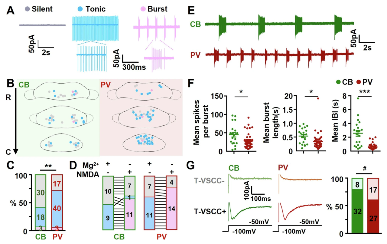
Figure 3. Differences in spontaneous firing between MMCB and MMPV neurons.


Segregated Outputs and Differential Inputs of MMCB and MMPV Neurons

To visualize the output projections of MMCB and MMPV  neurons, the researchers injected AAV-DIO-ChR2-RFP and AAV-fDIO-ChR2-GFP into the MM region of CBcre::PVFlp mice. The results showed that the axon terminals of MMCB and MMPV  neurons exhibited minimal overlap in downstream brain regions such as the anterior thalamus (AT), ventral tegmental nucleus (VTg), and retrotegmental pontine nucleus (RtTgP). This indicates that MMCB and MMPV neurons form anatomically segregated output circuits, each establishing topographically distinct subcircuits with their respective downstream targets.

Furthermore, the team used rabies virus-based tracing to label the inputs to MM neurons. In CBcre::PVFlp mice, helper viruses (AAV-DIO-GFP-TVA and AAV-DIO-G) were injected into the MM, and after two weeks, EnvA-RVΔG-RFP was injected into the AT region. The study found that compared to MMPV  neurons, MMCB neurons received relatively fewer inputs from the hippocampus and more from the hypothalamus. Both neuron types received their strongest input from the subiculum (SUB), but with distinct topographic preferences: MMCB favored the ventral subiculum (vSUB), while MMPV  favored the dorsal subiculum (dSUB), consistent with previously observed dorsoventral patterns of SUB-to-MM projections.


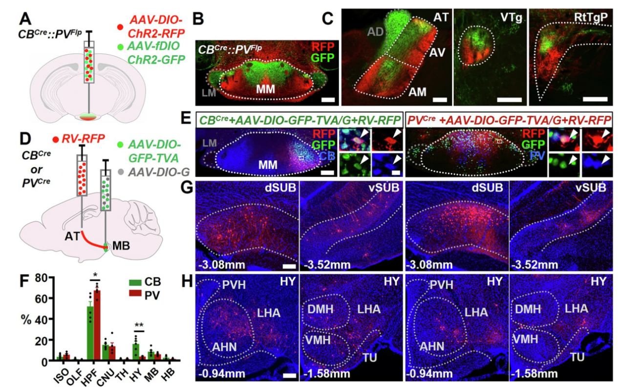
Figure 4. MMCB  and MMPV Project to Complementary Brain Regions and Receive Differential Inputs


MMCB and MMPV Mediate Distinct Behavioral Responses

To further determine whether MMCB and MMPV neurons elicit different behavioral effects, the researchers used optogenetic techniques by injecting AAV-DIO-ChR2-RFP into the MM region of CBcre::PVcre mice. Combining this with real-time place preference and open field tests, they found that only activation of MMCB induced avoidance behavior and enhanced locomotion, while MMPV activation had no such effect.

Additionally, by injecting a calcium imaging virus (AAV-DIO-GCaMP7) into the MM region, the researchers found that calcium activity in MMCB neurons significantly increased around movement onset, with movement-related intensity 65% higher than that of MMPV neurons, which showed no significant change. These results align with the optogenetic findings, further confirming that MMCB—but not MMPV—plays a key role in motor regulation, and highlighting the functional divergence of molecularly defined neuronal subtypes.


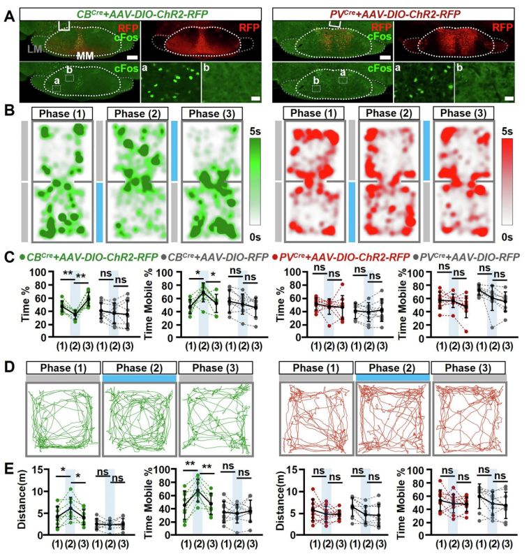
Figure 5. Optogenetic activation of MMCB—but not MMPV—induces place aversion and promotes locomotion.


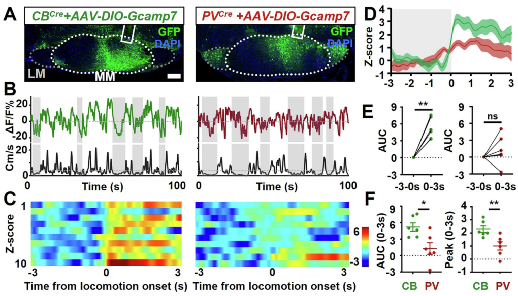
Figure 6. MMCB and MMPV mediate distinct movement-related activity.


MMCB and MMPV Form Parallel Circuits Without Local Interactions

To directly assess whether MMCB and MMPV neurons are interconnected, the researchers performed whole-cell patch-clamp recordings to measure postsynaptic responses following optogenetic activation of either MMCB or MMPV neurons. Interestingly, while direct activation could be readily detected in ChR2-expressing neurons, no optogenetically evoked postsynaptic currents (oPSCs) were observed in ChR2-negative neurons. To rule out potential NMDAR inactivation by Mg²⁺, Mg²⁺ was removed from the ACSF, yet still no oPSCs were detected in ChR2-negative neurons.

These findings further confirm that there are no synaptic connections or cross-activation between MMCB and MMPV neurons, indicating that they form independent, parallel neuronal subcircuits. Among the recorded ChR2-negative cells, some were located in the same genetically defined region as neighboring ChR2-positive cells, yet still showed no evidence of direct activation—suggesting that the lack of local connectivity among neurons within the MM is a general feature.


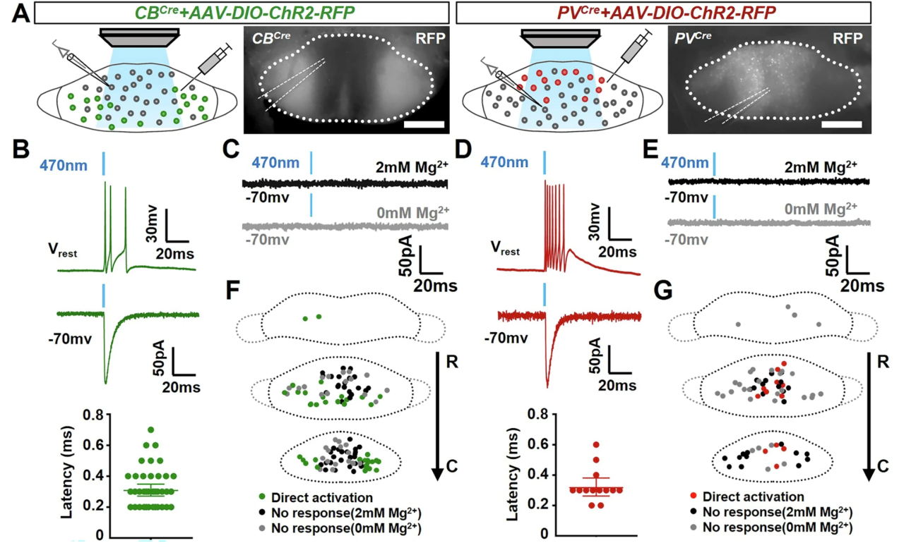
Figure 7. No Cross-Activation Observed Between MMCB and MMPV Neurons


Conclusion

This study is the first to systematically identify two distinct neuronal subtypes in the medial mammillary body, marked by CB and PV, and to reveal their independent, parallel circuit mechanisms and roles in behavioral regulation. The lack of local connectivity in the MM suggests that its functions are primarily mediated through long-range circuits. This research provides a cell type–specific framework for understanding the functional organization of the MM, the mechanisms underlying neuropsychiatric disorders, and potential targets for therapeutic intervention.

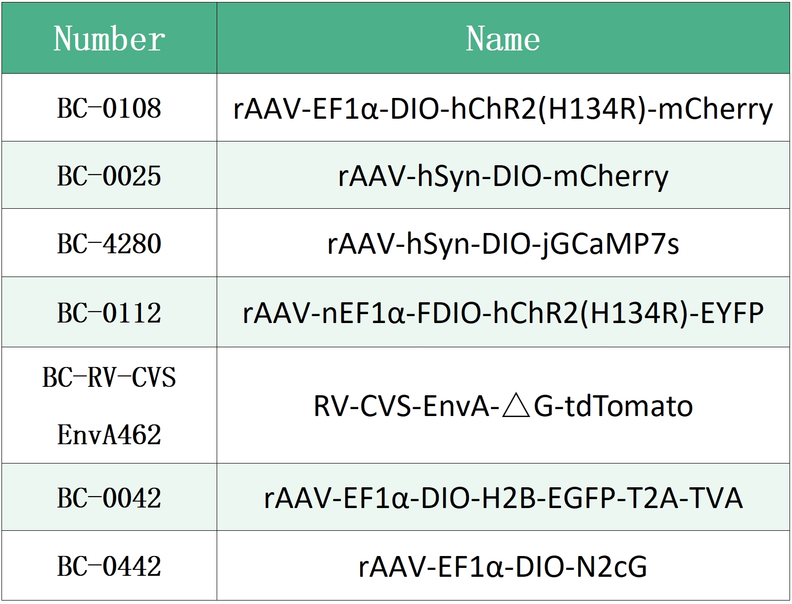
The viral tools used in this study were provided by Brain Case Biotech.


Contact Us

if you encounter any challenges during your neural circuit research, feel free to reach out to us at bd@ebraincase.com — we’d be happy to discuss and explore potential solutions with you.


💥Holiday

Special

Offers

Click to fill in the requirements and submit them to us!

Service Type :

Select the service you'd like to purchase.

Order Information(Premade-AAVs)

Please provide us some information about the service you'd like to order.

Detailed requirements:

scroll

Order Information(Custom AAV/Lentivirus)

Please provide us some information about the service you'd like to order.

Gene ID or gene information:

Selection of the reporting gene:

Special Instructions:

scroll

Order Information(Others)

Please provide us some information about the service you'd like to order.

Virus name/Detailed requirements:

scroll

Related products

Mol. Psychiatry | Team Led by Miao He and Ling Gong Reveals Independent Circuits and Behavioral Regulation Mechanisms of Two Neuronal Subtypes in the Medial Mammillary Body

Mol. Psychiatry | Team Led by Miao He and Ling Gong Reveals Independent Circuits and Behavioral Regulation Mechanisms of Two Neuronal Subtypes in the Medial Mammillary Body

Client Publication | Advanced Science | Discovery of Glial Cell Interaction Mechanism in Depression by the Teams of Jin Yu and Yuqiu Zhang

Client Publication | Advanced Science | Discovery of Glial Cell Interaction Mechanism in Depression by the Teams of Jin Yu and Yuqiu Zhang

Client Publication | Neuron | Liping Wang and Xuemei Liu’s Team at SIAT Uncovers Neural Mechanisms Behind Individual Differences in Innate Fear Responses

Client Publication | Neuron | Liping Wang and Xuemei Liu’s Team at SIAT Uncovers Neural Mechanisms Behind Individual Differences in Innate Fear Responses

Customer Publication | Sci. Adv. | Collaboration between Siyu Zhang from Shanghai Jiao Tong University and Min Xu from the Institute of Neuroscience, CAS reveals a whole-brain input-output map of basal forebrain cholinergic neuron subtypes.

Customer Publication | Sci. Adv. | Collaboration between Siyu Zhang from Shanghai Jiao Tong University and Min Xu from the Institute of Neuroscience, CAS reveals a whole-brain input-output map of basal forebrain cholinergic neuron subtypes.

map
{dede:global.cfg_webname/}

Virus product library

CRISPR
RNAi
Neurophilic virus
Optogenetics activation
Biosensors

News

Corporate News
New Product Launch
Media Activity
Investor News

Support

Literature interpretation
Customer article
Video Zone
FAQs
微信

WhatsApp Business Account

Tel: +8618971215294
E-mail: BD@ebraincase.com

Address:-

Address:-

  • Copyright © 2024 Brain Case All Rights Reserved.