Customer Publication | Sci. Adv. | Collaboration between Siyu Zhang from Shanghai Jiao Tong University and Min Xu from the Institute of Neuroscience, CAS reveals a whole-brain input-output map of basal forebrain cholinergic neuron subtypes.
Release time:2025-09-19 11:59:42
In the complex neural mechanisms underlying cognition and emotion, basal forebrain cholinergic neurons (BFCNs) play a pivotal role. These neurons regulate the function of brain regions such as the prefrontal cortex and amygdala by releasing acetylcholine (ACh). However, the whole-brain input-output network organization of distinct BFCN subtypes remains unclear.
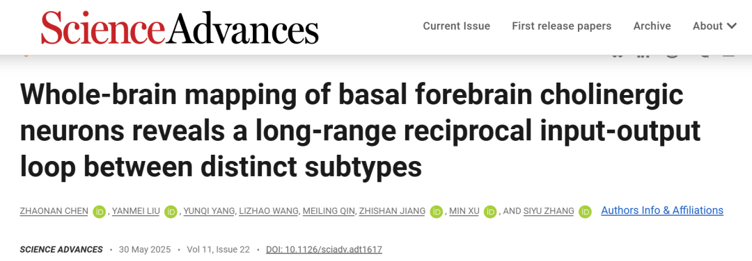
On May 30, 2025, Siyu Zhang from Shanghai Jiao Tong University School of Medicine, in collaboration with Min Xu’s team at the Center for Excellence in Brain Science and Intelligence Technology, Chinese Academy of Sciences, published a study in Science Advances titled “Whole-brain mapping of basal forebrain cholinergic neurons reveals a long-range reciprocal input-output loop between distinct subtypes.”This study systematically reveals for the first time the whole-brain connectivity maps of two BFCN subtypes: BFCN→ACA (projecting to the anterior cingulate cortex) and BFCN→BLA (projecting to the basolateral amygdala).
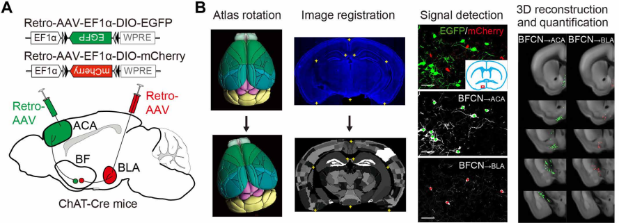
Using ChAT-Cre transgenic mice, the researchers combined retrograde adeno-associated virus (Retro-AAV) labeling, modified rabies virus (RV) monosynaptic tracing, triple RNAscope in situ hybridization, and whole-cell patch-clamp recording to establish a multi-scale analytical framework spanning from single-cell properties to whole-brain networks.
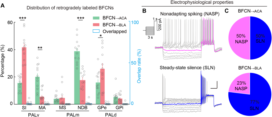
The study first used retrograde viral labeling to identify BFCN→ACA and BFCN→BLA as two subpopulations with distinct distribution patterns. Electrophysiological recordings revealed that these subgroups exhibit different firing properties: in BFCN→ACA, non-adapting spiking and steady low-frequency non-spiking (SLN) neurons each accounted for 50%, whereas in BFCN→BLA, SLN neurons dominated, making up 70%—suggesting a preference for rapid response modes in emotional processing.
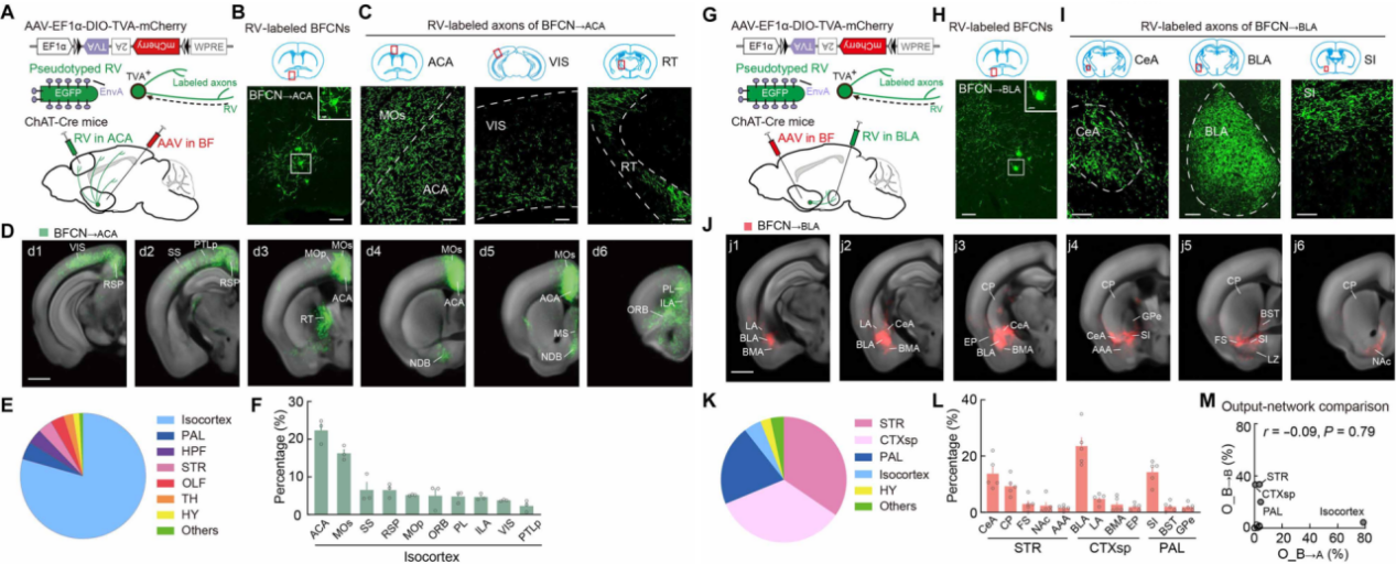
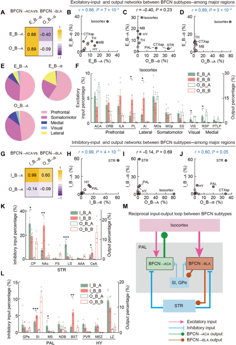
Whole-brain output mapping showed that BFCN→ACA primarily innervates isocortical regions (79% of total axonal projections), especially the prefrontal cortex, while BFCN→BLA predominantly projects to the striatum, subplate cortex, and globus pallidus, together accounting for 87%. This distinct projection pattern aligns well with their functional roles: the former in cognitive regulation and the latter in emotional processing.
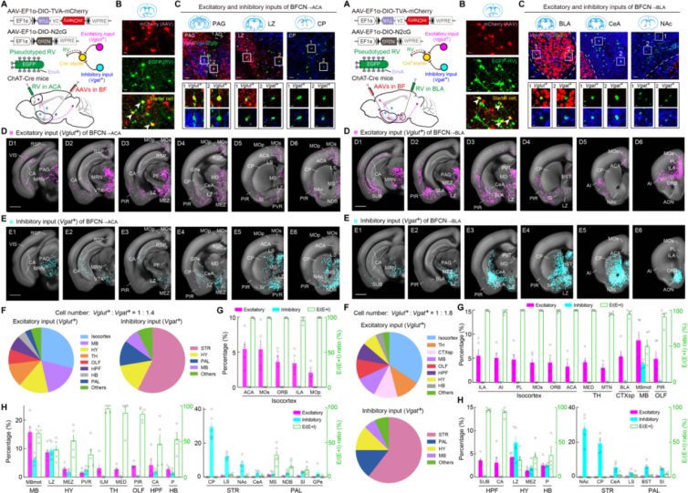
Using RV-assisted tracing to identify input neuron types, the researchers revealed for the first time the specificity of excitatory (Vglut+) and inhibitory (Vgat+) inputs to each BFCN subtype. Notably, while both subtypes receive shared inhibitory inputs from the striatum, hypothalamus, and globus pallidus, their excitatory inputs diverge significantly: BFCN→ACA mainly receives excitatory signals from the midbrain and hypothalamus, whereas BFCN→BLA receives more from the thalamus and amygdala.
Network interaction analysis uncovered a sophisticated mechanism of functional coupling between the two subtypes: BFCN→ACA modulates the activation of BFCN→BLA by dominating cortical areas (shared excitatory input sources), while BFCN→BLA regulates the inhibition of BFCN→ACA via projections to the striatum and globus pallidus (shared inhibitory input sources). This long-range reciprocal loop provides a structural foundation for understanding how the cholinergic system coordinates cognitive and emotional processing.
Summary
This study is the first to resolve the input-output networks of BFCN subpopulations at the single-cell type level, uncovering a novel interaction mechanism formed through shared input sources. Notably, the CA region of the hippocampusexhibits a strikingly distinct input preference—preferentially providing inhibitory input to BFCN→ACA and excitatory input to BFCN→BLA. This may represent the neural basis by which differences in memory context modulate cognitive and emotional processing. Beyond deepening our understanding of cholinergic regulation, the whole-brain, single-cell-type connectomics approach established here offers a valuable framework for investigating other complex neural circuits.
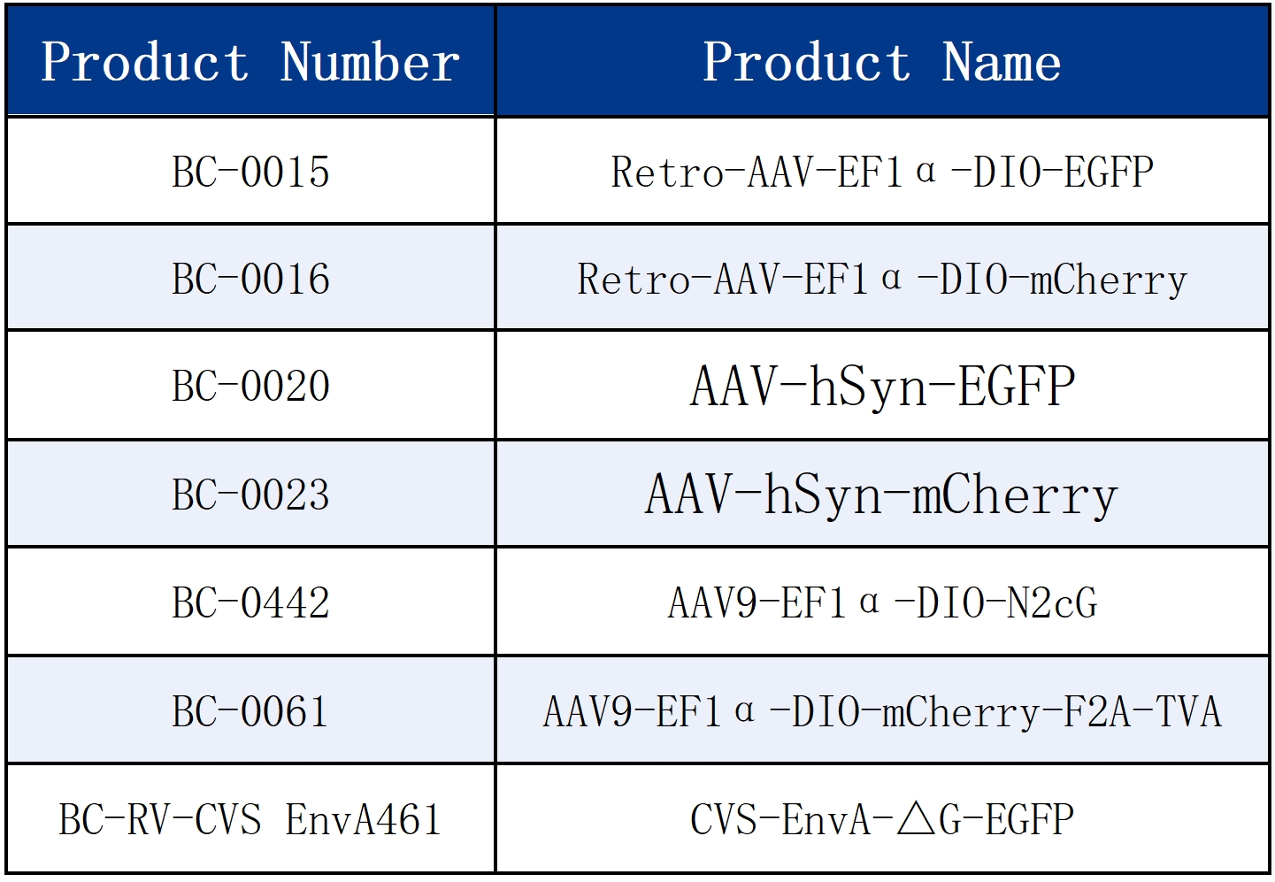
The viral tools used in this study are all available from Brain Case:
Interested in RV-assisted tracing? Brain Case provides vector combinations like BC-0442 + BC-0061 + BC-RV-CVS EnvA461, as featured in this publication. Reach out to us at bd@ebraincase.com for more details and support!
Service Type :
Select the service you'd like to purchase.
Order Information(Premade-AAVs)
Please provide us some information about the service you'd like to order.
Order Information(Custom AAV/Lentivirus)
Please provide us some information about the service you'd like to order.
Order Information(Others)
Please provide us some information about the service you'd like to order.