Client Publication | Nature Communications | USTC Team Led by Zhi Zhang, Juan Li, and Peng Cao Unveils Neural Circuit Mechanism of Male-Specific Conditioned Pain Hypersensitivity
Release time:2025-09-19 14:25:08
In both humans and rodents, males and females exhibit different responses to pain. For example, females are more susceptible to chronic pain, while males show specific pain hypersensitivity patterns, such as conditioned pain hypersensitivity. Testosterone plays a crucial role in the nociceptive system of male mice—castration (surgical removal of the gonads) alleviates pain hypersensitivity, whereas testosterone supplementation restores this phenotype. However, the underlying neural mechanisms have remained unclear.
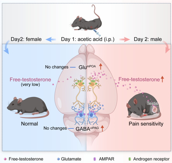
On April 17, 2025, a research team led by Zhi Zhang and Peng Cao from the School of Life Sciences and Medicine at the University of Science and Technology of China (USTC), along with Juan Li from the First Affiliated Hospital of USTC, published a study in Nature Communications titled “A neural circuit for sex-dependent conditioned pain hypersensitivity in mice.” This study reveals that in male mice, free testosterone activates androgen receptor signaling in glutamatergic neurons of the medial preoptic area (Glu-mPOA), leading to their hyperactivation. These neurons provide excitatory input to GABAergic neurons in the ventrolateral periaqueductal gray (GABA-vlPAG), thereby triggering conditioned pain hypersensitivity.
Conditioned Context Induces Hyperactivity of Glu-mPOA Neurons in Male Mice
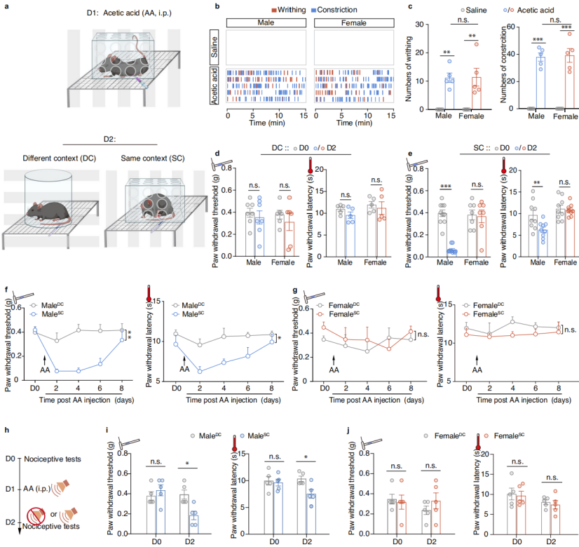
To investigate the neural mechanisms underlying sex-specific pain hypersensitivity (i.e., pain sensitization), the researchers established a mouse model of context-dependent conditioned pain hypersensitivity. Male and female mice received intraperitoneal injections of acetic acid (AA) in a specific context, which induced comparable abdominal writhing and contraction responses in both sexes (Fig. 1a–c). On the following day, the mechanical and thermal pain thresholds were measured as the mice were re-exposed to either the same or a different environment from the one used for AA injection (Fig. 1a). The results showed that male mice exhibited pronounced pain hypersensitivity when placed in the same context (male-SC), but not in a different one (male-DC). In contrast, female mice and control mice treated with saline did not display context-dependent pain hypersensitivity (Fig. 1d–e). This male-specific hypersensitivity persisted for at least five days (Fig. 1f–g). Furthermore, the same auditory cues associated with the pain-inducing environment were sufficient to trigger context-dependent pain hypersensitivity in males, highlighting the specificity of this conditioned response (Fig. 1h–j).
Figure 1. Enhanced Pain Sensitivity in Male Mice
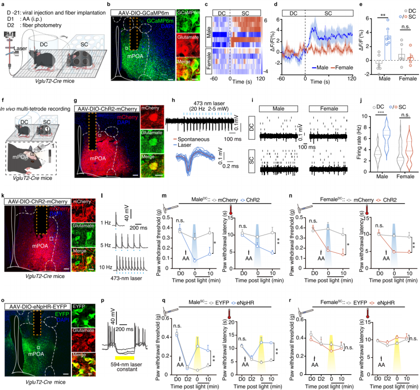
To determine whether increased Glu-mPOA neuronal activity results from the establishment of conditioned pain hypersensitivity, the researchers performed fiber photometry recordings in freely moving VgluT2-Cre mice. These mice received AAV-DIO-GCaMP6m injections into the mPOA (Fig. 2a–b). On the second day following acetic acid injection, GCaMP6m-labeled Glu-mPOA neurons in male-SC mice exhibited significantly higher activity compared to male-DC mice, while no such difference was observed between female groups (Fig. 2c–e). Similarly, no increased activity was found in Vgat-Cre male or female SC mice.
Subsequently, in vivo multi-electrode recordings were conducted in freely moving VgluT2-Cre mice after mPOA injection with AAV-DIO-ChR2 (Fig. 2f–h). Within 5 minutes of AA injection, both male and female mice showed a sharp increase in spontaneous Glu-mPOA neuron firing, which returned to baseline within 3 hours. However, on the following day, spontaneous firing rates in male-SC mice were significantly higher than in male-DC mice, whereas no difference was observed between female-SC and female-DC groups (Fig. 2i–j). The time-course dynamics of Glu-mPOA neuron hyperactivity in male-SC mice aligned closely with the behavioral progression of male-specific context-dependent pain hypersensitivity (Fig. 1f).
To confirm that Glu-mPOA neurons mediate pain hypersensitivity in male-SC mice, AAV-DIO-ChR2-mCherry was injected into the mPOA of VgluT2-Cre mice (Fig. 2k–l). Pain threshold testing and conditioned place aversion assays revealed that optogenetic activation of Glu-mPOA neurons induced pain hypersensitivity and avoidance behavior in both male-DC and female-DC mice (Fig. 2m–n). Conversely, optogenetic inhibition of Glu-mPOA neurons using AAV-DIO-eNpHR3.0-EYFP abolished context-dependent pain hypersensitivity in male-SC mice (Fig. 2o–q), while having no effect on the mechanical or thermal pain thresholds of female-SC mice (Fig. 2r). Chemogenetic inhibition of Glu-mPOA neurons by administering CNO on Day 2 also eliminated pain hypersensitivity in male-SC mice. These findings demonstrate that pain modulation in the context of conditioning selectively induces context-dependent hyperactivity in Glu-mPOA neurons of male mice.
Figure 2. Conditioned Context Induces Pain Hypersensitivity via Activation of Glu-mPOA Neurons
Free Testosterone Mediates Context-Dependent Pain Hypersensitivity in Male Mice
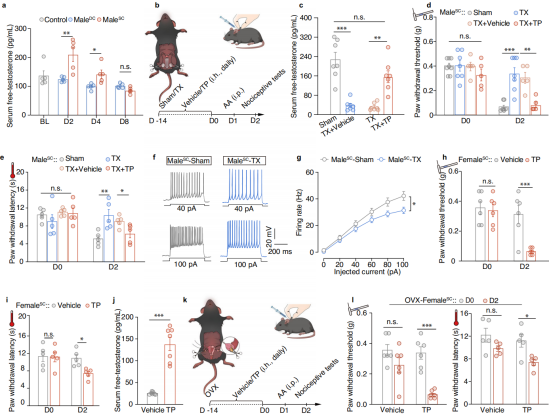
As the biologically active form of testosterone, free testosterone may regulate male-specific conditioned pain hypersensitivity via androgen receptor signaling. ELISA measurements of free testosterone in serum and plasma revealed that male-SC mice exhibited significantly elevated levels compared to male-DC mice and untreated males. This elevation persisted until Day 4 (D4), returning to baseline by Day 8 (D8) (Fig. 3a).
To assess the impact of artificially reducing free testosterone on context-dependent pain hypersensitivity in males, the researchers surgically castrated male mice to remove the gonads and subsequently administered daily subcutaneous injections of testosterone propionate to restore testosterone levels (Fig. 3b–c). Following castration, male-SC mice showed significantly increased mechanical pain thresholds (PWT) and thermal withdrawal latencies (PWL), indicating reduced pain hypersensitivity. This was accompanied by decreased Glu-mPOA neuronal activity (Fig. 3d–g). Upon testosterone supplementation, castrated male-SC mice exhibited a decrease in PWT and PWL, reflecting the return of pain hypersensitivity, suggesting that testosterone directly drives this pain phenotype (Fig. 3d–e).
In untreated female-SC mice, no pain hypersensitivity was observed. However, after testosterone propionate treatment, female-SC mice developed a male-like hypersensitivity phenotype, as evidenced by significantly reduced PWT compared to vehicle controls (Fig. 3h–j). This indicates that exogenous testosterone is sufficient to induce male-specific pain sensitivity in females, highlighting testosterone as a key factor underlying sex differences in pain response.
To rule out the influence of estrogen, a conditioned pain hypersensitivity model was established in ovariectomized (OVX) female mice (Fig. 3k). OVX female-SC mice did not exhibit hypersensitivity on their own, but developed it after testosterone supplementation, confirming the androgen-dependent, estrogen-independent nature of the response (Fig. 3l). In summary, free testosterone mediates male-specific, context-dependent pain hypersensitivity through the androgen receptor, and this effect is independent of estrogen.
Figure 3. Free Testosterone Contributes to Male-Specific Contextual Pain Hypersensitivity
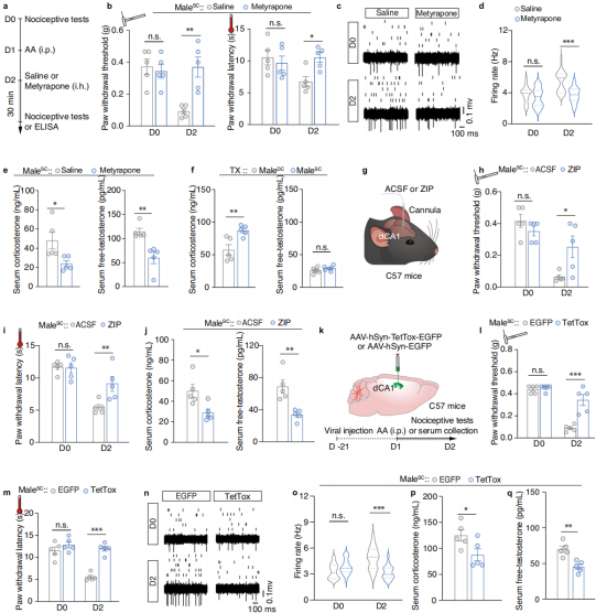
Acute stress produces analgesic effects by elevating corticosterone, whereas chronic stress induces pain sensitization. However, it remains unclear whether corticosterone is involved in male-specific contextual pain hypersensitivity. To investigate whether stress contributes to the formation of male-specific contextual pain hypersensitivity, researchers subcutaneously injected male-SC mice with a corticosterone synthesis inhibitor (metirapone) 30 minutes before testing on the following day (Figure 4a). They found that after metirapone treatment, the mechanical and thermal pain thresholds of male-SC mice returned to normal levels, and the overactivity of Glu-mPOA neurons was suppressed (Figures 4b–d). Male-SC mice treated with metirapone showed significantly reduced serum corticosterone and free testosterone levels, but castration experiments demonstrated that elevated corticosterone is not directly related to pain hypersensitivity (Figures 4e–f). These results indicate that free testosterone, rather than corticosterone, is required for male-specific contextual pain hypersensitivity.
Given that different pain-related environments can induce male-specific contextual pain hypersensitivity, it is speculated that the memory system might trigger this response. On the next day, the PKMζ inhibitor zeta inhibitory peptide (ZIP) was delivered to the dorsal hippocampal CA1 region (dCA1) of male-SC mice to suppress the formation of contextual memory (Figure 4g). The results showed that ZIP treatment abolished the conditioned contextual pain hypersensitivity in male-SC mice, and serum corticosterone and free testosterone concentrations were significantly lower than those in the vehicle-treated control group (Figures 4h–j). To inactivate synaptic transmission in the dCA1 region of male mice, an AAV virus expressing tetanus neurotoxin (AAV-hSyn-TetTox-EGFP) was directly injected (Figure 4k). It was found that the conditioned contextual pain hypersensitivity and overactivity of Glu-mPOA neurons in TetTox-infected male-SC mice were both reversed (Figures 4l–q). These findings suggest that contextual memory is indeed the trigger for male-specific contextual pain hypersensitivity.
Figure 4. Stress and Memory Systems Regulate Male-Specific Contextual Pain Hypersensitivity
Testosterone Induces Contextual Pain Hypersensitivity by Enhancing Glu-mPOA Neuronal Excitability
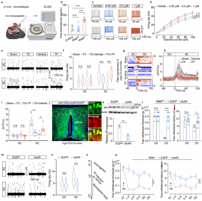
In vivo microdialysis combined with ELISA revealed that, compared to male-DC mice and untreated males, male-SC mice exhibited significantly elevated levels of free testosterone in the medial preoptic area (mPOA) on Day 2 (Fig. 5a–b). Whole-cell patch-clamp recordings on mPOA brain slices treated with 0.05 μM, 0.3 μM, or 1 μM testosterone propionate showed that 0.3 μM and 1 μM treatments increased the firing frequency of Glu-mPOA neurons (Fig. 5c–d).
To monitor activity changes in Glu-mPOA neurons in testosterone-manipulated male mice, in vivo multi-electrode recordings were conducted in castrated male-SC mice. Results showed that the firing frequency of Glu-mPOA neurons was significantly reduced in castrated mice compared to sham-operated controls, and daily injections of testosterone propionate restored firing rates to control levels (Fig. 5e–f). Fiber photometry in freely moving male mice confirmed that testosterone injection restored Glu-mPOA activity in castrated male-SC mice (Fig. 5g–i). Additionally, microinjection of testosterone propionate into the mPOA of untreated males lowered paw withdrawal threshold and latency compared to vehicle controls. These results indicate that increased levels of free testosterone can trigger contextual pain hypersensitivity by enhancing Glu-mPOA neuronal excitability.
Subsequently, AAV-shAR-EGFP virus was used to knock down androgen receptors (AR) specifically in Glu-mPOA neurons of VgluT2-Cre mice (Fig. 5j–k). Male-SC mice with AR knockdown in Glu-mPOA neurons displayed significantly increased paw withdrawal thresholds and latencies (Fig. 5l). In vivo multi-electrode recordings showed that on Day 2, AR knockdown mice had significantly lower spontaneous Glu-mPOA firing frequency compared to AAV-EGFP controls, while there was no difference between AR knockdown and control mice in the male-DC group (Fig. 5m–n). Furthermore, testosterone infusion into the mPOA of AAV-EGFP–infected untreated males significantly reduced their paw withdrawal thresholds and latencies, whereas no such effect was observed in AAV-shAR-EGFP–infected mice (Fig. 5o–p).
Conclusion: Elevated free testosterone induces male-specific contextual pain hypersensitivity by enhancing Glu-mPOA neuronal excitability through an androgen receptor–dependent mechanism.
Figure 5. The role of androgen receptors in contextual pain hypersensitivity.
Excitatory Glu-mPOA → GABA-vlPAG Circuit
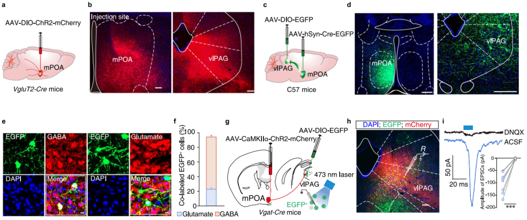
Previous studies have suggested that the medial preoptic area (mPOA) may be involved in pain regulation, although it has not been traditionally considered a classic pain-processing region. To explore the downstream targets of Glu-mPOA neurons, anterograde tracing was performed by injecting AAV-DIO-ChR2-mCherry into the mPOA of VgluT2-Cre mice (Figure 6a). The results revealed that mCherry+ axonal terminals from Glu-mPOA neurons were distributed across multiple brain regions, with the densest signals observed in the ventrolateral periaqueductal gray (vlPAG) (Figure 6b).
To identify the cell types in the mPOA→vlPAG projection, AAV2/1-Cre-EGFP was injected into the mPOA and AAV-DIO-EGFP into the vlPAG of C57 mice (Figure 6c-d). Three weeks later, most EGFP+ neurons in the vlPAG were co-labeled with GABA antibodies (Figure 6e-f). In addition, retrograde tracing using retroAAV-hSyn-EGFP injected into the vlPAG showed that EGFP+ neurons in the mPOA were primarily co-labeled with glutamate antibodies.
Further, modified rabies virus tracing was conducted by injecting EnvA-pseudotyped RV-ΔG-EGFP along with helper viruses (AAV-Ef1α-DIO-TVA-mCherry and AAV-Ef1α-DIO-RVG) into the vlPAG of male and female Vgat-Cre mice. A substantial number of EGFP+ neurons in the mPOA were co-labeled with glutamate antibodies, indicating the existence of Glu-mPOA→GABA-vlPAG connections in both sexes.
To further elucidate the functional relevance of the Glu-mPOA→GABA-vlPAG pathway, optogenetic experiments were performed (Figure 6g). Whole-cell patch-clamp recordings in brain slices showed that brief photostimulation of ChR2-expressing Glu-mPOA axon terminals in the vlPAG induced excitatory postsynaptic currents (EPSCs) in GABA-vlPAG neurons, which could be blocked by AMPA receptor antagonists (Figure 6h-i). These results demonstrate that GABA-vlPAG neurons receive functional monosynaptic excitatory glutamatergic input from Glu-mPOA neurons.
Critical Role of the Glu-mPOA → GABA-vlPAG Circuit in Male Contextual Pain Hypersensitivity
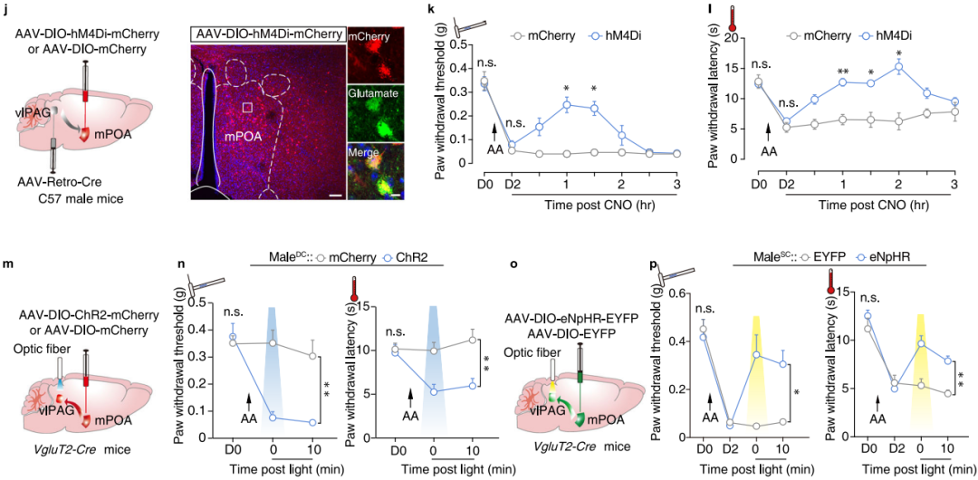
To determine whether the increased activity of GABA-vlPAG neurons in the contextual pain hypersensitivity model originates from enhanced excitatory input from Glu-mPOA neurons, AAV-DIO-hM4Di-mCherry was injected into the mPOA of C57 mice, and retroAAV-Cre was injected into the ipsilateral vlPAG (Figure 7j). Subsequent chemogenetic inhibition of Glu-mPOA neurons projecting to the vlPAG led to increased paw withdrawal thresholds and latencies in hM4Di-expressing male-SC mice (Figure 7k-l).
Furthermore, activation of Glu-mPOA axon terminals in the vlPAG induced pain hypersensitivity and place aversion in both male-DC and female-DC mice (Figure 7m-n). Conversely, inhibition of the Glu-mPOA → GABA-vlPAG circuit attenuated contextual pain hypersensitivity in male-SC mice (Figure 7o-p). Notably, since female-SC mice did not exhibit contextual pain hypersensitivity, optogenetic inhibition of this pathway had no significant effect on their nociceptive thresholds. These findings demonstrate that the Glu-mPOA → GABA-vlPAG circuit plays a key role in the development of contextual pain hypersensitivity in male mice.
Figure 7. Critical Role of the Glu-mPOA → GABA-vlPAG Circuit in Male Contextual Pain Hypersensitivity
Summary
This study is the first to reveal the neural circuit and hormonal regulatory mechanism underlying male-specific pain hypersensitivity. It provides a comprehensive explanatory chain of "memory–hormone–neural pathway" for understanding sex differences in pain perception. The findings suggest that targeting androgen receptors or the Glu-mPOA→vlPAG pathway could serve as potential strategies for treating male-specific pain. Furthermore, this work highlights the importance of considering sex as a critical factor in both pain research and clinical treatment outcomes.
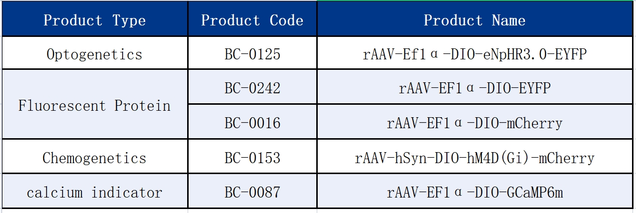
The following viral vector products used in this study were provided by Brain Case Biotech: