E-mail:BD@ebraincase.com
Tel:+8618971215294
English 中文版
Virus Vector - Viral Vector Production - BrainCaseVirus Vector - Viral Vector Production - BrainCase
  • Home
  • Virus product library
    CRISPRRNAiHSV-helperRV-helperNeurophilic virusCalcium SensorsOptogenetics activationOptogenetics inhibitionChemical geneticsSparse labelingFluorescent proteinBiosensorsRecombinaseApoptosis & AutophagyDisease ModelNeurotoxicityOther
  • Products & Service

    Product Center

    Virus

    VSV-circuit research-vaccine and gene therapy research-BrainCase
    Retrovirus-RCAS-TVA-BrainCase
    Lentivirus Vector - Lentivirus Production - BrainCase
    Rabies Virus Vector - RBV Vector - BrainCase
    Herpes simplex virus-Oncolytic and anterograde tracing-Brain Case
    PRV-retrograd multisynaptic-Peripheral-Btain Case
    AAV-gene therapy vectors-BrainCase

    Animal Model

    Neurological Disease Models-BrainCase
    Tumor animal models-anti-tumor-BrainCase
    Digestive System Disease Animal Model-Brain Case
    Cardiovascular System Disease Animal Models-Brain Case

    Plasmid Construction

    Bac
    Library Construction
    Plasmid design and construction

    Popular Applications

    Gene Regulation

    Gene Overexpression-Brain Case
    RNA interference(RNAi)-siRNA-Brain Case
    Gene Editing - CRSIPR cloning - BrainCase

    Neural Circuit Function Research

    Optogenetics - BrainCase
    Chemical genetics-DREADDs-Brain Case
    Calcium signal recording-Gels- Brain Case
    GRAB Neurotransmitter Fluorescent Probe- Brain Case
    Functional Magnetic Resonance Imaging Technology- Brain Case

    Research on the structure of neural circuits

    Direct Input and Output-viral vectors- Brain Case
    Anterograde Mono-synaptic Tracing -HSV- Brain Case
    Antrograde Muti-synaptic Tracing-HSV & VSV-Brain Case
    Retrograde Mono-synaptic Tracing-Rabies Virus-Brain Case
    Retrograde Muti-synaptic Tracing-PRV-Brain Case

    Featured Services

    Nervous System Disease Drug Effect

    Alzheimer's disease-AD-Brain Case
    Depression-mental disorders-Brain Case
    Parkinson's disease--PD-Brain Case
    Epilepsy-an ancient neurological disorder-Brain Case

    AAV Serotype Screening

    AAV Serotypes screening-gene therapy-Brain Case

    Tumorigenicity Test

    Tumorigenicity Testing-Evaluation of tumor models-Brain Case

    Efficacy of Oncolytic Virus

    Oncolytic virus-for cancer therapy-Brain Case
    Herpes Virus Vector-anti-tumor- BrainCase
    Vesicular stomatitis virus-killing tumor cells-Brain Case
  • News
    Corporate News New Product Launch Media Activity Investor News
  • Support
    Literature interpretation Customer article Video Zone FAQs
  • About Us
    Virus product library Products & Service News Support About Us Contact
  • Contact
    Contact Us Join us
  • 中文
    English 中文版
  • Home
  • Support
  • Customer article
  • Support
  • Literature interpretation
  • Customer article
  • Video Zone
  • FAQs

Customer Publication | Sci Adv | Team of Zhong Chen & Yi Wang from Zhejiang Chinese Medical University Reveals Astrocyte-Mediated Mechanism in Fear Extinction

Release time:2025-05-22 16:17:47
Fear extinction is a crucial process that enables organisms to readapt to their environment. Impairments in this process are associated with various psychiatric disorders, such as post-traumatic stress disorder (PTSD) and anxiety. Current exposure-based therapies offer limited effectiveness for fear- and anxiety-related conditions. While previous studies have mainly focused on neural circuits in fear extinction, the roles of astrocytes and acetylcholine (ACh) signaling remain unclear.

On April 4, 2025, the research team led by Professor Zhong Chen and Researcher Yi Wang from Zhejiang Chinese Medical University published a study in Science Advances, titled "Cholinergic signaling to CA1 astrocytes controls fear extinction." The study reveals that astrocytes in the hippocampal CA1 region exhibit novel Ca²⁺ dynamics during fear extinction. Suppression of these dynamics impairs extinction, while activation promotes it. The team further discovered that cholinergic inputs from the posterior basal forebrain (pBF), via nicotinic acetylcholine receptor subunits α4 and α7, drive the Ca²⁺ dynamics in CA1 astrocytes.

https://www.science.org/doi/10.1126/sciadv.ads7191
 

Responses of CA1 Astrocytes During Contextual Fear Extinction

To investigate whether astrocytes participate in the fear extinction process, a contextual fear extinction paradigm was employed.
Extinction Group: On Day 1, mice underwent contextual fear conditioning (CFC) with foot shocks. On Day 2, they were returned to the training chamber for 40 minutes without foot shocks to receive extinction training. On Day 3, they underwent a 5-minute extinction memory retrieval test in the same chamber (Figure 1C).
Non-Extinction Group: The procedures on Days 1 and 3 were the same as in the extinction group. However, on Day 2, the mice remained in their home cages without extinction training (Figure 1B).
Non-Conditioned Group: These mice underwent the same procedures as the extinction group on Days 2 and 3 but did not receive foot shocks on Day 1 (Figure 1A).
After the behavioral test on Day 2, immunohistochemistry was used to detect activity-dependent expression of the immediate early gene Fos in astrocytes. Fos expression was evaluated in astrocytes across multiple brain regions. The analysis showed that in the dorsal hippocampal CA1 region (dCA1), the proportion of Fos⁺GFAP⁺ double-positive cells among GFAP⁺ astrocytes was significantly higher in the extinction group compared to both the non-extinction and non-conditioned groups (Figures 1A–D).
There was no significant difference between the non-extinction and non-conditioned groups. These results suggest that during fear extinction, astrocytes in the dCA1 region of the hippocampus exhibit distinct activation patterns.

To dynamically record dCA1 astrocyte activity during CFC, fear extinction, and extinction memory retrieval, fiber photometry calcium imaging was performed (Figure 1E). AAV-GfaABC1D-GCaMP6f was injected into the dCA1 region. Immunostaining showed that 83.7% of GCaMP6f-positive cells co-labeled with GFAP, confirming they were astrocytes (Figures 1F–G). Additionally, 62.0% of GFAP-positive astrocytes expressed GCaMP6f. No GCaMP6f signal was detected in neurons labeled with NeuN (Figure 1F).

Behavioral results showed a significant increase in freezing behavior after CFC training and a decrease following extinction (Figure 1I). However, astrocytic Ca²⁺ responses did not immediately change after CFC training (Figure 1J). Notably, during extinction training on Day 2, when mice were re-exposed to the conditioning context, astrocytic calcium responses increased (Figures 1H and 1J).
Furthermore, a negative correlation was observed between astrocytic Ca²⁺ activity and freezing levels during fear extinction (Figure 1K), suggesting that astrocyte calcium dynamics may contribute to promoting fear extinction.

Figure 1. Ca²⁺ Responses of Hippocampal CA1 Astrocytes During Contextual Fear Extinction
 

CA1 Astrocytes Regulate Contextual Fear Extinction

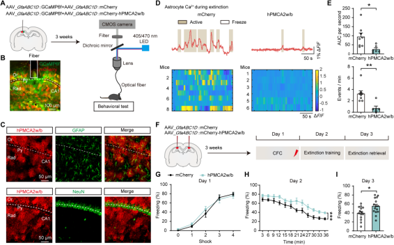
To test whether astrocytic Ca²⁺ activity is essential for fear extinction, AAV-GfaABC1D-hPMCA2w/b was injected into the hippocampus to suppress astrocyte activity (Figure 2A–B). Immunohistochemistry confirmed that hPMCA2w/b was specifically expressed in GFAP⁺ astrocytes, with no detectable expression in NeuN⁺ neurons (Figure 2C). Fiber photometry recordings of dCA1 astrocytes revealed that, compared to control mice, those expressing hPMCA2w/b showed significantly reduced Ca²⁺ responses during fear extinction (Figures 2D–E). Behaviorally, hPMCA2w/b mice exhibited a slower extinction curve on Day 2 and higher freezing levels on Day 3 (Figures 2F–I). These findings indicate that newly induced Ca²⁺ responses in hippocampal CA1 astrocytes are required for fear extinction.

Figure 2. Inhibiting Astrocytic Ca²⁺ Signaling Impairs Contextual Fear Extinction
 
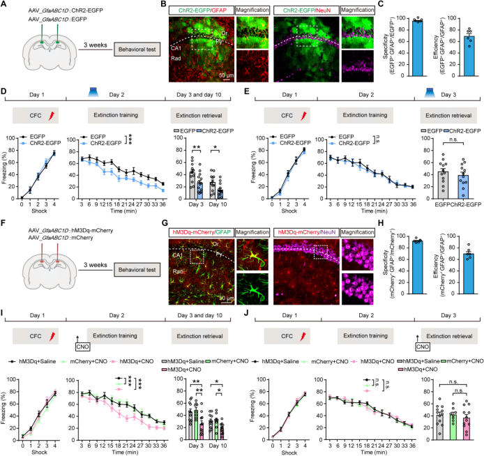
Because the continuous expression of hPMCA2w/b in astrocytes cannot temporally restrict Ca²⁺ signaling, AAV-GfaABC1D-ChR2-EGFP was injected into the dCA1 region (Figures 3A–B). Results showed that 95.5% of ChR2-EGFP⁺ cells were GFAP⁺ astrocytes, and 69.0% of GFAP⁺ astrocytes in CA1 expressed ChR2-EGFP (Figures 3B–C). Mice then underwent a 3-day contextual fear extinction protocol (Figure 3D). On Day 2, beginning 3 minutes after the start of extinction training, blue laser pulses (60 seconds every 60 seconds) were delivered to activate ChR2-expressing astrocytes in dCA1. Optogenetic activation of astrocytes enhanced fear extinction learning on Day 2 and reduced freezing levels on Days 3 and 10 (Figure 3D).

To distinguish the role of astrocytes in extinction training versus memory retrieval, optogenetic activation was applied during extinction memory retrieval on Day 3, but this did not affect freezing behavior (Figure 3E).

To further explore the effect of astrocyte activation on fear extinction, AAV-GfaABC1D-hM3Dq-mCherry was injected into dCA1 (Figure 3F). Results showed that 91.8% of hM3Dq-mCherry⁺ cells were GFAP⁺ astrocytes, and 69.8% of GFAP⁺ astrocytes expressed hM3Dq-mCherry (Figures 3G–H). Thirty minutes before extinction training on Day 2, mice received intraperitoneal injections of saline (1 mg/kg; hM3Dq + saline), or clozapine-N-oxide (CNO; 1 mg/kg; mCherry + CNO, hM3Dq + CNO). Chemogenetic activation of astrocytes during extinction training enhanced extinction learning on Day 2 and reduced freezing on Days 3 and 10 (Figure 3I). In contrast, chemogenetic activation during extinction memory retrieval had no effect on freezing behavior (Figure 3J). 
In summary, both optogenetic and chemogenetic activation of astrocytes in the dCA1 region facilitate the process of fear extinction.

Figure 3. Activation of Astrocytes Promotes Contextual Fear Extinction


Relationship Between ACh Signaling and Astrocytic Ca²⁺ Activity During Fear Extinction

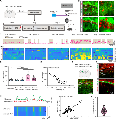
Astrocytes express a variety of G protein-coupled receptors and ion channels that respond to neurotransmitters or neuromodulators released and spilled over from synaptic activity, leading to elevations in astrocytic Ca²⁺ levels. To investigate this, researchers injected AAV-GfaABC1D-gACh4h, AAV-GfaABC1D-cATP1.0, or AAV-hSyn-5-HT3.0 into the CA1 region to record extracellular neurotransmitter signals via fiber photometry (Figures 4A–B). They measured extracellular levels of ACh, serotonin (5-HT), and adenosine triphosphate (ATP) in the CA1 region during the fear extinction process.

During extinction training on Day 2, ATP and 5-HT signals showed no significant changes. However, ACh levels during extinction memory retrieval were significantly lower than during extinction training, particularly when fear levels were high (Figures 4C–D). Further analysis revealed a negative correlation between freezing levels and ACh signals during extinction (Figure 4E).

To further examine the relationship between elevated extracellular ACh and Ca²⁺ activity in dCA1 astrocytes, researchers co-expressed the ACh sensor gACh4h and the red calcium indicator jRGECO1a in astrocytes, enabling simultaneous recording of both signals (Figures 4F–G). Results demonstrated a temporal and positive correlation between extracellular ACh signals and intracellular Ca²⁺ signals in astrocytes. Notably, the rise in extracellular ACh preceded the astrocytic Ca²⁺ increase by approximately 2 seconds (Figures 4H–J).

These findings suggest that
the elevation of extracellular ACh—likely due to synaptic activity and neurotransmitter spillover—stimulates Ca²⁺ dynamics in astrocytes during the process of fear extinction.

Figure 4. ACh Signaling Precedes Astrocytic Ca²⁺ Activity During Contextual Fear Extinction


pBF Cholinergic Projections Activate Astrocytes via nAChRs

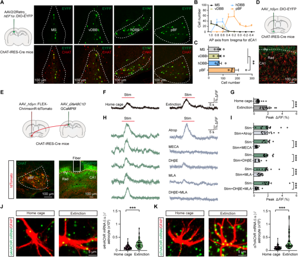
Previous studies have reported that cholinergic input to the hippocampus primarily originates from the anterior basal forebrain (aBF), including the medial septum (MS) and the diagonal band of Broca (DBB). To quantify cholinergic neurons projecting to the dCA1 region, a retrograde tracer virus (AAV2/2retro-hEF1a-DIO-EYFP) was injected into the hippocampus of ChAT-IRES-Cre mice. The results showed that only a few cholinergic neurons in the aBF were retrogradely labeled, whereas significantly more cholinergic neurons were labeled in the pBF (Fig. 5A–C).

To trace projections from the pBF to the hippocampus, an anterograde tracer virus (AAV-hSyn-DIO-EYFP) was injected into the pBF of ChAT-IRES-Cre mice. EYFP-labeled cholinergic projections were observed in the dCA1 region, with terminal boutons surrounding GFAP-labeled astrocytes,
indicating that pBF cholinergic terminals are anatomically adjacent to astrocytes in this area (Fig. 5D).

To test whether pBF cholinergic input drives astrocytic Ca²⁺ responses in dCA1, AAV-hSyn-FLEX-ChrimsonR-tdTomato (for optogenetic activation) and AAV-GfaABC1D-GCaMP6f (for Ca²⁺ imaging) were co-injected into the pBF and dCA1, respectively, of ChAT-IRES-Cre mice (Fig. 5E). Using 638 nm laser light (10 ms/pulse at 10 Hz for 10 s), optogenetic activation of cholinergic terminals in dCA1 evoked Ca²⁺ responses in astrocytes. This response was detectable on day 1 in mice at rest in their home cages, and was significantly enhanced on day 2 during fear extinction training (Fig. 5F–G), suggesting plasticity in this neuron-to-astrocyte signaling pathway.

To identify the type of acetylcholine receptors (AChRs) mediating this enhanced Ca²⁺ response on day 2, receptor antagonists were applied to the dCA1 region. The nicotinic ACh receptor (nAChR) antagonist MECA blocked ChrimsonR-induced astrocytic Ca²⁺ responses (Fig. 5H–I), while the muscarinic ACh receptor (mAChR) antagonist atropine did not. Furthermore, blocking either the α4 or α7 nAChR subunits using DHβE or MLA, respectively, also abolished the response, and their combined use showed an additive inhibitory effect.

RNAscope in situ hybridization confirmed a significant increase in α4 and α7 nAChR subunit mRNA levels in dCA1 astrocytes after day 2 fear extinction training, compared to mice that remained in their home cages on day 1 (Fig. 5J–K).
These findings indicate that fear extinction upregulates α4 and α7 nAChR subunits in astrocytes of the dCA1 region, and these receptors mediate the enhanced astrocytic Ca²⁺ activity driven by cholinergic input from nearby pBF terminals.

Figure 5. pBF Cholinergic Projections Activate Astrocytes via nAChRs


Cholinergic Input from the pBF to Hippocampal CA1 Astrocytes Mediates the Regulation of Fear Extinction

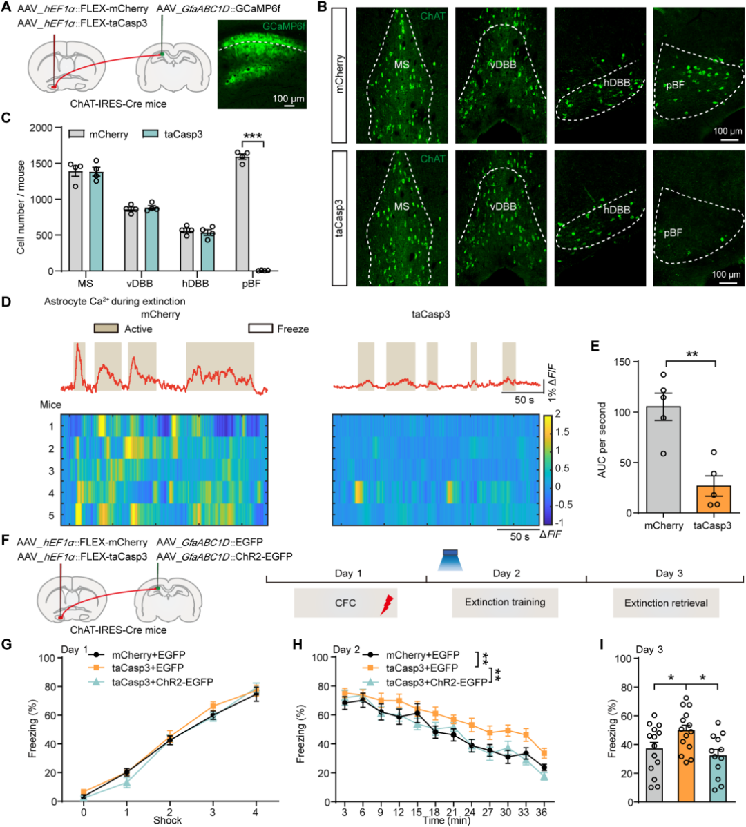
To assess whether cholinergic input is essential for the newly induced Ca²⁺ responses in astrocytes and their influence on fear extinction, AAV-hEF1a-FLEX-taCasp3 was injected into the pBF of ChAT-IRES-Cre mice to ablate cholinergic (ChAT-positive) neurons in the pBF (Figure 6A). Four weeks after virus injection, ChAT-positive neurons in the pBF of taCasp3+EGFP mice were nearly completely eliminated (Figure 6B–C).

Subsequently, Ca²⁺ dynamics in CA1 astrocytes during fear extinction were examined. Results showed a significant reduction in astrocytic Ca²⁺ activity in the CA1 region of taCasp3+EGFP mice (Figure 6D–E). In behavioral tests, compared to controls, taCasp3+EGFP mice exhibited a slower extinction curve on day 2 and a higher freezing level on day 3 (Figure 6F–I). However, after optogenetic activation of astrocytes, the freezing levels of taCasp3+EGFP mice on days 2 and 3 were restored to levels comparable to the control group (Figure 6H–I). 
These findings indicate that the enhanced Ca²⁺ dynamics in astrocytes are necessary for pBF cholinergic input-mediated regulation of fear extinction.

Figure 6. Ablation of pBF Cholinergic Neurons Reduces dCA1 Astrocyte Ca²⁺ Activity
 

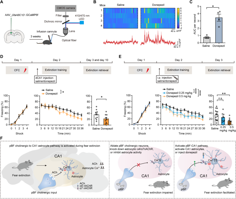
Donepezil Enhances Astrocytic Calcium Activity and Facilitates Fear Extinction

Enhancing cholinergic neurotransmission is a primary treatment strategy for mild to moderate Alzheimer’s disease. As a commonly used drug, donepezil's potential to boost astrocytic calcium activity and promote fear extinction is of significant interest. Local injection of the acetylcholinesterase inhibitor donepezil (0.1 mM) into the CA1 region significantly increased astrocytic Ca²⁺ activity compared to saline injection (Figure 7A–C). When donepezil (0.1 mM) was bilaterally administered into the CA1 region prior to fear extinction training, mice displayed accelerated fear extinction on day 2 and reduced fear memory on day 3. Notably, donepezil did not affect locomotor activity in the open field test, indicating that the effect was specific to fear extinction (Figure 7C).

Furthermore, intraperitoneal injection of donepezil (0.25 mg/kg) facilitated fear extinction on day 2 without affecting freezing levels on day 3. A higher dose (0.5 mg/kg) not only significantly promoted fear extinction on day 2 but also reduced freezing behavior on day 3. Importantly, intraperitoneal administration did not alter locomotor performance in the open field (Figure 7E).

These results demonstrate that pharmacologically enhancing the pBF-to-CA1 cholinergic-astrocytic signaling pathway promotes fear extinction, highlighting donepezil’s potential for treating disorders related to fear and anxiety, such as PTSD.

Figure 7. Donepezil Increases Astrocytic Ca²⁺ Activity and Promotes Fear Extinction
 

Summary

Astrocytes in the CA1 region play a crucial protective role in fear extinction. Cholinergic signaling from the pBF is coupled with astrocytic activity in CA1 to regulate fear extinction. The pBF-to-CA1 cholinergic-to-astrocyte signaling axis represents a promising therapeutic target for fear- and anxiety-related disorders.
 
 
The viral vectors used in this study are all available from Brain Case Biotech.
Product Category Product Name
Neurotransmitter Sensors rAAV9-hSyn-g5-HT3.0
rAAV5-GfaABC1D-cATP1.0
rAAV5-GfaABC1D-gACh4h
rAAV9-hSyn-rAChmut
rAAV5GfaABC1D-cATP1.0
Optogenetics  rAAV5-GfaABC1D-ChR2-EGFP
rAAV9-hEF1α-DIO-ChR2-EYFP
rAAV9-hSyn-DIO-ChrimsonR-tdTomato
Chemogenetics rAAV5-GfaABC1D-hM3Dq-mCherry
Fluorescent Proteins  rAAV5-GfaABC1D-mCherry
rAAV5-GfaABC1D-EGFP
Calcium Indicators rAAV5-GfaABC1D-GCaMP6f
rAAV5-GfaABC1D-jRGECO1a
rAAV9-hEF1α-DIO-axon-GCaMP6s
Others rAAV5-GfaABC1D-mCherry-hPMCA2w/b
rAAV9-hEF1α-DIO-taCasp3-TEVp
 

If you are interested in the details of the experiment or possible problems and causes during the experiment, please contact: BD@ebraincase.com


💥Holiday

Special

Offers

Click to fill in the requirements and submit them to us!

Service Type :

Select the service you'd like to purchase.

Order Information(Premade-AAVs)

Please provide us some information about the service you'd like to order.

Detailed requirements:

scroll

Order Information(Custom AAV/Lentivirus)

Please provide us some information about the service you'd like to order.

Gene ID or gene information:

Selection of the reporting gene:

Special Instructions:

scroll

Order Information(Others)

Please provide us some information about the service you'd like to order.

Virus name/Detailed requirements:

scroll

Related products

Customer Publication | Sci Adv | Team of Zhong Chen & Yi Wang from Zhejiang Chinese Medical University Reveals Astrocyte-Mediated Mechanism in Fear Extinction

Customer Publication | Sci Adv | Team of Zhong Chen & Yi Wang from Zhejiang Chinese Medical University Reveals Astrocyte-Mediated Mechanism in Fear Extinction

Client Article | Cell Stem Cell Teams Led by Jun Qin, Qi Han, Moubin Lin, and Xiao Su Reveal How Chronic Stress Induces Intestinal Stem Cell Aging via the Vagus Nerve–Cholinergic Pathway

Client Article | Cell Stem Cell Teams Led by Jun Qin, Qi Han, Moubin Lin, and Xiao Su Reveal How Chronic Stress Induces Intestinal Stem Cell Aging via the Vagus Nerve–Cholinergic Pathway

Fudan University's Institute of Brain Science Team Led by Xiaohong Xu Collaboratively Investigates and Deciphers the Organizational Patterns of Hypothalamic Neuronal Projections

Fudan University's Institute of Brain Science Team Led by Xiaohong Xu Collaboratively Investigates and Deciphers the Organizational Patterns of Hypothalamic Neuronal Projections

Client Article | Nat. Neurosci. | Breakthrough! First-ever whole-brain projection map of 7,180 hypothalamic peptidergic neurons unveiled

Client Article | Nat. Neurosci. | Breakthrough! First-ever whole-brain projection map of 7,180 hypothalamic peptidergic neurons unveiled

map
{dede:global.cfg_webname/}

Virus product library

CRISPR
RNAi
Neurophilic virus
Optogenetics activation
Biosensors

News

Corporate News
New Product Launch
Media Activity
Investor News

Support

Literature interpretation
Customer article
Video Zone
FAQs
微信

WhatsApp Business Account

Tel: +8618971215294
E-mail: BD@ebraincase.com

Address:-

Address:-

  • Copyright © 2024 Brain Case All Rights Reserved.