Fudan University's Institute of Brain Science Team Led by Xiaohong Xu Collaboratively Investigates and Deciphers the Organizational Patterns of Hypothalamic Neuronal Projections
Release time:2025-09-19 11:43:20
Eating, fighting, mating, fleeing... The "command center" for these instinctive animal behaviors lies deep within the brain — the hypothalamus. However, the neuronal networks in this critical region have long remained as elusive as a "black box." Recently, the research team led by Xiaohong Xu at the Institute of Brain Science, Fudan University, in collaboration with the Institute of Neuroscience of the Chinese Academy of Sciences and others, has, for the first time, systematically mapped the "projection atlas" of more than 7,000 hypothalamic neurons, categorizing them into 31 functional types! This groundbreaking study, titled "Projectome-based characterization of hypothalamic peptidergic neurons in male mice," was published in the top-tier journal Nature Neuroscience and represents a major milestone in the mapping of neural networks.
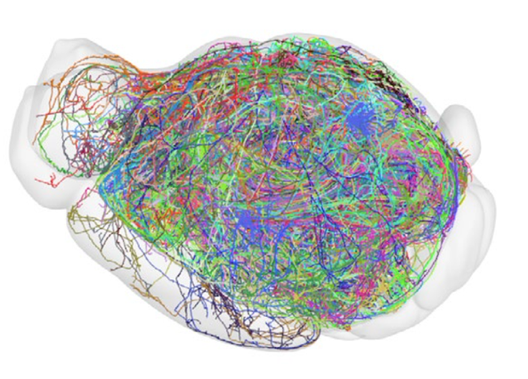
Figure 1. Schematic representation of the whole-brain projection map of hypothalamic projection neurons.
An Unprecedented "Whole-Brain Tracing" Effort
The research team employed high-resolution fMOST imaging technology to trace, one by one, neurons expressing 16 different neuropeptides in the brains of male mice. Each neuron — from the cell body to the axonal terminals — was precisely mapped, resulting in the reconstruction of the complete "journey" of 7,180 individual neurons across the brain. Through computational neuroanatomical analysis, the authors found that these neurons could be categorized into two major classes and 31 distinct projection types, each characterized by unique projection pathways, origins, and molecular markers.
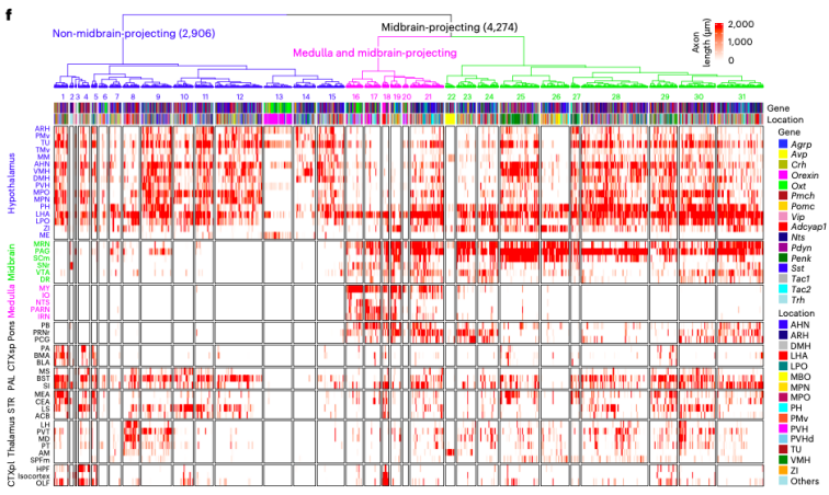
Figure 2. Heatmap of projection lengths for the 31 projection types.
One Neuron, Multiple "Assignments"
Surprisingly, many neurons utilize a single axon to serve multiple destinations, branching repeatedly along their journey and projecting to several distinct brain regions. For example:
💠Some Orexin neurons ascend to the cerebral cortex, regulating arousal and attention;
💠Other Orexin neurons descend to the brainstem and even the spinal cord, contributing to motor control;
💠A subset of Penk neurons projects precisely to the hippocampus, possibly involved in memory regulation.
This is akin to a delivery courier who, instead of delivering to just one address, distributes packages along a route to multiple destinations — a single neural fiber modulating multiple functional areas simultaneously!
Figure 3. Comparative whole-brain projection pathways of different Orexin neuron types.
Projection with "Topographic Matching"
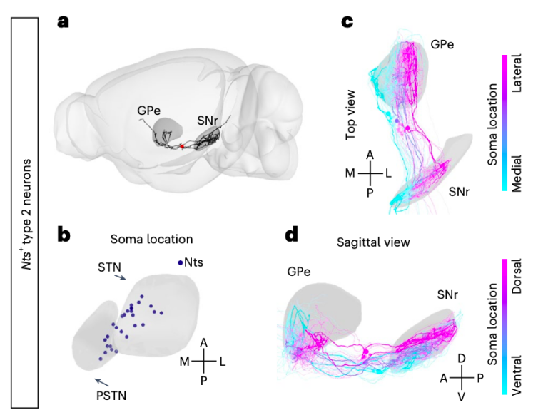
Not only can these neurons project to multiple areas, but they also exhibit remarkable structural topography in their connections. For example: ✨One class of neurons projects from the hypothalamus to the basal ganglia, with their spatial arrangement in the brain precisely matching their projection positions in the target area; ✨Another class projects from the hypothalamus to the hippocampus, where the dorsal-to-ventral distribution of their cell bodies aligns linearly with the anterior-to-posterior axis of their projections within the hippocampus.
This indicates that neural connections are not merely about linking regions — they are about ordered, organized linking.
Figure 4. Topographic arrangement of axons from different neurons in their target regions.
The Emerging "Identity Lineage" of Hypothalamic Neurons
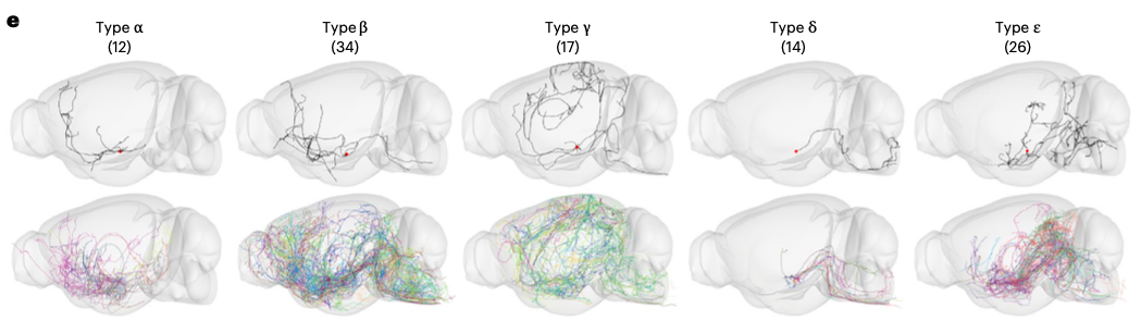
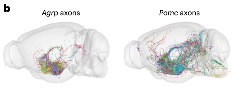
The study also revealed that each neuropeptide-labeled neuronal population does not correspond to a single function. For example: 🔅Neurons expressing Orexin can be further subdivided into five distinct types, each with different projection pathways (see Figure 3); 🔅Pomc-expressing neurons are more likely than Agrp-expressing neurons to project to the midbrain and brainstem, suggesting that Pomc neurons may have broader functions beyond appetite regulation.
These findings suggest that traditional molecular markers alone are insufficient to define the "duties" of neurons — structural projection patterns provide crucial complementary information.
Figure 5. Comparative projection ranges of Agrp and Pomc neurons.
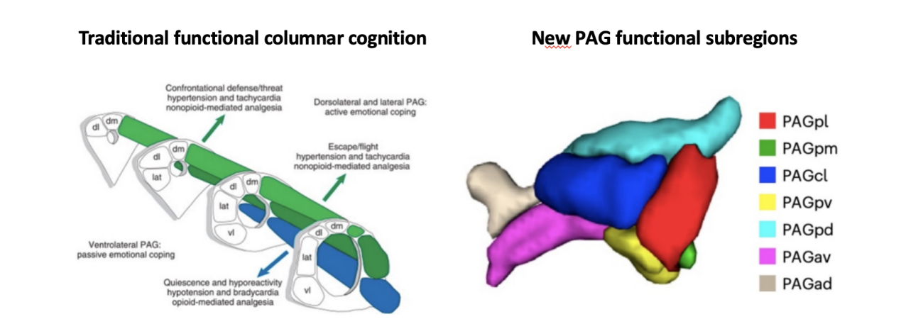
Redefining the PAG: Seven Functional "Subregions" Emerge
The midbrain periaqueductal gray (PAG) is often referred to as the "central hub for emotional regulation." This study found that: ✅Many hypothalamic neurons simultaneously project to multiple PAG subregions, and these projections exhibit a high degree of coordination; ✅By computationally analyzing the overlap of these projections, the authors redefined the PAG into seven functional subregions, challenging the traditional view of its "columnar" organization. This provides a new anatomical foundation for future studies aiming to unravel the central mechanisms underlying complex behaviors such as fear, aggression, and defense.
Figure 6. Schematic diagram of the newly defined PAG subregion structure.
Open Data, Shared Resources
This large-scale dataset is now publicly available to the global scientific community, including: 🔹3D whole-brain reconstruction files for 7,180 neurons; 🔹Neuron classification information, origin locations, axonal pathways, and target projection areas;
🔹Online access with options for visualization, browsing, downloading — and even running custom analyses.
This study, for the first time at the single-cell level, systematically classifies hypothalamic neurons based on their structural projection patterns. It not only provides a brand-new map of neural connectivity but also marks a crucial starting point for exploring the mechanisms underlying complex behaviors. "We hope that through these data, we can reveal how the brain uses a small number of neurons to orchestrate complex behavioral programs," said Professor Xiaohong Xu. Original article:https://www.nature.com/articles/s41593-025-01919-0