The hypothalamus plays a crucial role in regulating physiological homeostasis and innate behaviors, yet the axonal projection patterns of its peptidergic neurons have not been thoroughly studied. On March 26, 2025, a research team led by the Center for Excellence in Brain Science and Intelligence Technology (CEBSIT) of the Chinese Academy of Sciences and the National Key Laboratory of Brain and Cognitive Intelligence, in collaboration with Huazhong University of Science and Technology's Suzhou Institute of Brain Space Information and multiple domestic and international institutions, published a research article titled "Projection-based characterization of hypothalamic peptidergic neurons in male mice" in Nature Neuroscience.
The study performed single-neuron projection mapping of7,180 hypothalamic peptidergic neuronsand established a high-resolution single-neuron projection database (https://mouse.braindatacenter.cn/hy). The analysis revealed that these neurons can be categorized into two major groups and 31 distinct types, each exhibiting region-specific somatic distribution and unique neuropeptide enrichment patterns. Many of the neurons possess extensive axonal collaterals, with some showing topographically organized projection patterns. The study also identified distinct subregions within the periaqueductal gray of the midbrain and modular subnetworks within the hypothalamus.
1.Experimental Animals: Multiple transgenic mouse lines and wild-type C57BL/6J mice were used.
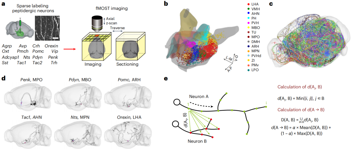
2.Viral Injection: Adeno-associated viruses (AAVs) carrying specific genes were injected into transgenic or wild-type mice to achieve sparse labeling of hypothalamic peptidergic neurons (AAVs purchased from Brain Case Biotech).
3.Imaging and Tracing: Fluorescence micro-optical sectioning tomography (fMOST) was employed for brain imaging, achieving a resolution of 0.32 μm × 0.32 μm × 1 μm. The researchers developed Fast Neurite Tracer (FNT) software for axon tracing and registered the data to the Allen Mouse Brain Common Coordinate Framework (CCFv3).
4.Data Analysis: Various neuronal parameters were quantified, including soma location, axon length, and number of axon terminals. Neurons were classified using a modified Hausdorff matching distance algorithm. Further analyses included enrichment analysis, topographical mapping, and more.
Research Results
1.Single-Neuron Projection Mapping: The study successfully reconstructed the whole-brain projection of 7,180 individual neurons from 16 peptidergic neuron populations in 166 adult male mice. The soma distribution of these neurons matched known neuropeptide expression patterns. Their axons projected extensively to both brain hemispheres, occupying approximately 21.8% of ipsilateral and 10.2% of contralateral brain voxels.
2.Neuron Classification: Based on axonal projection patterns, hypothalamic peptidergic neurons were classified into two major classes and 31 projection-defined types. Class I (Types 1–15): Non-midbrain-projecting neurons, primarily targeting non-midbrain regions. Class II (Types 16–31): Midbrain-projecting neurons, notably targeting areas such as the periaqueductal gray (PAG).
3.Axonal Projection Features: Each neuron type displayed distinct projection patterns with target specificity. Some neurons preferentially projected to particular nuclei of the amygdala or hippocampus. Comparison with the MouseLight dataset confirmed the reliability of these classifications.
4.Soma Distribution and Neuropeptide Enrichment: Midbrain-projecting neurons were enriched in the lateral hypothalamic area (LHA) and the ventromedial hypothalamic nucleus (VMH). In VMH, specific neuropeptides like Pdyn and Tac1 were highly enriched among midbrain-projecting neurons. Different neuron types showed complex correlations between soma distribution and neuropeptide expression.
5.Topographical Organization: Some peptidergic neurons exhibited topographic organization at their target sites. For instance, Type 2 neurons expressing Nts projected to the globus pallidus externus (GPe) and substantia nigra reticulata (SNr) along clear mediolateral (M-L) and dorsoventral (D-V) axes. In Type 4 neurons, Penk and Adcyap1-expressing subtypes showed distinct projection patterns to dorsal and ventral CA1 regions of the hippocampus.
6.Key Peptidergic Neuron Group Analysis: Detailed analysis of Orexin, Agrp, and Pomc neurons revealed:Orexin neurons comprise multiple projection types with varied axonal innervation patterns.Agrp and Pomc neurons exhibit markedly different projection profiles, with Pomc neurons projecting more extensively to midbrain, pons, and medulla regions.
7.PAG Subregions and Hypothalamic Networks: Seven distinct subregions within the PAG were identified, each receiving preferential input from specific hypothalamic neurons. Moreover, six modular subnetworks were discovered within the hypothalamus, partially corresponding to known "defensive" and "reproductive" circuits.
Single-Cell Projection of Hypothalamic Peptidergic Neurons
Research Significance
1.Revealing Projection Complexity: The study uncovers the intricate axonal projection patterns of hypothalamic peptidergic neurons, highlighting that extensive axonal collateralization is a fundamental feature of hypothalamic connectivity.
2.New Perspective on Neuron Typing: It demonstrates a lack of simple one-to-one correspondence between projection-based and transcriptome-based neuron classifications, offering new insights into the functional understanding of neuronal diversity.
3.Structural Basis for Functional Studies: The identified topographic organization and modular subnetworks provide a structural framework for future research into the neural mechanisms underlying hypothalamic functions. This paves the way for deeper exploration of how the hypothalamus regulates physiological processes and innate behaviors.