Customer Article|Effects of Nonthermal Radiofrequency Stimulation on Neuronal Activity and Neural Circuit in Mice
Release time:2024-10-15 17:20:38
Nonthermal radiofrequency (RFR) refers to the effects of radiofrequency radiation on biological tissues or systems without a significant temperature increase. There are reports that RFR may have adverse effects on the brain, including learning and memory impairments, sleep disturbances, anxiety and depression, and even tumors. Unlike thermal radiofrequency, nonthermal RFR primarily focuses on the impact of electromagnetic fields without involving temperature changes. Researchers exposed mice to 2856-MHz RFR to establish an animal model of spatial memory impairment. Let’s explore how nonthermal RFR stimulation regulates neuronal activity and affects nervous system function at the neural circuit level in this model.
1. RF Exposure Impairs Hippocampus-Dependent Spatial Learning and Memory in Mice
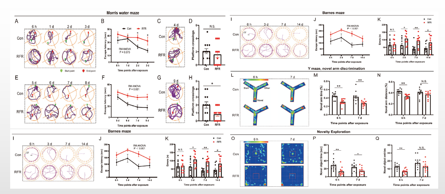
The researchers, through the Morris Water Maze (MWM), Barnes Maze, and hidden platform tests, found that radiofrequency radiation (RFR) led to a decline or impairment in spatial learning and memory abilities in mice. The Y-maze novel arm recognition test and novel object exploration (NOE) test suggested that RFR negatively affects spatial recognition memory and inhibits novelty-seeking behavior in mice. From these studies, they concluded that 2856 MHz RFR exposure below the thermal threshold (≤1°C) had adverse effects on hippocampus-dependent spatial and place memory abilities (both long-term and short-term) as well as novelty-seeking behavior.
Figure 1. Effects of radiofrequency radiation on cognitive performance in mice.
2. RF Exposure Causes No Obvious Change in Local Glutamate Release of Dorsal HPC CA1 in Mice but a Significant Decrease in Dopamine Release
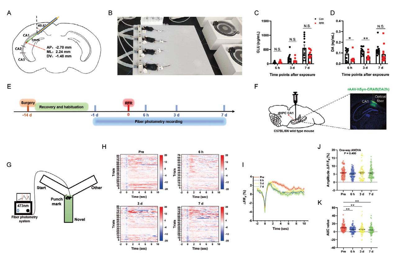
Exposure to radiofrequency radiation (RF) does not cause significant changes in glutamate (GLU) release in the dorsal hippocampal CA1 region of mice, but it does significantly reduce dopamine (DA) release. Researchers conducted microdialysis sampling in the dHPC CA1 region of anesthetized mice to investigate changes in neurotransmitter release caused by RF exposure. The results indicated that GLU release in the dHPC CA1 of RF-irradiated mice showed no significant change, while DA release was significantly decreased at 6 hours and 3 days post-radiation.
Additionally, the researchers utilized genetically encoded G protein-coupled receptor-activated dopamine (GRABDA) sensors (BC-0261 rAAV-hSyn-DA2h, packaged by Brain Case Biotech) to study DA release in the dHPC CA1 of awake mice. Fluorescence data collection revealed that during the period from 6 hours to 7 days after RF exposure, the intensity of DA signal waveforms decreased when mice performed behavioral tasks such as the Y-maze, novel object exploration, or open field tests, as evidenced by reductions in ΔF/F0 (%) and area under the curve (AUC).
Figure 2. Effects of radiofrequency exposure on the release of glutamate and dopamine in dorsal hippocampus CA1
3. RF Exposure Damages the Locus Coeruleus-to-dHPC Dopaminergic Axonal Projection Circuit, Which is the Main Source of DA Release in the dHPC
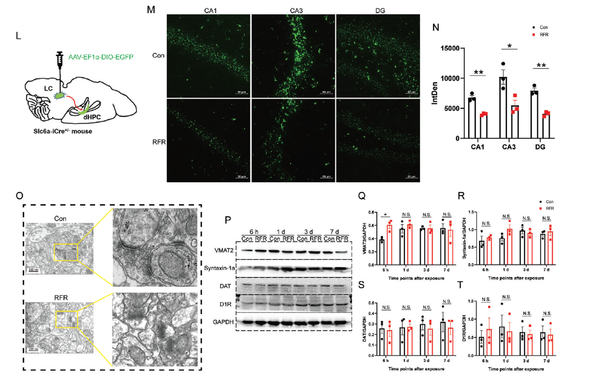
Numerous studies have shown that dopamine (DA) in the dorsal hippocampus (dHPC) primarily originates from the projections of tyrosine hydroxylase-expressing (TH+) neurons in the locus coeruleus (LC). Researchers recorded the calcium signaling activity of TH+ neurons projecting from the LC to the dHPC and found no significant differences in the signal amplitude (ΔF/F0 %) and area under the curve (AUC) of LC DAergic neurons before and after RF exposure during the Y-maze, novel object exploration (NOE), and open field testing (OFT) in mice. However, six hours after RF exposure, the density of DA projections from the LC to the dHPC significantly decreased in the dHPC CA1, CA3, and dentate gyrus, and the boundaries of synaptic structures in the dHPC CA1 were unclear. These results indicate that RF exposure did not significantly affect the activity of LC DAergic neurons, but rather led to a loss of their axonal projections in the dHPC and caused synaptic structural damage, resulting in abnormal DA release in the dHPC.
Fig 3. Effects of radiofrequency exposure on the locus coeruleus-to-dorsal hippocampus dopaminergic projection.
4. Optogenetic Activation of DAergic Terminals or Drug Activation of D1 Receptor in dHPC CA1 Improves RFR-Caused Spatial Memory Impairment in Mice
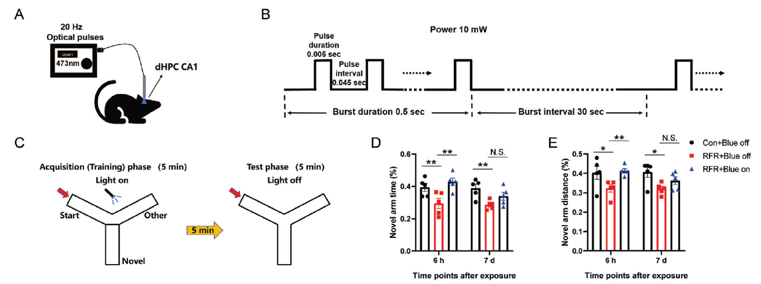
In the Y-maze and spatial object recognition tasks, researchers used Slc6aiCre+/−: Rose26-ChR2+/− model mice and activated DAergic neuronal cell bodies in the LC region and DAergic axons projecting to the CA1 of the dHPC through optogenetic ChR2 activation. Both methods improved short-term spatial memory impairment caused by RF exposure and facilitated long-term spatial memory in RF-exposed mice. Additionally, the researchers locally injected the DA D1 receptor agonist SKF38398 into the bilateral dHPC CA1 region using pre-implanted microtubules, finding that it significantly improved spatial learning and memory in mice after RF exposure. In summary, this part of the experiment indicates that the reduction of DA release in the dHPC CA1 region is an important cause of RF-induced short-term and long-term hippocampal-dependent spatial memory impairment.
Figure 4. Dopamine activation in dorsal hippocampus CA1 improved spatial learning and memory impairment caused by radiofrequency exposure.
The biological probe viruses used in this article to detect neuronal calcium signals, glutamate, and dopamine changes can be provided by Brain Case. Additionally, if you have any questions regarding the experiments that you would like to discuss with us, please feel free to contact BD@ebraincase.com
Service Type :
Select the service you'd like to purchase.
Order Information(Premade-AAVs)
Please provide us some information about the service you'd like to order.
Order Information(Custom AAV/Lentivirus)
Please provide us some information about the service you'd like to order.
Order Information(Others)
Please provide us some information about the service you'd like to order.