E-mail:BD@ebraincase.com
Tel:+8618971215294
English 中文版
Virus Vector - Viral Vector Production - BrainCaseVirus Vector - Viral Vector Production - BrainCase
  • Home
  • Virus product library
    CRISPRRNAiHSV-helperRV-helperNeurophilic virusCalcium SensorsOptogenetics activationOptogenetics inhibitionChemical geneticsSparse labelingFluorescent proteinBiosensorsRecombinaseApoptosis & AutophagyDisease ModelNeurotoxicityOther
  • Products & Service

    Product Center

    Virus

    VSV-circuit research-vaccine and gene therapy research-BrainCase
    Retrovirus-RCAS-TVA-BrainCase
    Lentivirus Vector - Lentivirus Production - BrainCase
    Rabies Virus Vector - RBV Vector - BrainCase
    Herpes simplex virus-Oncolytic and anterograde tracing-Brain Case
    PRV-retrograd multisynaptic-Peripheral-Btain Case
    AAV-gene therapy vectors-BrainCase

    Animal Model

    Neurological Disease Models-BrainCase
    Tumor animal models-anti-tumor-BrainCase
    Digestive System Disease Animal Model-Brain Case
    Cardiovascular System Disease Animal Models-Brain Case

    Plasmid Construction

    Library Construction
    Plasmid design and construction

    Popular Applications

    Gene Regulation

    Gene Overexpression-Brain Case
    RNA interference(RNAi)-siRNA-Brain Case
    Gene Editing - CRSIPR cloning - BrainCase

    Neural Circuit Function Research

    Optogenetics - BrainCase
    Chemical genetics-DREADDs-Brain Case
    Calcium signal recording-Gels- Brain Case
    GRAB Neurotransmitter Fluorescent Probe- Brain Case
    Functional Magnetic Resonance Imaging Technology- Brain Case

    Research on the structure of neural circuits

    Direct Input and Output-viral vectors- Brain Case
    Anterograde Mono-synaptic Tracing -HSV- Brain Case
    Antrograde Muti-synaptic Tracing-HSV & VSV-Brain Case
    Retrograde Mono-synaptic Tracing-Rabies Virus-Brain Case
    Retrograde Muti-synaptic Tracing-PRV-Brain Case

    Featured Services

    Nervous System Disease Drug Effect

    Alzheimer's disease-AD-Brain Case
    Depression-mental disorders-Brain Case
    Parkinson's disease--PD-Brain Case
    Epilepsy-an ancient neurological disorder-Brain Case

    AAV Serotype Screening

    AAV Serotypes screening-gene therapy-Brain Case

    Tumorigenicity Test

    Tumorigenicity Testing-Evaluation of tumor models-Brain Case

    Efficacy of Oncolytic Virus

    Oncolytic virus-for cancer therapy-Brain Case
    Herpes Virus Vector-anti-tumor- BrainCase
    Vesicular stomatitis virus-killing tumor cells-Brain Case
  • News
    Corporate News New Product Launch Media Activity Investor News
  • Support
    Literature interpretation Customer article Video Zone FAQs
  • About Us
  • Contact
    Contact UsJoin us
  • 中文
    English 中文版
  • Home
  • Support
  • Customer article
  • Support
  • Literature interpretation
  • Customer article
  • Video Zone
  • FAQs

Customer article|Super bright, super fast, super good! A new method for labeling cells in the sixth layer of cortex based on RV-CVS virus

Release time:2024-10-15 17:25:19

The mammalian cerebral cortex can be divided into six layers, which is an important structural basis for the brain to process sensory information. Compared with the superficial layers, the sixth layer of the cerebral cortex (layer 6, L6) is the earliest layer of structure that appears during development, but has been relatively rarely studied in depth. The cells of L6 are complex and diverse. There are two main types of pyramidal neurons in L6 of the sensory cortex of most species: corticothalamic (CT) neurons that project to the thalamus, and corticocortical (CC) neurons that project within the cortex. Although the specific markers and projection properties of L6 neurons have been partially revealed, how L6 neurons integrate inputs from different times and spaces on the dendrites to produce outputs remains unclear. This is mainly attributed to the high-quality function of L6 neurons. Technical difficulties in imaging.

On April 21, 2023, Zhang Jianxiong’s team from the Army Medical University jointly published a paper in the magazine "iScience" A research paper titled "Rabies virus-based labeling of layer 6 corticothalamic neurons for two-photon imaging in vivo" was published online. This study developed an RV-CVS virus-based labeling method that allows direct structural and functional imaging of projection-specific CT neurons in mouse auditory cortex L6 using conventional two-photon microscopy. This labeling method has the advantages of ultra-high brightness and ultra-fast expression within a week, and can be used for high-quality imaging of subcellular structures such as deep neuronal cell bodies and dendritic spines.

L6 CT neuron multi-scale structural imaging

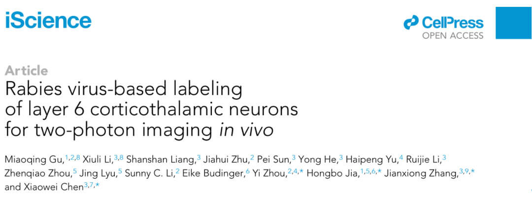
RV-CVS-labeled neurons have sufficiently high fluorescence intensity to enable high-contrast structural imaging of L6 neurons. The researchers used CVS-GFP to specifically label L6 neurons in the auditory cortex that project to the MGB. Compared with AAV-labeled neurons, the cell bodies, dendrites, and dendritic spines of RV-CVS-labeled neurons were more abundant in deeper layers. High fluorescence levels. Quantitative measurement of fluorescence intensity of multiscale structures in different layers showed that RV-CVS labeling was able to resolve individual dendritic spines of L5 and dendritic structures of L6. Therefore, RV-CVS labeling greatly improves the imaging quality of L6 CT neurons, enabling individual dendrites and dendritic spines to be studied in deeper layers.

L6 CT neuronal population functional Ca2+ imaging

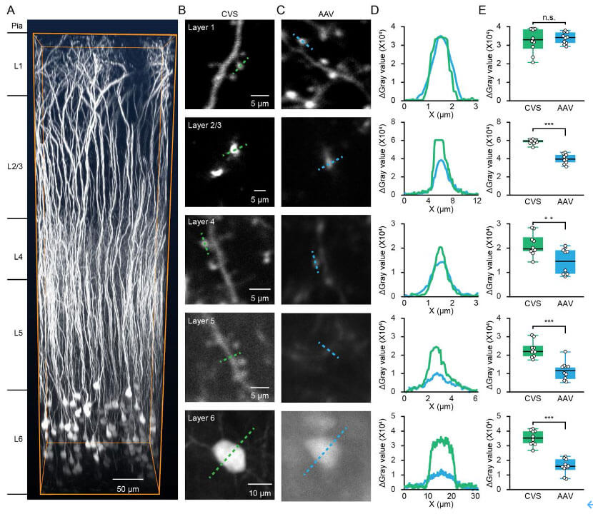
A common problem with two-photon in vivo population Ca2+ imaging is that neuronal signals are often interfered with by adjacent nerve fibers. To test whether RV-CVS labeling is subject to this interference, we used CVS-GCaMP6s to specifically label L6 neurons in the auditory cortex that project to the MGB and randomly sampled the signals from the neurons and their adjacent nerve fibers. The results showed that the response amplitude of neurons evoked by noise stimulation (BBN) was much higher than that of nearby nerve fibers. Next, the researchers performed functional Ca2+ imaging on neurons in the L6 CT population of awake mice, calculated the amplitude of each neuron's induction by BBN, and compared all 897 neurons based on their relative changes from pre-sound to post-sound. Sort by activity level. The above experiments show that CVS-GCaMP6s labeling can perform high-quality Ca2+ imaging of L6 CT neurons.

L6 CT neuronal subcellular structure functional Ca2+ imaging

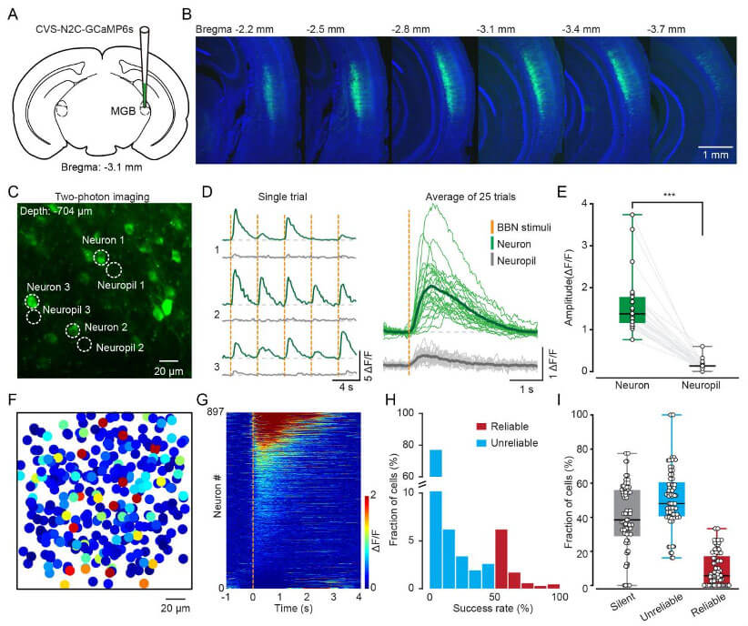
In addition to population imaging, RV-CVS labeling enables high-quality Ca2+ imaging of L6 single-cell structures spanning the entire cortex at subcellular resolution. The researchers recorded Ca2+ signals from different parts of the same neuron from superficial to deep layers and found that compared with the shallow layers, the amplitude and frequency of Ca2+ signals in the neuronal trunks located at L4 and L5 were greatly increased. The researchers recorded a total of 20 focal planes from shallow to deep on the same neuron, and completely reconstructed its functional map in the temporal and spatial dimensions to show the response characteristics of all structures. These suggest that RV-CVS labeling provides a suitable method to study dendritic information integration by imaging dendrites and somata of L6 CT neurons at single-cell resolution.

In summary, the researchers developed a neuron labeling strategy based on RV-CVS, which can perform high-contrast, multi-scale, high-quality structural and functional imaging of L6 CT neurons in the mouse auditory cortex.

Thanks to Dr. Gu for providing this document. Gu Miaoqing, a doctoral student at Guangxi University, is the first author of this paper. This work was strongly supported by the National Natural Science Foundation of China and the Scientific Instrument Development Project of the Chinese Academy of Sciences.

Original link: https://www.cell.com/iscience/fulltext/S2589-0042(23)00702-2#%20

All related products mentioned in the original article can be provided by Brain Case Biotech, as well as RV-CVS series products:

Product name Product number
RV-N2C(G)-ΔG-EGFP BC-RV-N2C862
CVS-N2c-ΔG-Gcamp6s BC-RV-CVS715
CVS-EnvA-ΔG-GCaMP6s BC-RV-CVS EnvA715
CVS-N2c-ΔG-tdTomato BC-RV-CVS462

Brain Case Biotech provided all viruses used in this study. If interested, please contact us at BD@ebraincase.com


💥Holiday

Special

Offers

Click to fill in the requirements and submit them to us!

Service Type :

Select the service you'd like to purchase.

Order Information(Premade-AAVs)

Please provide us some information about the service you'd like to order.

Detailed requirements:

scroll

Order Information(Custom AAV/Lentivirus)

Please provide us some information about the service you'd like to order.

Gene ID or gene information:

Selection of the reporting gene:

Special Instructions:

scroll

Order Information(Others)

Please provide us some information about the service you'd like to order.

Virus name/Detailed requirements:

scroll

Related products

Client Article | Cell Metabolism | Liu Danqian’s Team at the Institute of Neuroscience, CAS, Decodes the Oxidation-Sleep Code in Substantia Nigra Neurons

Client Article | Cell Metabolism | Liu Danqian’s Team at the Institute of Neuroscience, CAS, Decodes the Oxidation-Sleep Code in Substantia Nigra Neurons

Client Publication | Nature Communications | USTC Team Led by Zhi Zhang, Juan Li, and Peng Cao Unveils Neural Circuit Mechanism of Male-Specific Conditioned Pain Hypersensitivity

Client Publication | Nature Communications | USTC Team Led by Zhi Zhang, Juan Li, and Peng Cao Unveils Neural Circuit Mechanism of Male-Specific Conditioned Pain Hypersensitivity

Customer Publication | Sci Adv | Team of Zhong Chen & Yi Wang from Zhejiang Chinese Medical University Reveals Astrocyte-Mediated Mechanism in Fear Extinction

Customer Publication | Sci Adv | Team of Zhong Chen & Yi Wang from Zhejiang Chinese Medical University Reveals Astrocyte-Mediated Mechanism in Fear Extinction

Client Article | Cell Stem Cell Teams Led by Jun Qin, Qi Han, Moubin Lin, and Xiao Su Reveal How Chronic Stress Induces Intestinal Stem Cell Aging via the Vagus Nerve–Cholinergic Pathway

Client Article | Cell Stem Cell Teams Led by Jun Qin, Qi Han, Moubin Lin, and Xiao Su Reveal How Chronic Stress Induces Intestinal Stem Cell Aging via the Vagus Nerve–Cholinergic Pathway

map
{dede:global.cfg_webname/}

Virus product library

CRISPR
RNAi
Neurophilic virus
Optogenetics activation
Biosensors

News

Corporate News
New Product Launch
Media Activity
Investor News

Support

Literature interpretation
Customer article
Video Zone
FAQs
微信

WhatsApp Business Account

Tel: +8618971215294
E-mail: BD@ebraincase.com

Address:-

Address:-

  • Copyright © 2024 Brain Case All Rights Reserved.