- E-mail:BD@ebraincase.com
- Tel:+8618971215294
Cardiovascular diseases (CVDs) remain one of the leading causes of death worldwide. In recent years, increasing evidence has shown that many cardiac disorders are associated with genetic abnormalities, making gene delivery technologies an important tool for both mechanistic studies and therapeutic development.
Among the currently available viral vectors, recombinant adeno-associated virus (rAAV) has become one of the most commonly used platforms for cardiac gene delivery. Its relatively low immunogenicity, broad tissue tropism, multiple serotype options, and ability to support long-term transgene expression make it particularly suitable for in vivo studies.
In addition to gene delivery, researchers have become increasingly interested in understanding how the nervous system regulates cardiac function. Neurotropic viral tracers provide powerful tools for mapping the neural circuits connecting the heart and the brain, helping reveal the anatomical basis of autonomic regulation and cardiovascular disease.
In this article, Xiaobu summarizes several commonly used viral strategies for cardiac research, including AAV serotype selection, promoter selection, and neural circuit tracing approaches.
Different AAV serotypes exhibit distinct tissue tropisms because of differences in their capsid structures. AAV1, AAV6, AAV8, and AAV9 have demonstrated relatively efficient systemic delivery and are capable of crossing vascular endothelial barriers after intravenous administration.
For heart-targeted studies, AAV8 and AAV9 are generally considered the most effective options. In particular, AAV9 has become one of the most widely used serotypes for cardiac gene transfer.
Please see a dataset from the following study.
Virus Vector: rAAV-cTnT-EGFP
Injection Method: Jugular vein injection
Animal Model: One-week-old mouse
Expression Period: 4 weeks
Injection Dose: See Figure 1
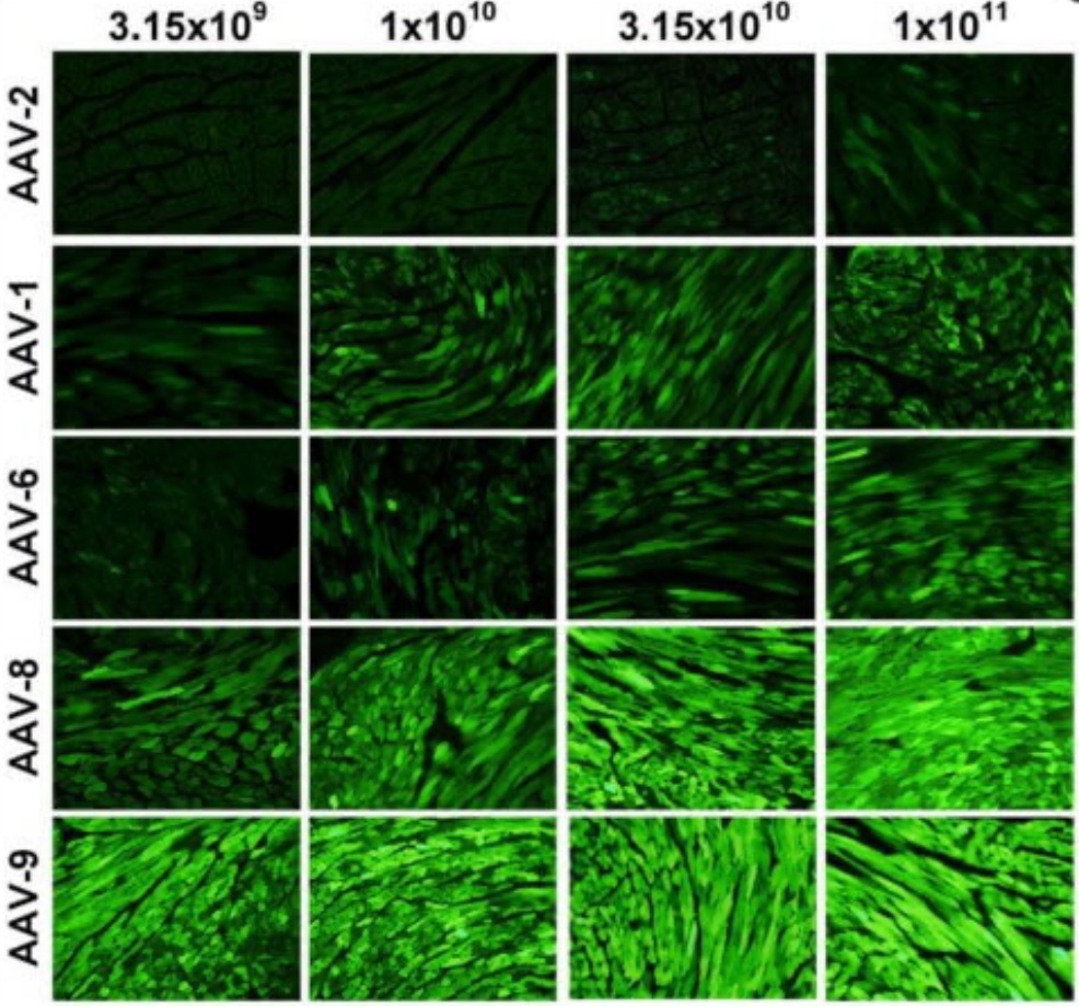
Figure 1. Cardiac transduction efficiency of different AAV serotypes at various doses (PMID: 20703310).
The results indicate that AAV9 achieves the highest transduction efficiency in cardiac tissue, followed by AAV8, while AAV1 and AAV2 show substantially lower levels of cardiac expression.
Serotype selection determines where a virus can reach, while promoter selection largely determines where the transgene is expressed. Several promoters have been developed for cardiac applications, including: cTnT (Cardiac Troponin T), αMHC (Alpha-Myosin Heavy Chain), Desmin (Des), MLC2v. Among these, cTnT and αMHC are the most frequently used promoters in cardiac gene delivery studies.
-cTnT Promoter
Compared with ubiquitous promoters such as CMV, cTnT drives expression primarily in cardiomyocytes and is often used when higher cardiac specificity is desired.
For an application example of the cTnT promoter, please see the data from the study below.
Virus Vector: rAAV9-cTnT-Luciferase
Injection Method: Jugular vein injection
Animal Model: One-week-old mouse
Expression Period: 4 weeks
Dose: 1 × 10¹¹ GC/mouse
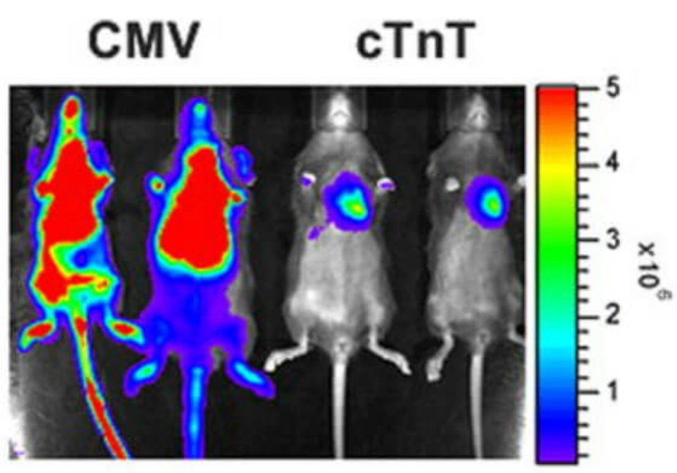
Figure 2. Comparison of cardiac expression driven by cTnT and CMV promoters (PMID: 20703310).
The study demonstrated selective cardiac expression when using the cTnT promoter.
-αMHC Promoter
αMHC is another commonly used cardiac promoter and is often selected when highly restricted cardiac expression is required.
Virus Vector: rAAV2/9-CMV/αMHC/cTnC-LacZ
Injection Method: Superficial temporal vein injection
Animal Model: Neonatal mouse
Expression Period: 4 weeks
Dose: 5 × 10¹⁰ GC/mouse
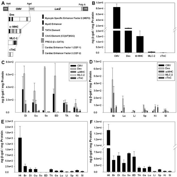
Figure 3. Tissue-specific expression driven by different promoters (PMID: 18811960).
The results showed that αMHC exhibited the strongest cardiac specificity. Desmin-mediated expression was detected in both cardiac and skeletal muscle, while CMV drove widespread expression across multiple tissues.
When the goal is selective gene expression in the heart, cTnT and αMHC remain two of the most commonly used promoter options.
The heart is under continuous regulation by both sympathetic and parasympathetic pathways. Understanding these neural networks has become an important area of cardiovascular research.
Neurotropic viruses possess the ability to spread through synaptically connected neurons and are therefore widely used for neural circuit tracing. For studies focused on identifying upstream neural inputs to the heart, pseudorabies virus (PRV) remains one of the most commonly used retrograde trans-synaptic tracers.
Researchers injected PRV into ventricular myocardium to investigate central neural circuits associated with cardiac regulation.
Virus Vector: PRV-CAG-EGFP
Injection Site: Multiple ventricular myocardial injection sites
Experimental Animal: Adult male SD rat
Dose: 1 × 10⁷ PFU
Expression Time: 5 days
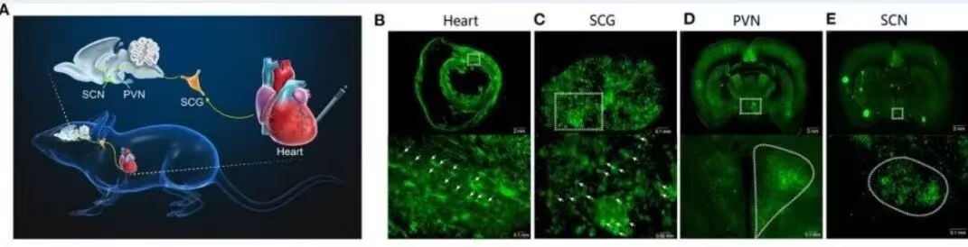
Figure 4. PRV-based tracing of the SCN–PVN–SCG–heart pathway (PMID: 33842566).
Tracing results revealed polysynaptic neural connections linking the heart to the suprachiasmatic nucleus (SCN), the central circadian pacemaker. The identified pathway included the SCN, paraventricular nucleus (PVN), superior cervical ganglion (SCG), and sympathetic innervation of the heart.
These findings provide anatomical evidence supporting communication between the circadian system and cardiac function.
For most in vivo cardiac gene delivery experiments, AAV9 remains one of the preferred serotypes because of its robust cardiac tropism. Combining AAV9 with cardiac-specific promoters such as cTnT or αMHC can further improve expression specificity within the heart.
For researchers interested in heart–brain interactions, neurotropic tracers such as PRV offer valuable tools for mapping cardiac neural circuits and identifying upstream regulatory pathways.
The optimal viral strategy ultimately depends on the experimental objective, target cell population, delivery route, and desired expression pattern. The examples discussed here may serve as useful references when designing future cardiac studies.
For technical consultation, custom vector design, or viral packaging services, please contact:bd@ebraincase.com


WhatsApp Business Account
Address:-
Address:-