- E-mail:BD@ebraincase.com
- Tel:+8618971215294

Understand the Major Light- and Calcium-Gated Transcription Tools in One Article
Researchers working on neural circuits, activity-dependent labeling, and functional manipulation are likely familiar with four major tools: FLARE, scFLARE, FLiCRE, and Cal-Light. These systems were all developed by the Alice Ting laboratory and collaborators, evolving from 2017 to 2020 into a powerful toolkit for activity-dependent neuronal labeling and manipulation.
All four systems operate based on the same logic: Calcium elevation + blue light = gene expression. Only neurons that are active (high intracellular calcium) during blue light illumination undergo protease-mediated transcription factor release, leading to reporter or optogenetic protein expression.
Plasmid Backbone (Dual-Plasmid System)
• Plasmid 1:
AAV-hSyn-TM-CaM-NES-TEV-N-AsLOV2-TEVseq-tTA
• Plasmid 2:
AAV-hSyn-M13-TEV-C-P2A-tdTomato
Design Principle
1. Calcium elevation induces binding between CaM (calmodulin) and M13 (CaM-binding peptide), allowing reconstitution of TEV-N and TEV-C into an active TEV protease. 2. Blue light illumination triggers conformational opening of the AsLOV2 photosensitive domain, exposing the TEV cleavage site. 3. The active TEV protease then cleaves the linker, releasing the transcription factor into the nucleus and initiating reporter gene expression.
Features
One of the earliest practical calcium/light dual-gated systems. The split-TEV reconstitution strategy is conceptually elegant but limited in overall performance.
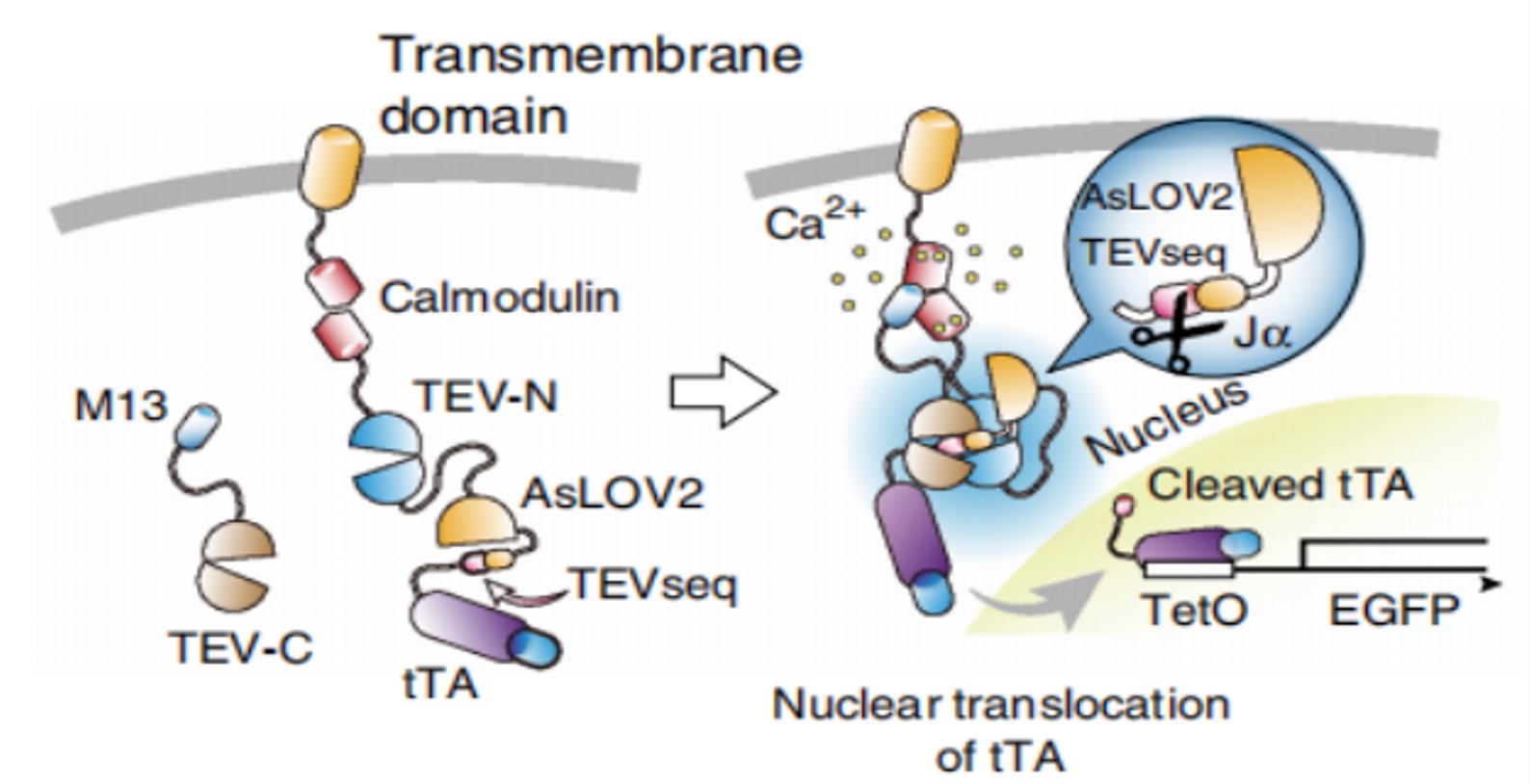
FIG1. Schematic Diagram of Cal-Light. Original article: https://www.nature.com/articles/nbt.3902
Plasmid Backbone (Dual-Plasmid System)
• Plasmid 1:
AAV-hSyn-TM-NaV1.6-M2-eLOV-TEVcs-FLAG-tTA-VP16
• Plasmid 2:
AAV-hSyn-CaM-V5-TEVp
Design Principle
1. Upon calcium elevation, cytoplasmic CaM-TEVp complexes are recruited by MKII to the membrane-localized TEV cleavage site. 2. Blue light illumination opens the optimized eLOV domain, exposing the cleavage site. 3. TEV protease cleavage releases the transcription factor, which translocates into the nucleus.
Features
The major innovation is the optimized eLOV photosensitive domain, which significantly reduces background leakage and establishes the foundational backbone for calcium/light dual-gated systems.
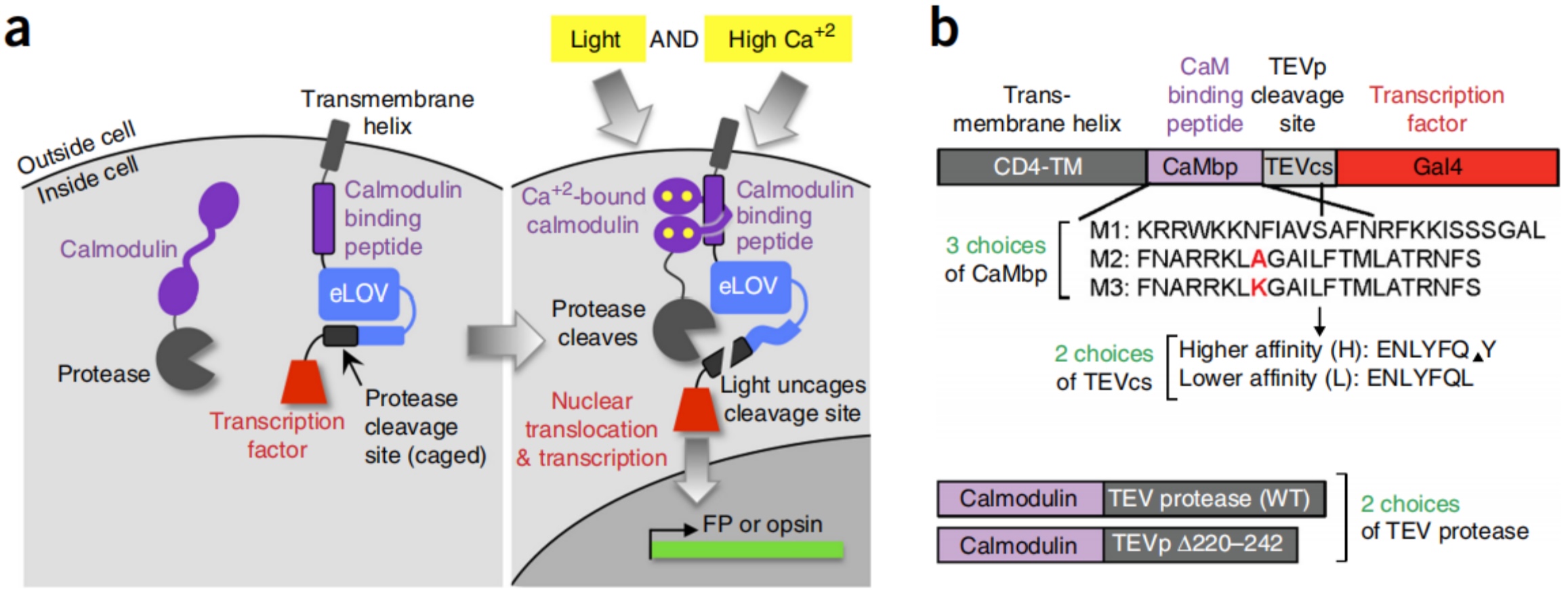
FIG2. Schematic Diagram of FLARE. Original article: https://www.nature.com/articles/nbt.3909
Plasmid Backbone (Single-Plasmid System)
• Plasmid:
AAV-hSyn-NRX-TM-CaTEV-hLOV-TEVcs-tTA-VP16
Design Principle
1. scFLARE adopts a self-contained single-chain design integrating all functional modules into one polypeptide. 2. Calcium elevation activates the CaM-CKK module within CaTEV, while blue light opens the hLOV domain to expose the TEV cleavage site. 3. CaTEV then performs intramolecular cleavage, releasing the transcription factor into the nucleus.
Features
The single-plasmid architecture eliminates expression ratio dependency between separate plasmids, making scFLARE highly robust for in vivo applications.
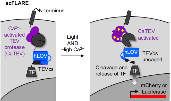
FIG3. Schematic Diagram of scFLARE. Original article: https://pmc.ncbi.nlm.nih.gov/articles/PMC7777206/
Plasmid Backbone (Optimized Dual-Plasmid System)
• Plasmid 1:
AAV-hSyn-Nrxn3b-Nav1.6-MKII-f-hLOV1-TEVcs(ENLYFQ/M)-tTA-VP16 or AAV-hSyn-Nrxn3b-Nav1.6-MKII-hLOV1-TEVcs(ENLYFQ/M)-tTA-VP16
• Plasmid 2:
AAV-hSyn-GFP-CaM-uTEVp
Design Principle
1. Calcium elevation rapidly recruits CaM-uTEVp complexes to membrane-localized MKII. 2. Blue light induces rapid conformational changes in hLOV1/f-hLOV1 domains with extremely low dark-state leakage. 3. The highly optimized uTEVp protease then performs rapid cleavage, enabling ultrafast activity labeling.
Features
Currently regarded as the highest-performing platform, combining high temporal resolution, excellent signal-to-noise ratio, and compatibility with single-cell sequencing workflows.
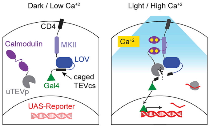
FIG4. Schematic Diagram of FLiCRE. Original article: https://www.cell.com/cell/fulltext/S0092-8674(20)31532-4
1. Viral Injection
Inject AAV vectors into the target brain region (e.g., NAc, VTA, motor cortex).
2. Expression Period
Allow 1–2 weeks for sufficient expression of the tool components.
3. Optical Stimulation Window
Illuminate the target region using 470 nm blue light for a tool-dependent duration (approximately 30 seconds to 15 minutes).
4. Labeling and Analysis
Only neurons that are active during the illumination window become permanently labeled. Subsequent analyses may include fluorescence imaging, single-cell sequencing, optogenetic manipulation, or circuit tracing.
Core Value
These tools convert transient neuronal activity occurring within minutes into stable genetic labels for downstream molecular profiling and functional studies.
| Parameter | FLARE | scFLARE | FLiCRE | Cal-Light |
| Plasmid System | Dual-plasmid | Single-plasmid | Dual-plasmid | Dual-plasmid |
| Activation Time | 10–15 min | 5–10 min | 30 s–1 min | 5–10 min |
| Temporal Resolution | ~1 min | 1–5 min | ≤1 min | Limited |
| Signal-to-Noise Ratio | ~120:1 | Higher than FLARE | Up to 1103-fold | Higher background leakage |
| Expression Dependency | Dependent on plasmid ratio | Minimal dependency | Low dependency | Dependent on plasmid ratio |
| In Vivo Robustness | Good | Excellent | Excellent | Moderate |
| scRNA-seq Compatibility | Compatible | Compatible | Fully compatible | Difficult |
| Key Features | Low background leakage | Single-chain robust design | Ultrafast and highly sensitive | Early-generation split-TEV system |
Cal-Light
• Successfully labeled lever-press-associated neurons in the mouse motor cortex.
• Optogenetic inhibition of labeled neurons reversibly impaired behavioral performance.
• Temporal resolution remains limited, making it difficult to distinguish sequential versus simultaneous calcium/light events.
FLARE
• Achieved a light/dark signal ratio of approximately 120:1 with minimal background.
• Successfully labeled behaviorally activated neurons in cortical and hippocampal regions.
• Limitation: relatively slow activation kinetics and dependency on dual-plasmid expression balance.
scFLARE
• Single-chain design greatly improves robustness.
• Successfully labeled seizure-activated neurons in epilepsy models, including indirectly activated neurons in the contralateral hemisphere.
• Demonstrated strong calcium specificity and low background.
FLiCRE
• Combines ultrafast labeling with exceptional signal-to-noise performance.
• Compatible with scRNA-seq for identification of previously inaccessible neuronal subtypes.
• Functional validation experiments demonstrated causal behavioral modulation via optogenetic reactivation of labeled neurons.
| Tool | Suitable Applications |
| FLARE | In vitro neuronal studies, mechanistic validation, introductory calcium/light-gated experiments |
| scFLARE | In vivo experiments, epilepsy and pathological models, uneven viral expression conditions, high-background brain regions |
| FLiCRE | Behavioral circuit studies, single-cell sequencing, short-timescale neuronal activity labeling, functional optogenetic validation |
| Cal-Light | Educational demonstrations, introductory studies, simple in vitro activity-labeling experiments |
For experiments involving short-timescale neuronal activity labeling, circuit tracing, single-cell sequencing, or behavioral functional validation, FLiCRE and scFLARE are generally the most reliable choices.For more information regarding these activity-dependent indicators, please contact: bd@ebraincase.com
References
1.Lee et al., Nature Biotechnology (2017)
2. Wang et al., Nature Biotechnology (2017)
3. Sanchez et al., PNAS (2020)
4. Kim et al., Cell (2020)
Tips | Series on Neuronal Activity-Dependent Tools: Tet-on/off System
Tips | Series on Neuronal Activity-Dependent Tools: scFLARE2
New Tools | Next-Generation NE Probes nLightG2 & nLightR2 Unveiled: Improved In Vivo Detection Performance


WhatsApp Business Account
Address:-
Address:-