Literature Review | Major Nature Discovery: GABAergic Neurons Drive Diffuse Midline Glioma Growth via Synaptic Signaling
Release time:2025-08-12 11:04:34
High-grade gliomas (HGGs) are a leading cause of brain cancer–related mortality, with subtypes that differ markedly in clinical presentation, anatomical location, and molecular characteristics. These subtypes can be further stratified based on their biological features. For instance, diffuse midline gliomas (DMGs) are a deadly pediatric central nervous system cancer, particularly the H3K27M-mutant diffuse intrinsic pontine glioma (DIPG), which is highly aggressive with extremely poor prognosis. Another category is hemispheric high-grade gliomas, such as IDH-wildtype glioblastoma, which typically arise in the cerebral hemispheres and have distinct molecular signatures. This classification aids in more precise diagnosis and treatment, while also laying a foundation for research. Neuronal activity can drive glioma progression through paracrine signaling and neuron–glioma synapses, but studies on neurotransmitters other than glutamate in neuron–glioma synaptic interactions are scarce. Previous research has shown that normal oligodendrocyte precursor cells (OPCs) receive GABAergic synaptic input from neurons, and that GABA has a depolarizing effect on OPCs, suggesting that GABAergic synapses might play a role in DMG progression. On February 19, 2025, researchers from Stanford University published an article in Nature titled “GABAergic neuron-to-glioma synapses in diffuse midline gliomas”, revealing that in DMGs, GABAergic neurons establish growth-promoting synaptic connections with glioma cells. This finding provides important evidence for understanding the neurophysiological mechanisms of brain cancer and for advancing therapeutic research.
Research Methods
Single-cell RNA sequencing analysis: Analyzed single-cell RNA sequencing datasets of different glioma types and tumor-associated non-malignant oligodendrocytes to examine the expression of genes related to GABAergic synapses.
Animal model construction: Established patient-derived orthotopic xenograft models by transplanting labeled DMG tumor cells into regions such as the hippocampus of mice. These models were used for immunoelectron microscopy, immunohistochemistry, and other experiments to observe structural and functional features.
Electrophysiological recording: Performed whole-cell patch-clamp and perforated patch-clamp recordings on transplanted DMG cells to study their electrophysiological responses to stimulation, assessing GABAergic synaptic function and effects on membrane potential.
Drug treatment experiments: Treated tumor-bearing mice with the benzodiazepine drug lorazepam to evaluate its effects on glioma cell proliferation, tumor growth, and mouse survival.
The research team first performed single-cell RNA sequencing analysis on various types of gliomas and tumor-associated non-malignant oligodendrocytes. The results showed that H3K27M+ DMG cells broadly express genes encoding GABA receptor subunits as well as genes for proteins associated with postsynaptic GABAergic structures. The expression levels of these genes were much higher in H3K27M+ DMGs and IDH-mutant high-grade gliomas than in IDH-wildtype high-grade gliomas. Further analysis revealed that the GABAergic synapse–related gene expression profile was present across all malignant cell compartments in H3K27M+ DMGs, with a notable enrichment in OPC-like cell compartments, and with marked heterogeneity among patient samples.
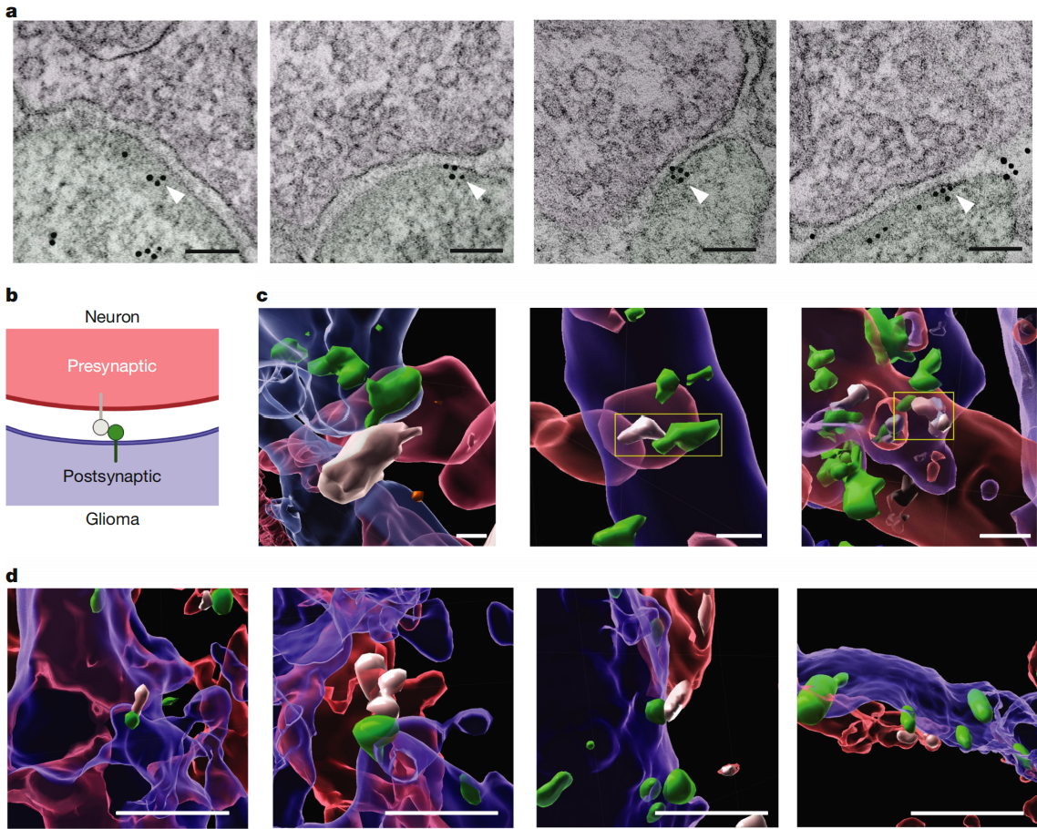
To directly “see” the presence of GABAergic synapses, the researchers validated their findings through multiple experiments. They transplanted labeled DMG tumor cells into the hippocampus of mice and, using immunoelectron microscopy, observed synaptic structures between GABAergic neurons and DMG cells. Both in vitro co-culture of DMG cells with mixed neurons and in vivo mouse models confirmed the structural presence of GABAergic synapses between the two, with about 20% of glioma cell gephyrin puncta co-localizing with presynaptic VGAT.
Figure 1. Structural GABAergic neuron–glioma synapses in DMG
Functional GABAergic Synapses in DMG
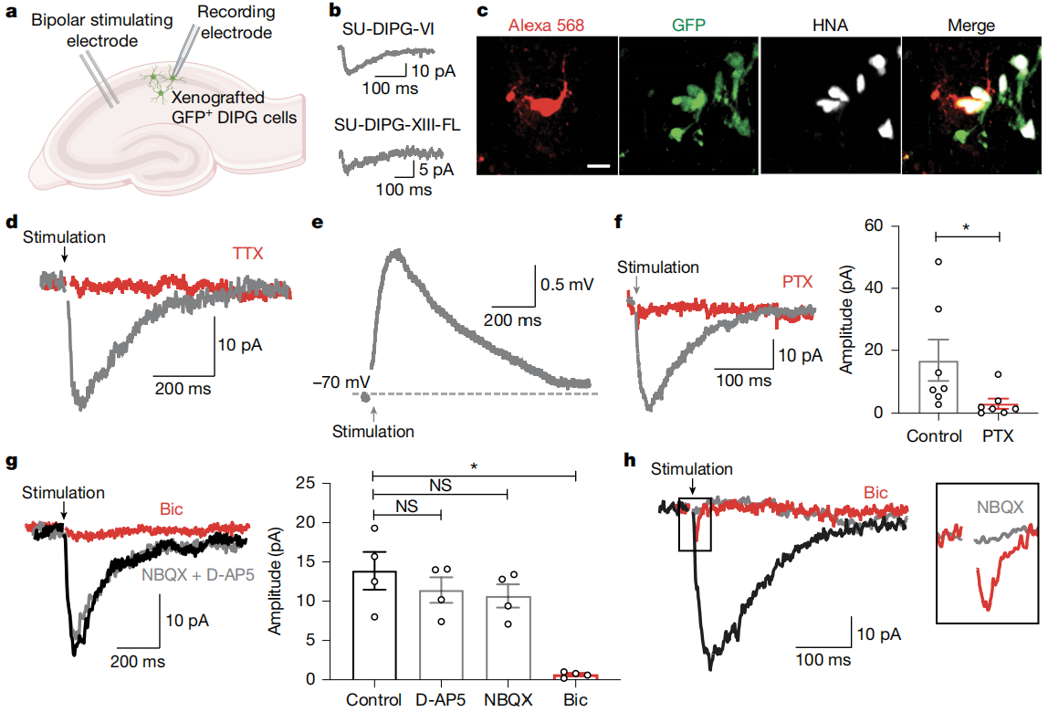
With the structural basis established, the next question was: do these synapses have functional activity? The researchers performed whole-cell patch-clamp recordings on transplanted DMG cells. They found that about 40% of DMG cells exhibited GABAergic synaptic responses to low-intensity electrical stimulation of local neurons, with specific rise times, decay times, and latencies. These GABAergic currents were triggered by neuronal action potentials, could be blocked by GABA receptor antagonists, and were unaffected by glutamate receptor antagonists.
What effect does GABA have on the membrane potential of DMG cells? Using perforated patch-clamp recordings, the researchers discovered that GABA induced inward currents in DMG cells, leading to membrane depolarization. This occurs because DMG cells have a higher intracellular Cl⁻ concentration compared to mature neurons. The study confirmed that the NKCC1 chloride transporter plays a key role in maintaining this high Cl⁻ concentration in DMG cells. Inhibiting NKCC1 altered the reversal potential of GABA currents.
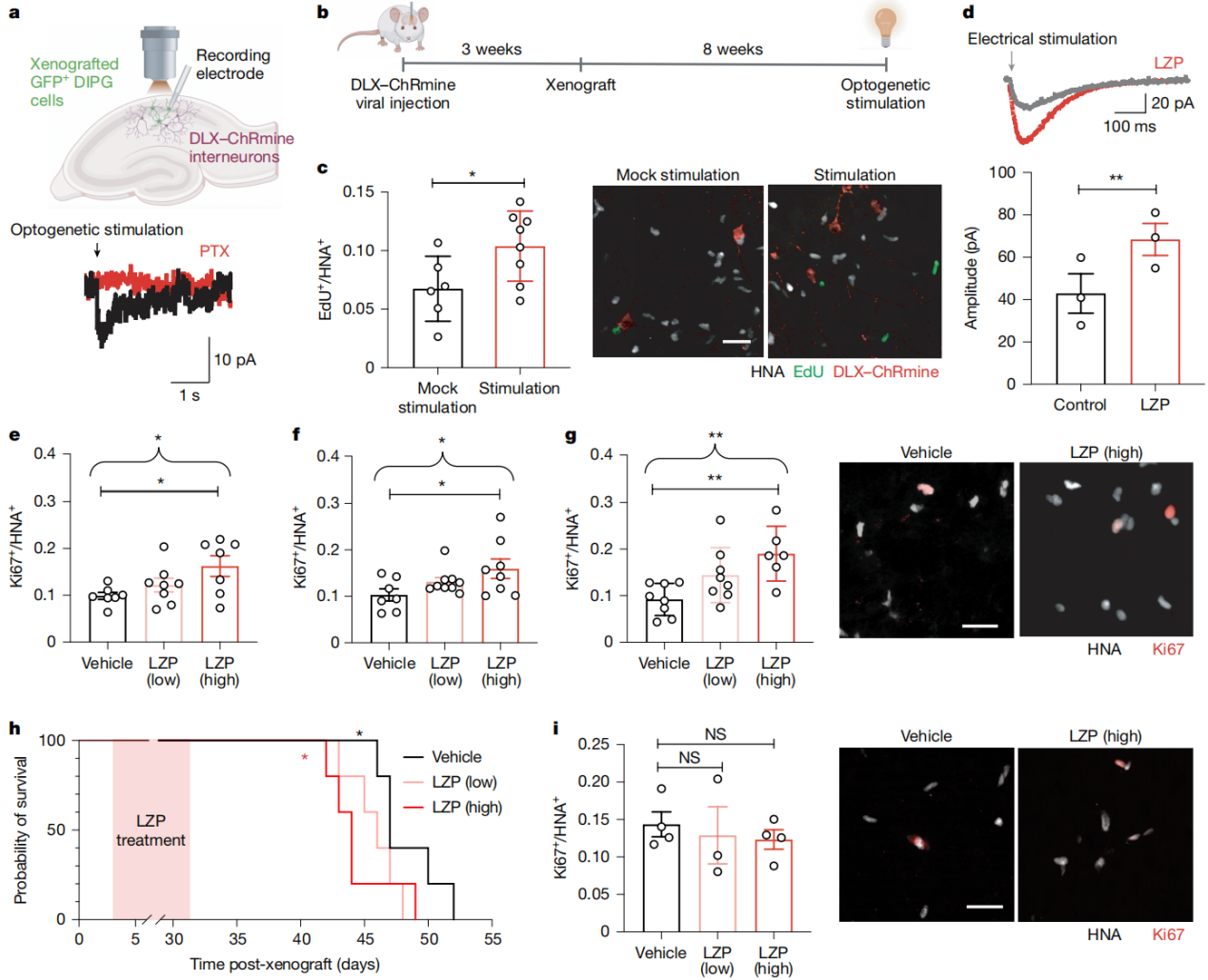
In in vivo experiments, the researchers used optogenetic stimulation of GABAergic interneurons and found that this significantly promoted the proliferation of DMG cells. This was accompanied by increased expression of the Fos protein within gliomas, indicating that neuronal activity can indeed influence tumor cell behavior. Moreover, unlike in IDH-wildtype glioblastomas, the number of GABAergic interneurons in the DMG microenvironment is relatively preserved, providing favorable conditions for their interaction with tumor cells.
Finally, the research team turned their attention to lorazepam, a clinically used benzodiazepine drug. As a positive allosteric modulator of GABA receptors, lorazepam enhances GABA-mediated signaling. Experiments revealed that lorazepam increased the amplitude of postsynaptic GABAergic currents in DMG cells, promoted the proliferation and tumor growth of H3K27M+ DMG cells, and shortened the survival of tumor-bearing mice. In contrast, lorazepam had no such effect on H3/IDH-wildtype hemispheric high-grade gliomas, further highlighting the differences in GABAergic signaling responses across glioma subtypes.
This study is the first to clearly demonstrate the presence of growth-promoting synaptic communication between GABAergic neurons and H3K27M+ DMG cells—a brain cancer neurophysiological mechanism specific to this tumor subtype—which may help guide the development of safer and more effective therapies.
Service Type :
Select the service you'd like to purchase.
Order Information(Premade-AAVs)
Please provide us some information about the service you'd like to order.
Order Information(Custom AAV/Lentivirus)
Please provide us some information about the service you'd like to order.
Order Information(Others)
Please provide us some information about the service you'd like to order.