Breaking Tool | Cell | Allen Institute for Brain Science Releases Over 1,000 Cortical Cell Targeting Tools, Marking a New Breakthrough in Decoding Brain Function!
Release time:2025-06-05 11:27:36
The mammalian brain, as a highly complex organ, has a cortex composed of hundreds of distinct cell types. These cells can be classified along multiple dimensions based on shared characteristics such as morphology, function, and molecular expression profiles. Understanding the contribution of each cell type to the physiological processes governed by the cortex is essential for elucidating their roles in both health and disease.
On May 29, 2025, a major milestone of the U.S. National Institutes of Health's BRAIN Initiative was published in Cell, in a paper titled “A suite of enhancer AAVs and transgenic mouse lines for genetic access to cortical cell types.” The researchers used transcriptomic and epigenomic classifications of mouse and human cortical cell types to define marker genes and candidate enhancers, ultimately creating a large toolkit comprising 15 transgenic driver mouse lines, 2 reporter mouse lines, and over 1,000 different enhancer AAV vectors. These tools enable specific targeting of cortical cell subpopulations, with some demonstrating targeting specificity greater than 90%.
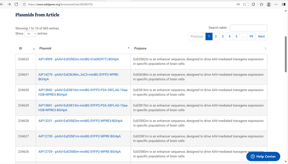
All tools are publicly available through Addgene and Jackson Labs. Experimental data related to the SYFP2 expression vectors in this study can be accessed via the Genetic Tools Atlas portal. The Allen Institute has launched new public online resources: the Genetic Tools Atlas (RRID:SCR_025643;https://portal.brain-map.org/genetic-tools/genetic-tools-atlas) and BioFileFinder (BFF; RRID:SCR_026930;https://bff.allencell.org/), which also include data from individual scRNA-seq experiments.
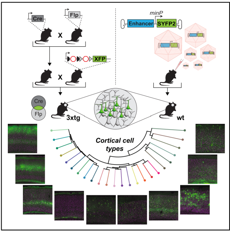
Figure 1. Strategies for generating and characterizing the two types of genetic tools.
Enhancer AAV Vector Screening and Optimization
Using both historical genomic datasets and newly generated single-nucleus multi-omics data (snMultiome), the researchers developed a cross-species (mouse/human) classification system for transcriptomic and epigenomic features at subtype resolution. Mouse data were derived from the primary somatosensory (SSp), primary motor (MOp), and primary visual (VISp) cortices, while human data came from the middle temporal gyrus (MTG). These methods enabled the profiling of tens of thousands of molecular features at the single-cell level, allowing classification into molecularly defined cell types. They also facilitated the identification of marker genes and genomic enhancers aligned with specific cell types, which were then used to create tools for targeting cortical cells.
A total of 1,164 unique enhancer AAV vectors were designed and tested, including 802 candidate sequences (643 mouse-derived and 159 human-derived). Approximately 40% (313 vectors) demonstrated cell-type targeting specificity. Each candidate enhancer sequence was assigned a unique ID beginning with "AiE" (for Allen Institute–discovered enhancers) or "ExE" (for sequences based on coordinates reported by other research groups), followed by a four-digit number and a species identifier (“m” for mouse, “h” for human).
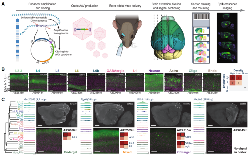
Candidate enhancer sequences were PCR-amplified from their respective genomes and cloned upstream of a minimal promoter (minBG, minRho, minCMV, or endogenous promoter) within an AAV plasmid backbone to drive expression of the yellow fluorescent protein SYFP.
The plasmids were packaged into PHP.eB AAV vectors, which are capable of crossing the blood-brain barrier, and delivered to C57BL/6J mice via retro-orbital (RO) injection. Approximately four weeks post-injection, initial screening was conducted by visually inspecting fluorescence in brain slices. Whole-brain labeling was then assessed using serial two-photon tomography (STPT), and cell-type specificity was confirmed through single-cell RNA sequencing (scRNA-seq). The experiments focused on the mouse visual cortex to ensure data consistency. Through core deletion and tandem optimization strategies, the SYFP2 mRNA expression from the enhancer AAVs was increased by an average of 839%, while maintaining stable specificity. Recombinase variants such as Cre and FlpO were also developed. Specificity was further improved by introducing a Cre(R297T) mutation and optimizing the WPRE sequence to reduce off-target expression.
Figure 3. Screening of enhancer AAVs in the mouse brain
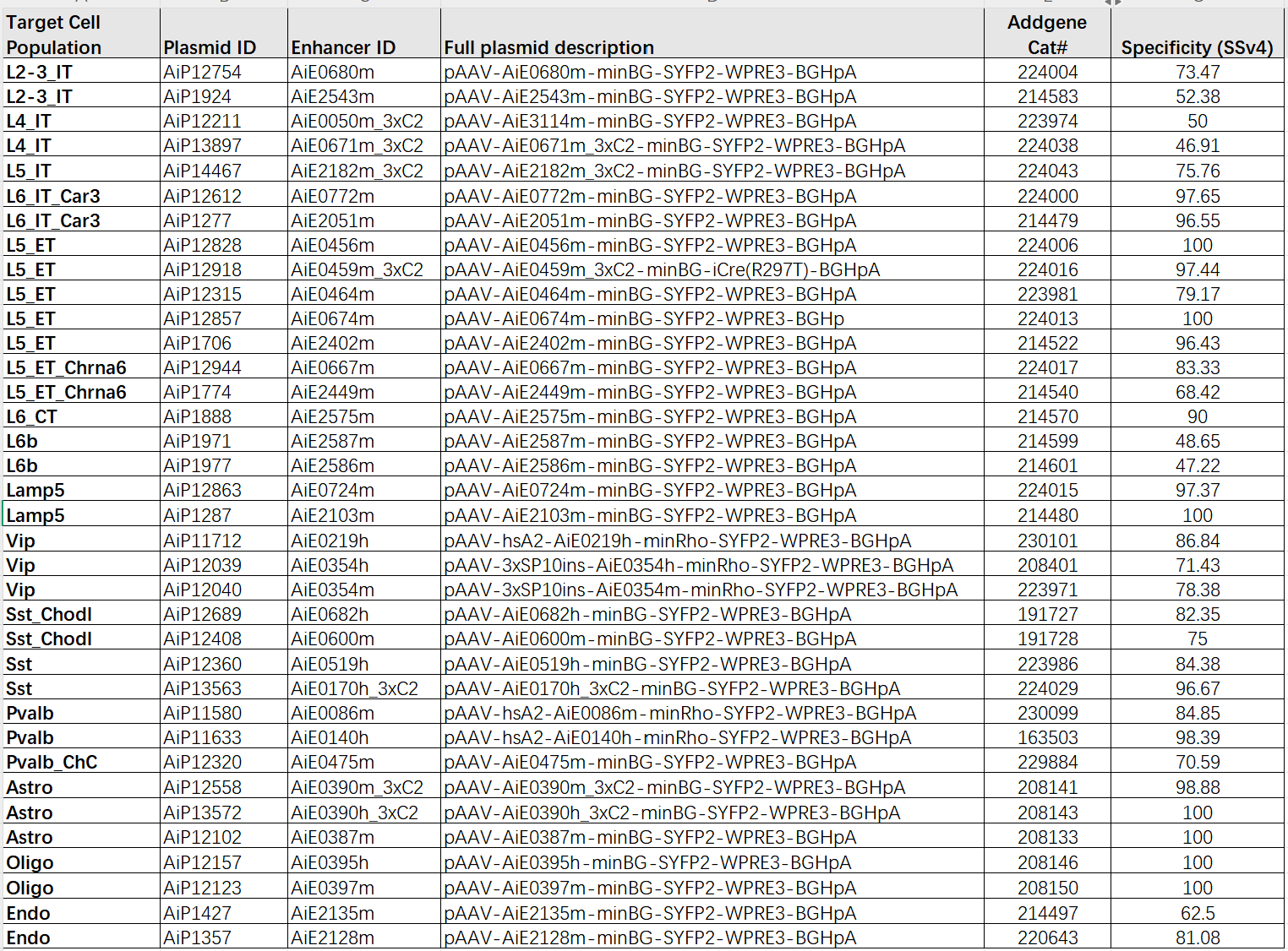
The researchers designated the most valuable tools for genetically accessing specific subtypes or clusters with the “Hall of Fame” (HoF) label. This designation was manually assigned by scientists at the Allen Institute based on multiple factors, such as specificity determined from scRNA-seq data and the signal intensity of the enhancer AAVs. The goal of the HoF label is to help researchers quickly identify the most effective tools for targeting particular cell populations.
Figure 4. Hall of Fame (HoF) enhancer AAVs
Establishment of Transgenic Driver or Reporter Mouse Lines
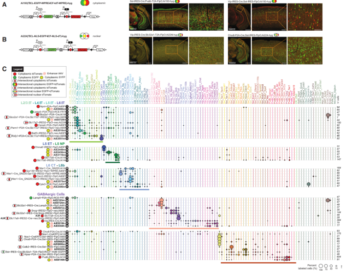
Finally, the researchers established and evaluated 15 transgenic driver mouse lines and 2 reporter mouse lines. These lines can be used alone or in combination with enhancer AAVs to genetically access specific cortical populations even at the finest subclassification levels. The two reporter lines (Ai193 and Ai224) are as follows: Ai193, which is TICL-EGFP-WPRE-ICF-tdT-WPRE-hyg, and Ai224, which is TICL-NLS-EGFP-ICF-NLS-dT-hyg. They contain independent transcriptional units that report Cre and Flp expression by expressing different fluorescent proteins: GFP is expressed in the presence of Cre, tdTomato is expressed in the presence of Flp, and when both are present, both fluorescent proteins are expressed simultaneously. Based on transcriptomic classification of mouse cortical cell types, marker genes capable of labeling specific cell types (subclasses, supertypes, or clusters) were selected. Fifteen genes were chosen to generate transgenic driver Cre or Flp lines: for glutamatergic cell type-based drivers, 11 marker genes (Batf3, Parm1, Rxfp1, Chrna6, Slco2a1, Cplx3, Cpne4, Ctxn3, Gpr139, Npnt, Slc17a7) were used to construct driver lines. For example, Batf3-IRES2-FlpO-WPRE-neo primarily targets L5_IT clusters in L5_IT_VISp_Batf3 but also labels some L2/3 IT, other L5_IT, L6_CT types, and microglia; Chrna6-IRES2-FlpO specifically labels the L5_ET_Chrna6 cell type. For GABAergic cell type-based drivers, genes such as Lamp5, Sncg, and Chodl were selected to construct driver lines. Lamp5-P2A-FlpO labels Lamp5 GABAergic cells but also marks glutamatergic cells in layers L2-3, L5, and L6; Sncg-IRES-FlpO;Ai65F labeling of the Sncg neuronal population also marks endothelial cells.
Figure 5: Comparison of Transgenic Reporter and Driver Lines with Selected Enhancer AAVs
Significance of the Study
This article created and publicly released over 1,000 enhancer AAVs and 17 transgenic mouse lines along with other genetic tools. Their specificity was validated using single-cell sequencing and imaging techniques. The tools were optimized to enhance efficacy and used to construct a cross-species cortical cell taxonomy. This provides a standardized toolkit and data resources for cortical cell type research, advancing the study of brain function and disease mechanisms. The large-scale development pipeline also establishes a methodological foundation for future innovations in neuroscience tools.
To support your research on GABAergic neurons, Brain Case has selected a set of enhancers derived from specific GABAergic neuron subtypes for the construction of a GABAergic-specific promoter library. In addition,Brain Case Biotech offers various custom and viral packaging services. Please contact bd@ebraincase.com
Service Type :
Select the service you'd like to purchase.
Order Information(Premade-AAVs)
Please provide us some information about the service you'd like to order.
Order Information(Custom AAV/Lentivirus)
Please provide us some information about the service you'd like to order.
Order Information(Others)
Please provide us some information about the service you'd like to order.