Breakthrough | WESTLAKE UNIVERSITY TEAM DEVELOPS ADVANCED RED GENETICALLY ENCODED CALCIUM SENSOR “SOMAFRCAMPI” WITH SENSITIVITY MATCHING TO GCAMP FOR NEURONAL IMAGING IN VIVO
Release time:2025-08-25 10:56:53
Research Background
Genetically encoded calcium indicators (GECIs) have become essential tools for monitoring neuronal activity in vivo. GECIs based on the green fluorescent protein (GFP) family, such as the GCaMP series, have been extensively optimized in various model organisms, enabling efficient detection of neuronal activity. However, the development of red fluorescent calcium indicators has lagged behind, with fewer available variants and relatively weaker performance. On Apirl 29, 2025, Professor Kiryl Piatkevich's team at Westlake University published a study on PLOS BIO titled "A Sensitive Soma-localized Red Fluorescent Calcium Indicator for Multi-Modality Imaging of Neuronal Populations In Vivo." The research involved re-engineering a former red GECI, FRCaMP. By using topological inversion and soma-targeting strategies, it resulted in FRCaMPi and its soma-localized variant, SomaFRCaMPi, with optimized performance. Compared to the commonly used jRGECO1a, SomaFRCaMPi exhibits a broader dynamic range, higher peak ΔF/F₀, and an improved signal-to-noise ratio (SNR).Notably, its overall performance is now comparable to the best-performing green soma-localized GECIs, highlighting its potential advantage in enhancing the accuracy of neuronal activity imaging.
Main Findings
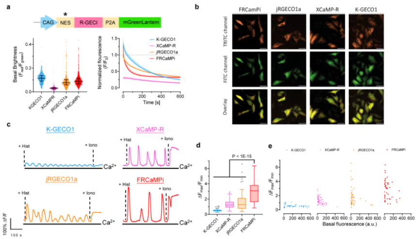
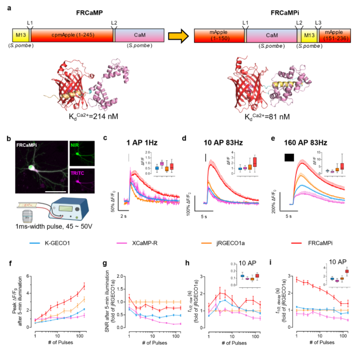
By "inverting" the topology of the original FRCaMP, the researchers developed a novel red GECInamed FRCaMPi. Compared to its parental design, FRCaMPi exhibits a threefold increase in calcium-binding affinity and a 2.36-fold increase in peak fluorescence in neurons. In experiments with HeLa cells and cultured hippocampal neurons, FRCaMPi demonstrated higher sensitivity and a broader dynamic range than jRGECO1a (Figures 1 and 2). Additionally, fiber photometry imaging in vivo confirmed its stability and applicability for up to three months.
Figure 1 Characteristics of FRCaMPi in HeLa cells
Figure 2 Design of FRCaMPi and its characterization in primary mouse hippocampal neurons
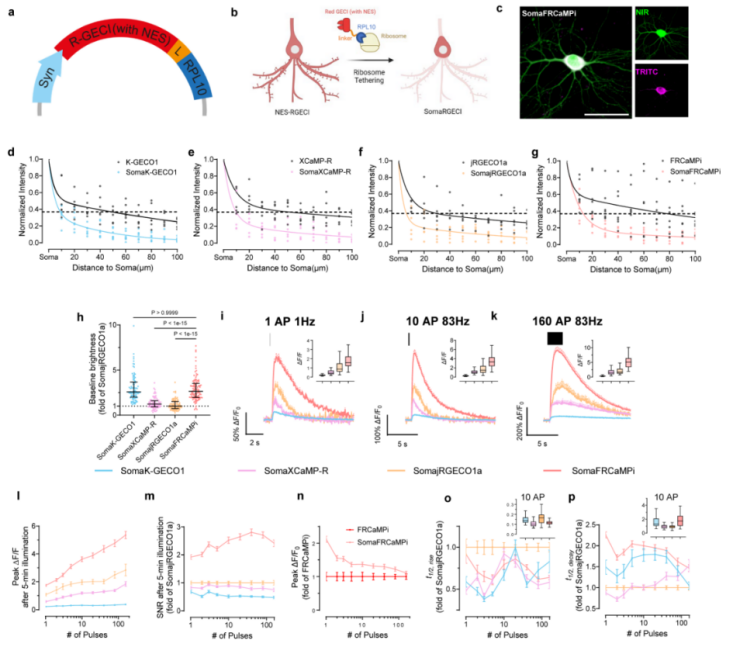
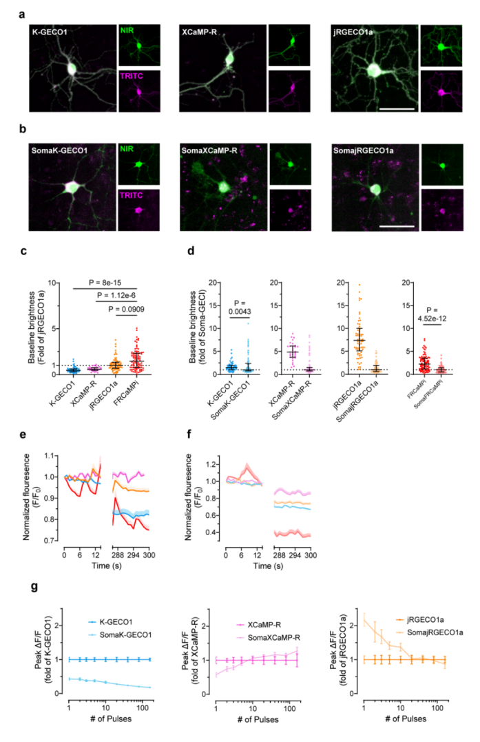
To achieve soma targeting and explore the properties of soma-localized red sensors, the authors added an RPL10-mediated ribosomal targeting peptide to the C-terminus of the sensor, creating SomaFRCaMPi. Compared to other soma-targeted red sensors, SomaFRCaMPi exhibited significantly improved sensitivity and dynamic range, with a SNR at least twice that of other red sensors. Cultured neuron experiments further demonstrated that SomaFRCaMPi had higher baseline brightness, faster kinetics, and superior sensitivity compared to its non-soma-targeted counterpart. These findings establish SomaFRCaMPi as a powerful tool for in vivo neuronal calcium imaging, outperforming other red GECIs (Figures 3 and 4).
Figure 3 Optimization of SomaFRCaMPi and its characterization under electrical field stimulation in primary hippocampal neurons
Figure 4 Baseline brightness and photobleaching characteristics of red GECIs in primary hippocampal neurons
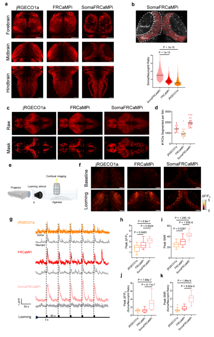
For neuronal imaging in vivo, the authors first tested SomaFRCaMPi in a transgenic zebrafish model (with pan-neuronal expression) and compared it with non-targeted jRGECO1a and FRCaMPi. Results showed that SomaFRCaMPi fluorescence was confined to the soma region, significantly improving cell segmentation accuracy. Using Cellpose for automated segmentation, SomaFRCaMPi identified twice as many neurons as FRCaMPi. Regarding soma localization and signal response, the fluorescence ratio between somata and neurites was more than twice that of non-targeted indicators. Under looming visual stimuli, SomaFRCaMPi exhibited significantly higher peak ΔF/F0 and SNR, while neurite signal responses were reduced threefold. These findings highlight SomaFRCaMPi's superior performance in extracting soma signals in zebrafish calcium imaging (Figure 5).
Figure 5 Expression and characterization of jRGECO1a, FRCaMPi, and SomaFRCaMPi in zebrafish neurons
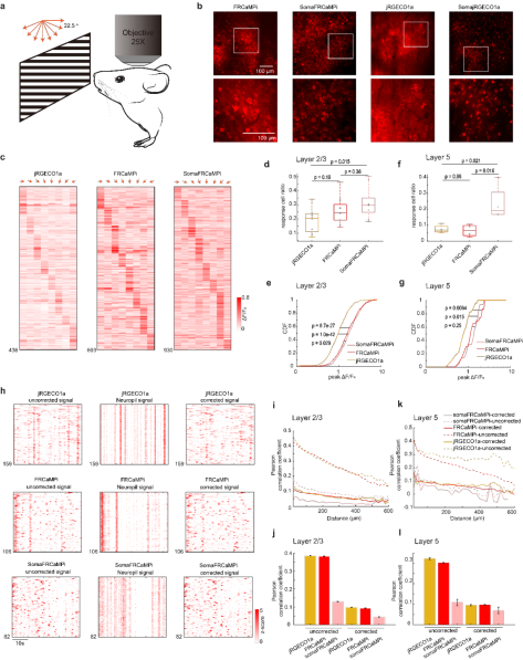
Next, the authors conducted two-photon calcium imaging of neuronal populations in the primary visual cortex (V1) of mice. SomaFRCaMPi exhibited exceptional soma localization, effectively reducing fluorescent interference from neurites, whereas soma-targeted jRGECO1a showed apparent toxicity. Under visual grating stimulation, the average number of neurons detected in layer 2/3 using SomaFRCaMPi (58.1 neurons/FOV) was 2.6 times higher than with jRGECO1a (21.9 neurons/FOV). Compared to jRGECO1a, SomaFRCaMPi exhibited a peak ΔF/F0 at least 77% higher, with a 50% increase in responsive neurons. Signal correlation (Pearson coefficient) was reduced by 2-7 times depending on distance before neurite signal correction. Furthermore, SomaFRCaMPi maintained high sensitivity and stable SNR even after four months of expression. In deep cortical layers (L5), SomaFRCaMPi displayed similarly high peak amplitudes and low signal correlations, demonstrating its ability to achieve high-sensitivity and low-background neuronal activity detection in deep brain regions, making it a high-performance red GECI for two-photon in vivo imaging (Figure 6).
Figure 6 In vivo neuronal population imaging in the mouse V1 cortex
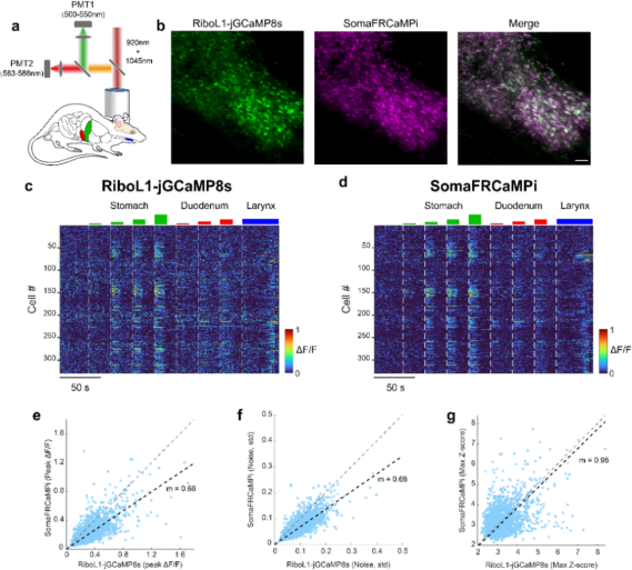
To further evaluate whether SomaFRCaMPi could match high-performance green indicators, the authors co-expressed SomaFRCaMPi with the green soma-targeted RiboL1-GCaMP8s (one of the most sensitive green GECIs) in the mouse brainstem nucleus tractus solitarius (NTS). During digestive tract distension stimulation, two-color two-photon imaging revealed that SomaFRCaMPi maintained clear neuronal fluorescence signals in this densely packed neural region while exhibiting lower noise levels. The Z-score SNR of SomaFRCaMPi was nearly identical to that of RiboL1-GCaMP8s, indicating that its sensitivity is comparable to the best green soma-targeted GECI while leveraging the advantages of red-wavelength imaging (Figure 7).
Figure 7 Two-color in vivo calcium imaging in the NTS during gastric distension
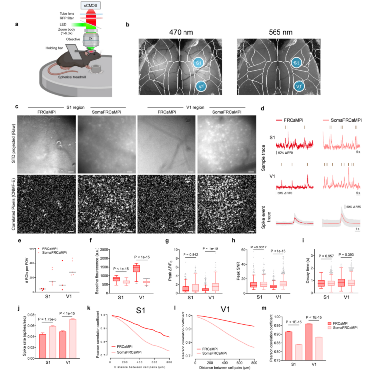
Finally, the authors tested SomaFRCaMPi using one-photon wide-field imaging in the primary somatosensory cortex (S1) and V1 cortex of awake mice. Compared to FRCaMPi, SomaFRCaMPi exhibited significant advantages: when 80-90% of cortical neurons were labeled, it detected 3.3 times more neuronal ROIs than FRCaMPi. SomaFRCaMPi also recorded 50% more calcium events than FRCaMPi, with peak SNRs 10% and 40% higher in different conditions. More importantly, despite the higher neuronal density and greater number of detected calcium events, SomaFRCaMPi effectively reduced neuronal activity correlation, particularly over long distances (800 µm), where correlation was 20-30% lower than with FRCaMPi. These results confirm the efficacy of SomaFRCaMPi in one-photon wide-field imaging, reducing noise while enhancing the number of detectable ROI, signal sensitivity, and accuracy, under an imaging modality with a large FOV, low resolution and high noise level. (Figure 8).
Figure 8 Dynamic changes recorded in S1 and V1 using wide-field imaging with FRCaMPi and SomaFRCaMPi
Conclusion
By employing topology inversion and soma-targeting strategies, researchers redesigned the mApple-based red calcium indicator FRCaMP, enhancing its calcium affinity and developing SomaFRCaMPi. This sensor exhibited superior sensitivity, SNR, and reduced signal correlation in neuronal population recordings in both mice and zebrafish, performing on par with the most sensitive soma-targeted green GECI. The development of SomaFRCaMPi introduces a novel method to enhance sensitivity without requiring extensive mutagenesis screening. Furthermore, it underscores the potential of topology inversion (converting conventional circularly permuted structures into non-circularly permuted structures) in sensor optimization and targeted integration, providing new design strategies for future single-fluorophore GECIs.
Brain Case is honored to have received authorization from Professor Kiryl’s laboratory to produce various tool vectors for FRCaMPi and SomaFRCaMPi. For inquiries, please contact Kiryl’s laboratory (kiryl.piatkevich@westlake.edu.cn and zhoushihao@westlake.edu.cn), or reach out to Brain Case at BD@ebraincase.com.
Service Type :
Select the service you'd like to purchase.
Order Information(Premade-AAVs)
Please provide us some information about the service you'd like to order.
Order Information(Custom AAV/Lentivirus)
Please provide us some information about the service you'd like to order.
Order Information(Others)
Please provide us some information about the service you'd like to order.