E-mail:BD@ebraincase.com
Tel:+8618971215294
English 中文版
Virus Vector - Viral Vector Production - BrainCaseVirus Vector - Viral Vector Production - BrainCase
  • Home
  • Virus product library
    CRISPRRNAiHSV-helperRV-helperNeurophilic virusCalcium SensorsOptogenetics activationOptogenetics inhibitionChemical geneticsSparse labelingFluorescent proteinBiosensorsRecombinaseApoptosis & AutophagyDisease ModelNeurotoxicityOther
  • Products & Service

    Product Center

    Virus

    VSV-circuit research-vaccine and gene therapy research-BrainCase
    Retrovirus-RCAS-TVA-BrainCase
    Lentivirus Vector - Lentivirus Production - BrainCase
    Rabies Virus Vector - RBV Vector - BrainCase
    Herpes simplex virus-Oncolytic and anterograde tracing-Brain Case
    PRV-retrograd multisynaptic-Peripheral-Btain Case
    AAV-gene therapy vectors-BrainCase

    Animal Model

    Neurological Disease Models-BrainCase
    Tumor animal models-anti-tumor-BrainCase
    Digestive System Disease Animal Model-Brain Case
    Cardiovascular System Disease Animal Models-Brain Case

    Plasmid Construction

    Bac
    Library Construction
    Plasmid design and construction

    Popular Applications

    Gene Regulation

    Gene Overexpression-Brain Case
    RNA interference(RNAi)-siRNA-Brain Case
    Gene Editing - CRSIPR cloning - BrainCase

    Neural Circuit Function Research

    Optogenetics - BrainCase
    Chemical genetics-DREADDs-Brain Case
    Calcium signal recording-Gels- Brain Case
    GRAB Neurotransmitter Fluorescent Probe- Brain Case
    Functional Magnetic Resonance Imaging Technology- Brain Case

    Research on the structure of neural circuits

    Direct Input and Output-viral vectors- Brain Case
    Anterograde Mono-synaptic Tracing -HSV- Brain Case
    Antrograde Muti-synaptic Tracing-HSV & VSV-Brain Case
    Retrograde Mono-synaptic Tracing-Rabies Virus-Brain Case
    Retrograde Muti-synaptic Tracing-PRV-Brain Case

    Featured Services

    Nervous System Disease Drug Effect

    Alzheimer's disease-AD-Brain Case
    Depression-mental disorders-Brain Case
    Parkinson's disease--PD-Brain Case
    Epilepsy-an ancient neurological disorder-Brain Case

    AAV Serotype Screening

    AAV Serotypes screening-gene therapy-Brain Case

    Tumorigenicity Test

    Tumorigenicity Testing-Evaluation of tumor models-Brain Case

    Efficacy of Oncolytic Virus

    Oncolytic virus-for cancer therapy-Brain Case
    Herpes Virus Vector-anti-tumor- BrainCase
    Vesicular stomatitis virus-killing tumor cells-Brain Case
  • News
    Corporate News New Product Launch Media Activity Investor News
  • Support
    Literature interpretation Customer article Video Zone FAQs
  • About Us
    Virus product library Products & Service News Support About Us Contact
  • Contact
    Contact Us Join us
  • 中文
    English 中文版
  • Home
  • Support
  • Literature interpretation
  • Support
  • Literature interpretation
  • Customer article
  • Video Zone
  • FAQs

Brain Case’s interpretation | An article to help you understand the construction of stable cell lines

Release time:2024-10-15 17:11:50
Introduction: In biomedical research and drug development, cell models are essential foundational tools that play a crucial role. Among them, stable cell lines—specifically those that can stably express exogenous genes over the long term—have become indispensable resources in scientific exploration due to their exceptional ability to continuously express genes or proteins. The construction of stable cell lines is a significant technology in the biotechnology field, playing a vital role in drug development, gene function research, and disease model creation. Next, let's learn about the principles, methods, and theoretical applications of constructing stable cell lines with Brain Case!
 

I. Principles of Stable Cell Line Construction

The construction of stable cell lines relies on techniques from both genetic engineering and molecular biology. This process involves cloning exogenous DNA into a vector with specific resistance properties. The vector is then transfected into host cells and integrates into the host chromosome. Cells are selected using the resistance marker contained in the vector, identifying positive clones (cells with higher transfection efficiency), and ultimately ensuring that the exogenous DNA is stably inherited and expressed during cell division. This process involves the following key steps:
1. Selection and Cloning of the Target Gene: Based on the research objective, select an appropriate gene sequence and clone it into an expression vector with a specific resistance marker. Common resistance markers in expression vectors include neomycin, blasticidin, puromycin, and geneticin (G418).
2. Vector Design: Design an expression vector that can integrate into the host cell genome, typically including elements such as promoters, enhancers, and selection markers.
3. Transfection: Use physical or chemical methods to introduce the expression vector into host cells, enabling them to express the exogenous gene.
4. Screening and Identification: Use antibiotic selection or other screening markers to initially identify positive cells that have successfully integrated the exogenous gene. Perform RNA and protein level analysis to further confirm the successful construction and functional expression of the stable cell line.


II. Methods for Constructing Stable Cell Lines 

1. Plasmid-Mediated Cell Transfection

Transient transfection refers to the introduction of plasmids or other vectors carrying exogenous DNA into cells, where they exist in a non-integrated form and express the contained genes for a limited period. This approach is often used for short-term gene expression studies and can be achieved using standard plasmid vectors like pcDNA3.1 and pUC57.
In contrast, stable transfection involves integrating the exogenous gene into the host cell's genome to achieve long-term and stable gene expression. Constructing stable cell lines requires gene editing technologies, which primarily include the following two techniques:

DNA Transposon Technology: In nature, DNA transposon systems include transposase, an enzyme responsible for recognizing and catalyzing the movement of transposons within the genome. Transposons have terminal inverted repeat sequences (TRs) at both ends. Transposase can introduce double-strand breaks at the TRs and genomic sites, facilitating the transposition of the entire transposon region. For gene editing applications, transposase components can be separated into:
1. 
A helper plasmid encoding the transposase.
2.
 A transposon plasmid containing optimized TRs at both ends, with the transposon region in between where the gene of interest can be inserted.

Commonly used transposon systems include PiggyBac, Sleeping Beauty, and Tol2, each with its specific characteristics as detailed in the table below.

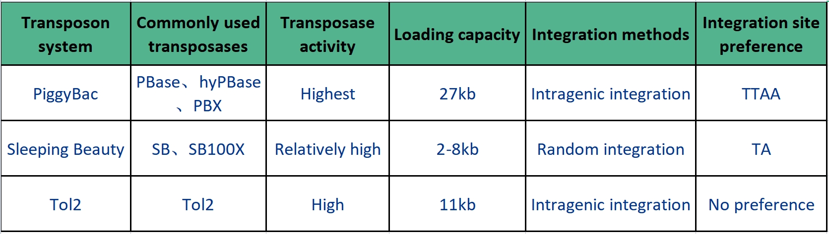
Table 1: Characteristics of PiggyBac, Sleeping Beauty, and Tol2
 
Among them, PiggyBac is one of the most popular transposon systems, used for delivering transgenes, shRNA, recombinant antibodies, and more. Since its discovery, PiggyBac transposons have undergone a series of optimizations and modifications. The optimized transposase (hyPBase) enhances transposition efficiency, with cutting activity but lacking integration activity. This ensures that once the integrated sequence is removed, the position will not be reinserted by the transposon, achieving seamless cutting.
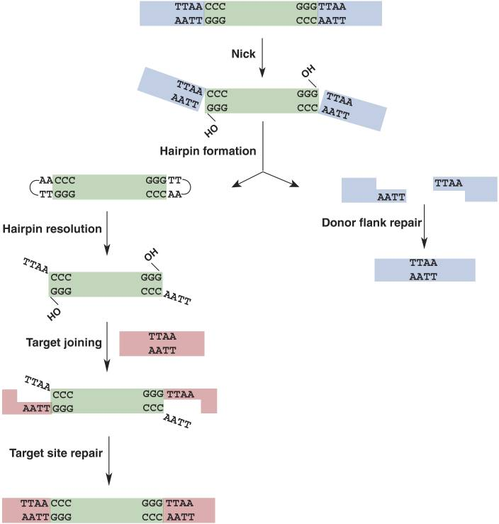
Additionally, a significant advantage of the PiggyBac transposon is its large cargo capacity, able to carry up to 27 kb of DNA between the 5' and 3' terminal repeats (TRs)
. During gene integration mediated by PiggyBac transposons, DNA is cut at the chromosomal TTAA sites, with a preference for integrating into gene regions. The specific integration process for the PiggyBac system is illustrated in the figure below:

 
Figure 1: PiggyBac Transposon Cleavage and Repair Mechanism
 
CRISPR/Cas9 Technology: CRISPR/Cas9 is a type II CRISPR system. It only requires one protein, the Cas9 protein, for the cleavage of foreign genes. Due to its straightforward design and operation, it has been widely used in gene editing to achieve gene knockout, knock-in, and transcriptional activation. When constructing a gene knockout stable cell line, the process begins with plasmid design, which includes a guide RNA (sgRNA, targeting the gene of interest), the Cas9 enzyme gene, and an EGFP or other marker protein. After plasmid transfection into cells, gene editing occurs within the cell: guided by the sgRNA, the Cas9 nuclease cleaves the DNA double strand at a specific position adjacent to the PAM sequence (i.e., at the 5' end), resulting in double-strand breaks in the DNA.
 

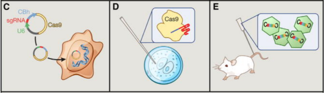
Figure 2:Application of Cas9 as a Gene Editing Tool in Cells (Left: Cell Lines; Center: Zygotes; Right: Somatic Cells)
 

2. Lentivirus-Mediated Cell Infection: 

Direct plasmid transfection and selection is one of the earliest methods used for constructing cell lines, but it has relatively low efficiency and poor stability. In contrast, lentiviruses can infect almost all types of cells (both dividing and non-dividing) and efficiently integrate genetic material into the host cell genome, achieving long-term stable expression. Due to its high infection efficiency and ease of use, lentivirus-mediated methods have become the mainstream technology for constructing stable cell lines. The construction of stable cell lines using lentivirus involves three main steps: constructing the lentivirus expression vector, packaging the lentivirus, and establishing the stable cell line.
 

Ⅲ.   Advantages of Stable Cell Lines

1.Long-Term Stable Expression: Stable cell lines can express foreign genes consistently over long periods, facilitating extended experiments and research, with most research cell lines capable of stable, repeated passaging.
2. Reproducibility and Accuracy: The consistent genetic background of stable cell lines provides high experimental reproducibility and accuracy of results.
3. Functional Research: Stable cell lines are used for in-depth studies of specific gene functions by continuously expressing the target gene, allowing researchers to investigate its role in cellular physiological and pathological processes.
4. Gene Editing Validation: Stable cell lines are useful for validating gene editing techniques, such as CRISPR/Cas9, by evaluating editing efficiency through stable expression of reporter genes. They can also be used to achieve gene knockout, knock-in, transcriptional activation, or repression for functional exploration of genes.
5. Disease Model Construction: Stable cell lines can simulate gene expression changes related to diseases, helping in the construction of disease models and providing a platform for studying disease mechanisms and developing therapeutic strategies.
6. Drug Screening and Development: In drug screening, stable cell lines serve as model systems for high-throughput screening and evaluation of compound efficacy and safety.
7. Production of Biopharmaceuticals: In the biotechnology industry, stable cell lines are used for the production of recombinant proteins, antibodies, and other biopharmaceutical products.

Brain Case Biotech offers a complete suite of stable cell line construction services (including transposon and lentivirus methods). If you have any needs related to stable cell line development, please feel free to contact us for more details or to place a custom order!


References:

[1]Mitra R, Fain-Thornton J, Craig NL. piggyBac can bypass DNA synthesis during cut and paste transposition. EMBO J. 2008 Apr 9;27(7):1097-109. 
[2]Hsu PD, Lander ES, Zhang F. Development and applications of CRISPR-Cas9 for genome engineering. Cell. 2014 Jun 5;157(6):1262-1278.
[3]Wu Y, Wang J, Zhao T, Chen J, Kang L, Wei Y, Han L, Shen L, Long C, Wu S, Wei G. Di-(2-ethylhexyl) phthalate exposure leads to ferroptosis via the HIF-1α/HO-1 signaling pathway in mouse testes. J Hazard Mater. 2022 Mar 15;426:127807.
[4]Yu L, Sui B, Zhang X, Liu J, Hao X, Zheng L. miR-92a-1-5p enriched prostate cancer extracellular vesicles regulate osteoclast function via MAPK1 and FoxO1. J Exp Clin Cancer Res. 2023 May 2;42(1):109.


If you are interested in the details of the experiment or possible problems and causes during the experiment, please contact: BD@ebraincase.com


💥Holiday

Special

Offers

Click to fill in the requirements and submit them to us!

Service Type :

Select the service you'd like to purchase.

Order Information(Premade-AAVs)

Please provide us some information about the service you'd like to order.

Detailed requirements:

scroll

Order Information(Custom AAV/Lentivirus)

Please provide us some information about the service you'd like to order.

Gene ID or gene information:

Selection of the reporting gene:

Special Instructions:

scroll

Order Information(Others)

Please provide us some information about the service you'd like to order.

Virus name/Detailed requirements:

scroll

Related products

Literature Review | Nature | Adenosine Signaling in Astrocytes Coordinates Brain Metabolism and Function

Literature Review | Nature | Adenosine Signaling in Astrocytes Coordinates Brain Metabolism and Function

Breakthrough | WESTLAKE UNIVERSITY TEAM DEVELOPS ADVANCED RED GENETICALLY ENCODED CALCIUM PROBE“SOMAFRCAMPI”WITH SENSITIVITY MATCHING TO GCAMP FOR NEURONAL IMAGING IN VIVO

Breakthrough | WESTLAKE UNIVERSITY TEAM DEVELOPS ADVANCED RED GENETICALLY ENCODED CALCIUM PROBE“SOMAFRCAMPI”WITH SENSITIVITY MATCHING TO GCAMP FOR NEURONAL IMAGING IN VIVO

IF=85 Cutting-edge Review | CRISPR’s Three-Dimensional Breakthrough: A Comprehensive Overview of Genome, Epigenome, and Transcriptome Editing

IF=85 Cutting-edge Review | CRISPR’s Three-Dimensional Breakthrough: A Comprehensive Overview of Genome, Epigenome, and Transcriptome Editing

Literature Insight | Nature Methods | A “Magic Tool” for Real-Time Observation of Synaptic Structural Dynamics — SynapShot

Literature Insight | Nature Methods | A “Magic Tool” for Real-Time Observation of Synaptic Structural Dynamics — SynapShot

map
{dede:global.cfg_webname/}

Virus product library

CRISPR
RNAi
Neurophilic virus
Optogenetics activation
Biosensors

News

Corporate News
New Product Launch
Media Activity
Investor News

Support

Literature interpretation
Customer article
Video Zone
FAQs
微信

WhatsApp Business Account

Tel: +8618971215294
E-mail: BD@ebraincase.com

Address:-

Address:-

  • Copyright © 2024 Brain Case All Rights Reserved.