Customer Publication | Nat. Biomed. Eng. | Tongji University Team Led by Rongrong Zhu and Liming Cheng Achieves Spinal Cord Injury Repair and Motor Function Recovery Through Thoracic Spinal Cord Organoid Transplantation
Release time:2025-11-20 16:21:13
Thoracic (T3–T12) spinal cord injury (SCI) often leads to the loss of lower-limb motor neurons (MNs), causing paralysis, urinary incontinence, and fecal incontinence, and remains highly challenging to treat clinically. Existing spinal cord organoids (sOrgs) fail to recapitulate: thoracic segmental heterogeneity (such as the absence of HOXC9⁺ thoracic MNs); dorsoventral (D–V) spatial organization (disordered arrangement of dorsal sensory neurons and ventral motor neurons); and functional integration (difficulty forming mature synapses and neural circuits, resulting in the inability to restore sensorimotor function).
On October 24, 2025, the team of Rongrong Zhu and Liming Cheng from Tongji University published an article titled “Engineered thoracic spinal cord organoids for transplantation after spinal cord injury” in Nature Biomedical Engineering (IF = 26.6). Using fibroblast-derived induced pluripotent stem cells (iPSCs), along with a layered double hydroxide (LDH) matrix and a basement membrane hydrogel (Matrigel), the researchers constructed thoracic-specific engineered spinal cord organoids (enTsOrg). These organoids precisely match the transplantation site and, in a mouse model of complete thoracic transection SCI, significantly restored hindlimb motor function in paralyzed mice by promoting the maturation and functionalization of motor neuron and interneuron subtypes, establishing synaptic connections, and enhancing neural electrophysiological transmission. This study provides a new strategy for designing region-specific organoids in neural injury repair.
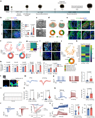
Using fibroblast-derived iPSCs as the starting cells, the researchers performed an LDH–Matrigel embedding experiment during the neural differentiation stage. iPSC spheroids were embedded in an LDH–Matrigel composite whose physicochemical properties were validated via X-ray photoelectron spectroscopy and X-ray diffraction, and whose surface morphology was examined by scanning electron microscopy. This matrix demonstrated excellent neuronal growth compatibility and biocompatibility.During culture, organoid morphology was monitored, showing that by day 21 the organoids reached ~1.5 mm in diameter, and by day 42 developed structures containing abundant mature neurons (NeuN⁺) and dense nerve fibers (NF200⁺).
In the characterization phase, spatial transcriptomics sequencing (ST-seq), RNA sequencing (RNA-seq), quantitative PCR (qPCR), and immunofluorescence staining confirmed that enTsOrgs exhibited strong thoracic identity (high expression of thoracic marker genes such as HOXC9 and HOXB9, and increased proportions of ChAT⁺ cholinergic neurons). Electrophysiological activity assessed by multi-electrode array (MEA) recording and whole-cell patch-clamp showed significantly higher peak firing rates, spontaneous action potential frequencies, and sodium current amplitudes compared with standard sOrgs.Calcium imaging further demonstrated stronger intracellular Ca²⁺ responses to KCl stimulation.
Taken together, these results confirm that enTsOrgs display a “central neuronal soma – peripheral nerve fiber” spinal cord–like architecture and show strong potential for improving motor dysfunction caused by spinal cord injury.
Figure 1. Experimental workflow for reconstructing thoracic neural tube segment morphogenesis and electrophysiological properties using enTsOrg technology.
enTsOrg Transplantation Promotes Hindlimb Motor Function Recovery After Complete Thoracic Spinal Cord Injury
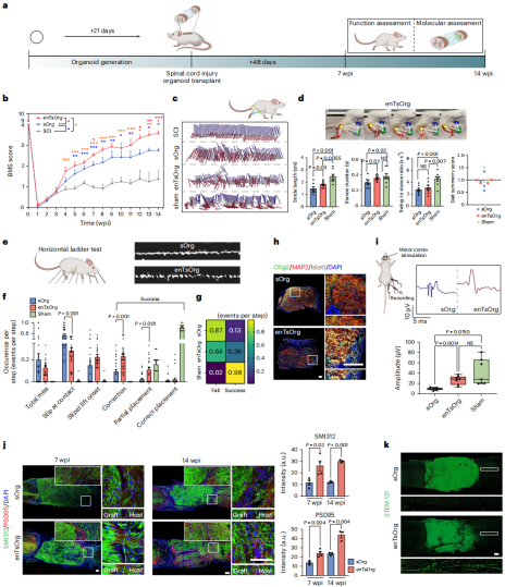
The researchers identified day-21 enTsOrgs as the optimal stage for transplantation (lower apoptosis rate compared with day-42 organoids, higher stem cell content, better functional restoration, and no excessive proliferation or tumorigenicity after transplantation; LDH also protects cells from hypoxia-induced apoptosis). Subsequent behavioral assessments—including BMS scoring, digital footprint analysis, and horizontal ladder tests—demonstrated that enTsOrg transplantation significantly improved hindlimb motor function in mice (e.g., gait symmetry approaching 1, motor task success rate of 0.36), with superior outcomes compared with standard sOrgs.
Further analyses using motor-evoked potentials (MEP), nerve fiber visualization, synaptic protein detection, and neural tracing (biotinylated dextran amine, BDA; pseudorabies virus, PRV) confirmed that enTsOrgs form neural connections with the host and reconstruct neural circuits (e.g., MEP amplitude 26.70 ± 8.46 μV). Additionally, by enhancing the expression of sensory neuron–related markers, enTsOrgs helped restore mouse responses to mechanical and thermal stimuli, indicating recovery of sensory function. These findings demonstrate that enTsOrg transplantation is an effective strategy for repairing thoracic spinal cord injury.
Figure 2. Transplantation of enTsOrgs into thoracic spinal cord injury sites significantly restores motor and neural function.
Enhanced Neuronal Maturation in Transplanted Organoids
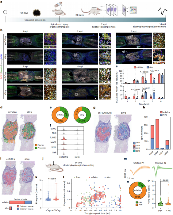
Immunostaining and ST-seq confirmed that after enTsOrg transplantation, neural progenitor cells (Nestin⁺/SOX2⁺) first undergo rapid proliferation, followed by a high proportion of mature neurons (NeuN⁺), reaching 67.76%—substantially higher than in sOrgs. Genes associated with neuronal maturation—such as TUBB3 (neurite formation), MAP2 (axon formation), and synapse development genes (SYP, SYN1)—were also highly expressed.Compared with human spinal cord developmental stages, enTsOrgs progressed more rapidly, corresponding to Carnegie stage (CS) 14, whereas sOrgs corresponded to CS12.
Moreover, enTsOrgs exhibited broad and physiologically balanced distributions of excitatory (SLC17A6⁺) and inhibitory (GAD1⁺) neurons, supporting more coordinated signal transmission with the host. Finally, in vivo electrophysiological recordings showed more active neuronal firing in enTsOrgs (approaching levels seen in sham-operated mice). They differentiated into excitatory principal neurons (broad-spike) and inhibitory interneurons (narrow-spike), whereas sOrgs lacked clear inhibitory neuronal signals. These results indicate that enTsOrgs achieve superior neuronal maturity and functional integrity in vivo.
Figure 3. Spatial transcriptomics and in vivo electrophysiology reveal advanced neuronal maturation within enTsOrgs.
Diverse Cellular Composition and Functional Maturation in Transplanted Organoids
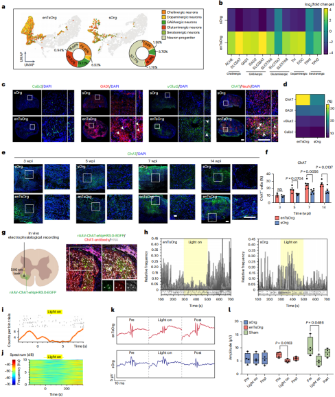
Uniform manifold approximation and projection (UMAP) analysis and KEGG enrichment analysis demonstrated that enTsOrgs can differentiate into multiple neurotransmitter neuron types (including cholinergic and GABAergic neurons). The proportions of GABAergic and dopaminergic neurons, as well as the enrichment of neurodevelopment-related pathways (such as axon guidance), were all higher in enTsOrgs than in sOrgs. Immunofluorescence further confirmed that mature cholinergic neurons accounted for 32.94% of cells in enTsOrgs (compared with 18.28% in sOrgs), and their numbers continued to increase from 7 to 14 weeks after transplantation.
Combined optogenetics and electrophysiology experiments revealed that cholinergic neurons within enTsOrgs can function as spinal motor neurons. When ChAT⁺ neurons were optogenetically inhibited, the firing frequency of putative principal neurons (P-PNs) and the amplitude of MEPs significantly decreased, accompanied by reduced locomotor activity in mice—an effect not observed with sOrgs. These results indicate that enTsOrgs outperform sOrgs in both cellular diversity and functional maturation, and possess the capability to participate in host neural regulation as functional spinal motor neurons.
Figure 4. enTsOrg grafts exhibit the cellular composition and functional maturity of key neuron types involved in motor function.
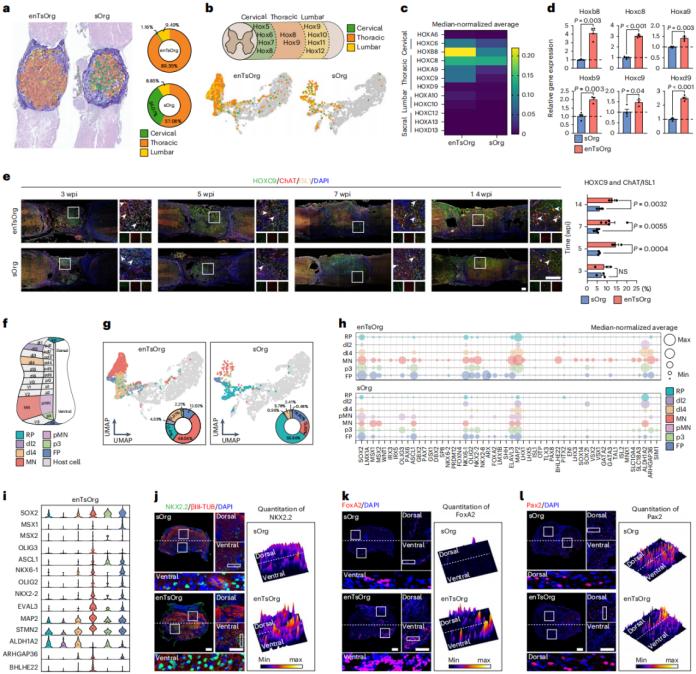
Transplanted enTsOrgs Exhibit Corresponding Segmental Identity and Dorsoventral (D–V) Patterning
Focusing on the segmental identity and D–V axis patterning after enTsOrg transplantation: ST-seq and RT-qPCR analyses showed that 89.35% of loci within enTsOrgs displayed thoracic spinal cord characteristics (vs. 57.08% in sOrgs), with high expression of thoracic marker genes such as Hoxb8 and Hoxc9, matching the segment of the injury site.
Clustering along the D–V axis revealed that motor neuron–feature loci accounted for 53% of positions in enTsOrgs (only 13% in sOrgs). Ventral genes (NKX2.2, FoxA2) and dorsal genes (Pax2) exhibited region-specific expression corresponding to their physiological positions, forming a spatial distribution consistent with neural tube organization. In contrast, sOrgs were dominated by immature cells with disordered gene expression patterns.
These findings demonstrate that transplanted enTsOrgs maintain thoracic specificity and form a mature D–V spatial architecture, providing a structural foundation for neural repair.
Figure 5. Developmental patterning of enTsOrg grafts shows characteristic thoracic spinal segment identity and dorsoventral axis organization.
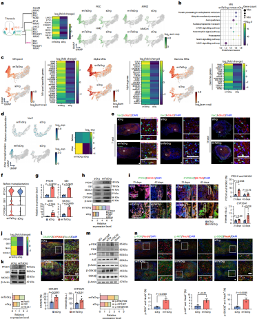
enTsOrg Transplantation Promotes the Formation of Specialized Neurons for Spinal Cord Repair
Gene expression analysis, heatmaps, UMAP clustering, and immunofluorescence experiments confirmed that enTsOrgs can differentiate into specialized motor neuron subtypes matching the thoracic region (PGC, HMC, MMC), as well as key interneurons (Vsx2⁺/vGlut2⁺). Motor neuron–related functional genes (SV2) and regeneration-associated genes were highly expressed, with levels significantly superior to those in sOrgs.
KEGG pathway enrichment analysis (focusing on motor neuron–related pathways), gene expression assays, Western blotting, and immunostaining revealed that LDH promotes thoracic specialization and motor neuron differentiation in enTsOrgs by regulating the SHH and RA signaling pathways.
After transplantation, neuroprotective pathways such as PI3K/AKT were activated, and the ubiquitin-mediated proteolysis pathway was enriched, supporting enTsOrg functionality. Gene expression profiling further confirmed regulatory roles for neuron-maturation genes such as UCHL1 and MFN2.
Using species-based cell-type classification, host–graft integration analyses, and gene expression studies, researchers found that enTsOrgs integrate more efficiently with host cells, improving the host microenvironment by reducing barrier-forming cell populations and upregulating regeneration-associated genes. In contrast, sOrgs showed poor integration due to obstruction by host barrier cells.
These findings indicate that enTsOrgs support spinal cord repair by promoting specialized neuron generation, modulating key signaling pathways, and achieving efficient host integration.
Figure 6. Transplanted enTsOrgs facilitate the production and functional maturation of key neuron populations, thereby restoring motor function.
Conclusion
This study for the first time generated spinal cord organoids with thoracic segment–specific identity and a defined dorsoventral axis, addressing the long-standing challenge of regional matching in organoid-based therapy. The work provides a potential therapeutic strategy for SCI—which accounts for a large proportion of SCI cases—achieving significant restoration of hindlimb motor function with a favorable safety profile. It also establishes a paradigm for designing region-specific organoids for neural injury repair, underscoring the importance of anatomical matching for functional integration of transplanted organoids.
All viral tools used in this study were supplied by
Brain Case Biotech(BD@ebraincase.com):
Product Category
Product Name
Optogenetics
rAAV-ChAT-eNpHR3.0-EGFP
PRV
PRV-CAG-3Ms
Service Type :
Select the service you'd like to purchase.
Order Information(Premade-AAVs)
Please provide us some information about the service you'd like to order.
Order Information(Custom AAV/Lentivirus)
Please provide us some information about the service you'd like to order.
Order Information(Others)
Please provide us some information about the service you'd like to order.