Client Article | J Hazard Mater | Hui-Li Wang’s Team at Hefei University of Technology Reveals the Mechanism of Pb-Induced Non-Spatial Memory Deficits
Release time:2025-09-19 15:30:24
Lead(Pb), a common environmental pollutant, poses significant health risks—especially to children. It is associated with neurodevelopmental and behavioral impairments, with blood lead levels negatively correlated with cognitive development. Lead exposure also damages the hippocampus, leading to emotional problems; however, the mechanisms underlying susceptibility to lead-induced memory deficits remain unclear.
The hippocampus is responsible for processing and storing learning and memory, which can be divided into spatial and non-spatial memory. Pyramidal neurons in the hippocampal CA1 region play a critical role in information processing and memory encoding, particularly in associative memory. Reduced neuronal activity in this region is linked to memory deficits. Epigenetic reprogramming mechanisms allow cells to integrate environmental signals, but the specific epigenetic pathways through which environmental risk factors induce memory impairment are still poorly understood. Previous studies have shown that lead exposure reduces histone acetylation levels (H3K9ac) in the brains of monkeys and mice, involving histone acetyltransferases and histone deacetylases (HDACs). Follow-up research further demonstrated that HDAC1 and HDAC2 are directly responsible for the lead-induced reduction of H3K9ac, with HDAC2 additionally regulating neuronal memory-related proteins and participating in spatial memory reprogramming.
On June 2, 2025, Professor Hui-Li Wang’s research team at Hefei University of Technology published a study in Journal of Hazardous Materials entitled Epigenetic reprogramming of HDAC2 in CA1 excitatory neurons determines Pb-induced non-spatial memory deficits. This study is the first to reveal that lead exposure increases susceptibility to non-spatial memory deficits by impairing CA1 excitatory neurons, with epigenetic reprogramming of HDAC2 identified as a key trigger of this vulnerability.
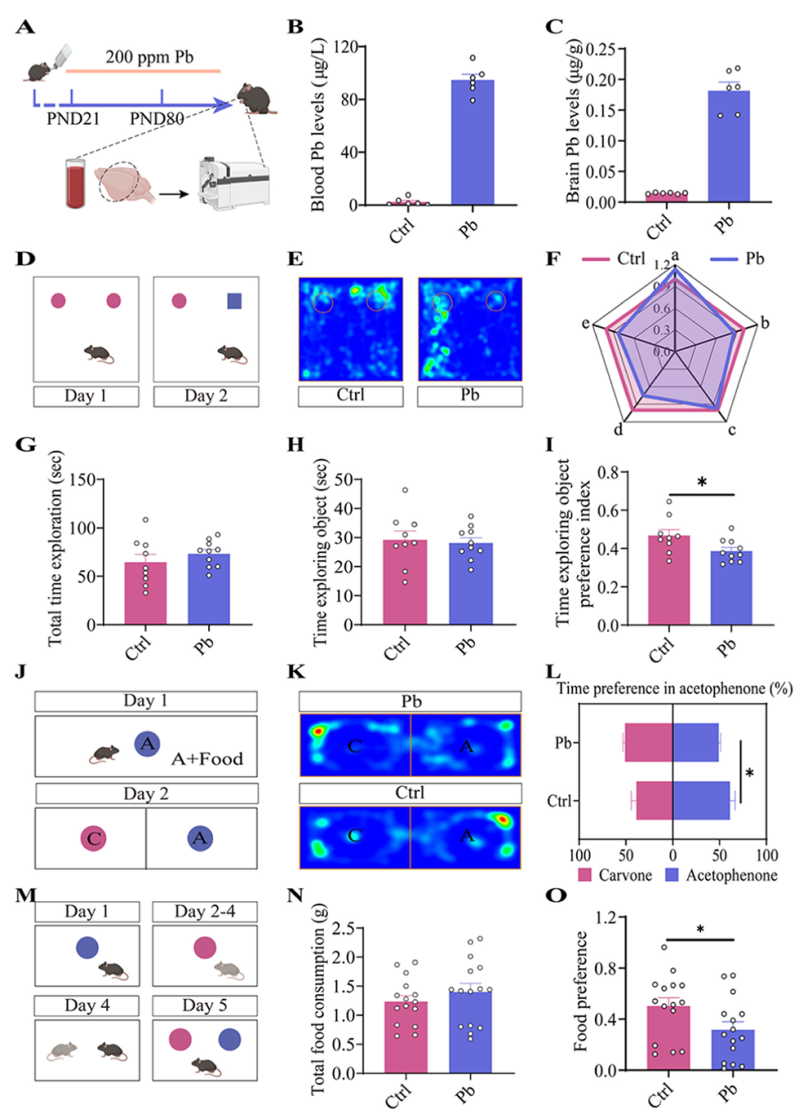
01 Lead Increases Susceptibility to Non-Spatial Memory Deficits
To investigate the impact of environmental risk factors on non-spatial memory, the study used C57 mice at postnatal day 21 (PND21), which were given lead-containing drinking water to establish a memory deficit model (excluding transgenerational effects). At PND80, blood lead and brain lead levels in Pb-exposed mice were 94.618 ± 9.833 μg/L and 0.182 ± 0.031 μg/g, respectively.
Behavioral assessments using the Novel Object Recognition (NOR) test, the Olfactory Reward Memory (ORM) test, and the Social Transmission of Food Preference (STFP) task revealed that Pb-exposed mice (Pb group) showed a significantly reduced preference index for time spent with and entries to novel objects, as well as markedly lower preference indices for phenylacetone and food preference (with total food consumption unchanged).In summary, lead exposure increases the susceptibility of mice to non-spatial memory deficit phenotypes.
Figure 1. Lead increases susceptibility to non-spatial memory deficits.
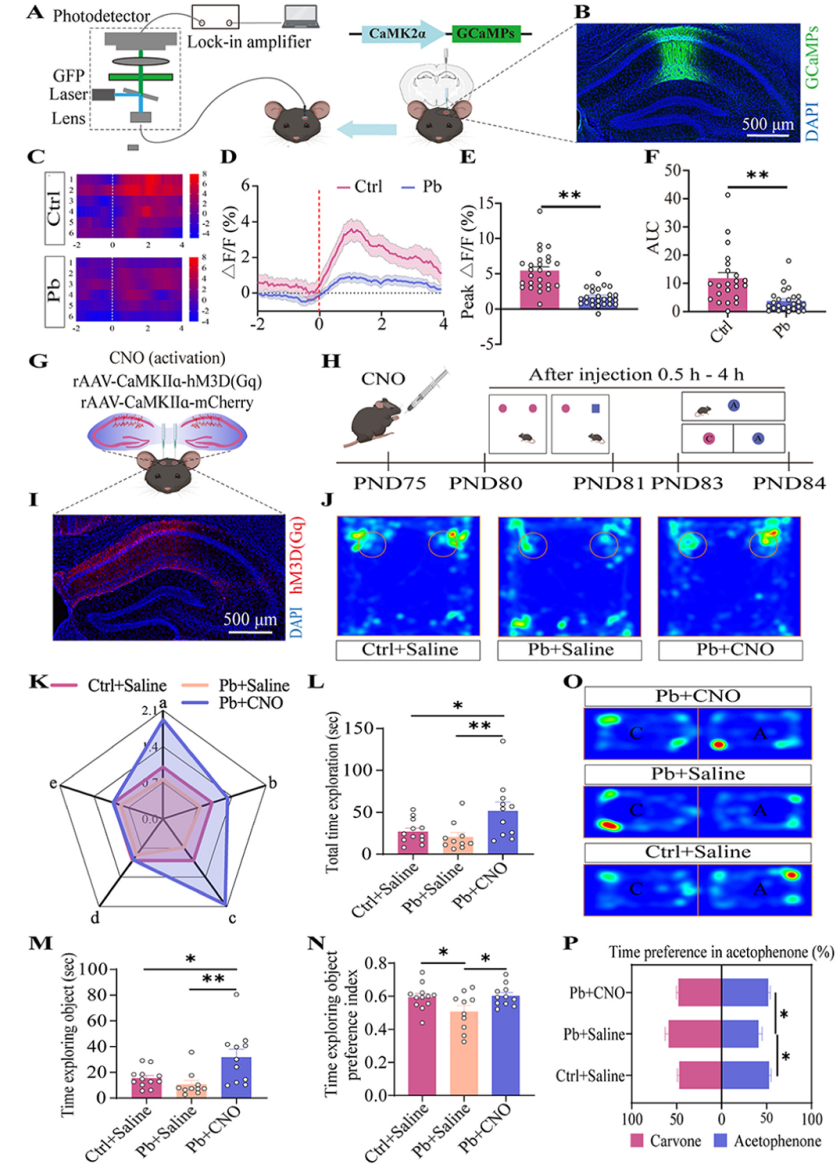
02 Lead Increases Susceptibility to Non-Spatial Memory Deficits via Hippocampal Excitatory Neurons
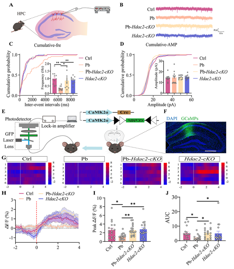
Pyramidal neurons in the hippocampal CA1 region are involved in information processing and encoding for associative memory. By injecting rAAV-CaMKIIɑ-GCaMP6s into this region and recording with fiber photometry, the study found that compared with the Ctrl group, Pb-exposed mice showed significantly reduced calcium signals, peak amplitudes, and area under the curve (AUC) in CA1 excitatory neurons.
Using chemogenetic activation of CA1 excitatory neurons in Pb-exposed mice (via rAAV-CaMKIIɑ-hM3D(Gq)-mCherry injection), combined with NOR and ORM behavioral tests, results showed that compared with the Pb+Saline group, Pb+CNO mice exhibited significantly increased total exploration time of two objects, exploration time of the novel object, novel object preference index, and phenylacetone preference index. This indicates that activation of CA1 excitatory neurons partially rescues non-spatial memory deficits induced by Pb exposure.
Furthermore, compared with the Ctrl+Saline group, Pb+CNO mice also showed significantly increased total exploration time of two objects and exploration time of the novel object, suggesting that CA1 excitatory neurons play a decisive role in susceptibility to non-spatial memory deficits induced by environmental risk factors such as Pb exposure.
Figure 2. Lead increases susceptibility to non-spatial memory deficits via CA1 excitatory neurons.
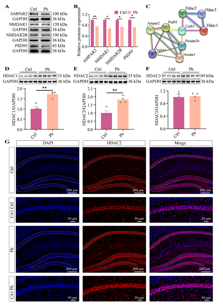
03 Lead Elevates HDAC2 Levels in Hippocampal Excitatory Neurons
Because classical calcium channels and their receptors determine neuronal excitability, the study examined protein expression of AMPAR2, NMDAR1, NMDAR2B, and PSD95 in the hippocampus. Results showed that the levels of all four proteins were significantly reduced in Pb-exposed mice compared with the Ctrl group. Epigenetic reprogramming of HDAC1 and HDAC2 was identified as the key factor responsible for changes in H3K9ac and the above proteins. Further analysis revealed that hippocampal levels of HDAC1 and HDAC2 were significantly elevated in Pb-exposed mice, while HDAC3 showed no change. Immunohistochemistry demonstrated that HDAC2 levels were markedly increased in hippocampal CA1 excitatory neurons of Pb-exposed mice, with widespread distribution in this region, whereas HDAC1 expression was relatively low. These findings indicate that Pb abnormally activates epigenetic reprogramming of HDAC2 in CA1 excitatory neurons, which may underlie the altered susceptibility to non-spatial memory deficits.
Figure 3. Lead promotes upregulation of HDAC2 expression in hippocampal excitatory neurons.
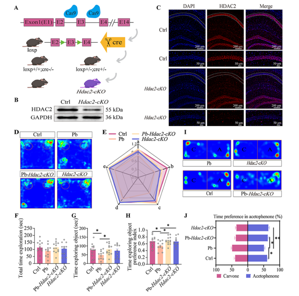
Hdac2 conditional knockout (Hdac2-cKO) mice were generated by crossing Hdac2^flox/flox mice with CaMKIIa-Cre transgenic mice. qRT-PCR, Western blot, and immunohistochemistry confirmed successful deletion of HDAC2 in hippocampal CA1 neurons, and further revealed that Hdac2-cKO increased H3K9ac protein levels in both Pb-Hdac2-cKO and Hdac2-cKO mice.
Non-spatial memory was assessed using NOR and ORM tests. Results showed no difference in the total exploration time of objects between groups. Compared with Pb-exposed mice, both Pb-Hdac2-cKO and Hdac2-cKO mice exhibited significantly increased time preference indices for novel objects and phenylacetone. Additionally, Pb-Hdac2-cKO mice showed a significant increase in exploration time of novel objects compared with Pb-exposed mice, whereas Hdac2-cKO mice did not show this change. These findings indicate that conditional knockout of Hdac2 determines the response to susceptibility of Pb-induced non-spatial memory deficits.
05 Hdac2-cKO Restores Lead-Induced Reduction of Calcium Signals in Hippocampal Excitatory Neurons
Whole-cell patch-clamp recordings revealed that compared with the Ctrl group, Pb-exposed mice showed a significant decrease in the frequency of miniature excitatory postsynaptic currents (mEPSCs) in CA1 excitatory neurons. In contrast, both Pb-Hdac2-cKO and Hdac2-cKO mice exhibited significantly increased mEPSC frequency in CA1 excitatory neurons compared with Pb-exposed mice. These results indicate that conditional knockout of Hdac2 alleviates synaptic transmission deficits in CA1 excitatory neurons following Pb exposure.
Furthermore, by injecting a Cre-dependent GCaMP6s virus into the hippocampal CA1 region to record calcium signals, it was found that compared with the Ctrl group, Pb-exposed mice exhibited significantly reduced calcium signals, peak amplitudes, and area under the curve (AUC) in CA1 excitatory neurons. However, these indices were significantly increased in Pb-Hdac2-cKO and Hdac2-cKO mice compared with Pb-exposed mice. These findings suggest that Pb reduces CA1 excitatory neuronal activity via epigenetic reprogramming that elevates HDAC2, whereas HDAC2 knockout restores this impaired neuronal activity.
Figure 5. Hdac2-cKO restores lead-induced reduction of calcium signals in hippocampal excitatory neurons.
06 Hdac2-cKO Restores Lead-Induced Structural Deficits in Hippocampal Excitatory Neurons
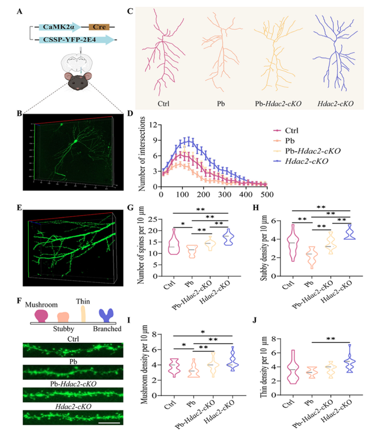
The study used a Cre-dependent sparse labeling virus (CSSP-YFP-2E4) to achieve cell-type–specific sparse labeling of CA1 excitatory neurons. Sholl analysis showed that compared with the Ctrl group, Pb-exposed mice exhibited a reduced number of dendritic intersections in CA1 excitatory neurons, indicating that lead decreases dendritic complexity in this region. In contrast, Pb-Hdac2-cKO and Hdac2-cKO mice displayed more complex dendritic branching in CA1 excitatory neurons.
Dendritic spines were categorized into four types: stubby, mushroom, thin, and branched. Results showed that lead exposure reduced total spine density, mature stubby spine density, and mature mushroom spine density in CA1 excitatory neurons, while the density of immature thin spines remained unaffected. Compared with Pb-exposed mice, Pb-Hdac2-cKO and Hdac2-cKO mice exhibited significant increases in the three affected spine types. Moreover, Hdac2-cKO mice showed significantly increased dendritic spine density in CA1 excitatory neurons compared with Ctrl mice. These findings indicate that lead-induced epigenetic reprogramming via elevated HDAC2 leads to structural and dendritic spine impairments in CA1 excitatory neurons.
07 Hdac2-cKO Restores Lead-Induced Reduction of Neuroplasticity Molecules in Hippocampal Excitatory Neurons
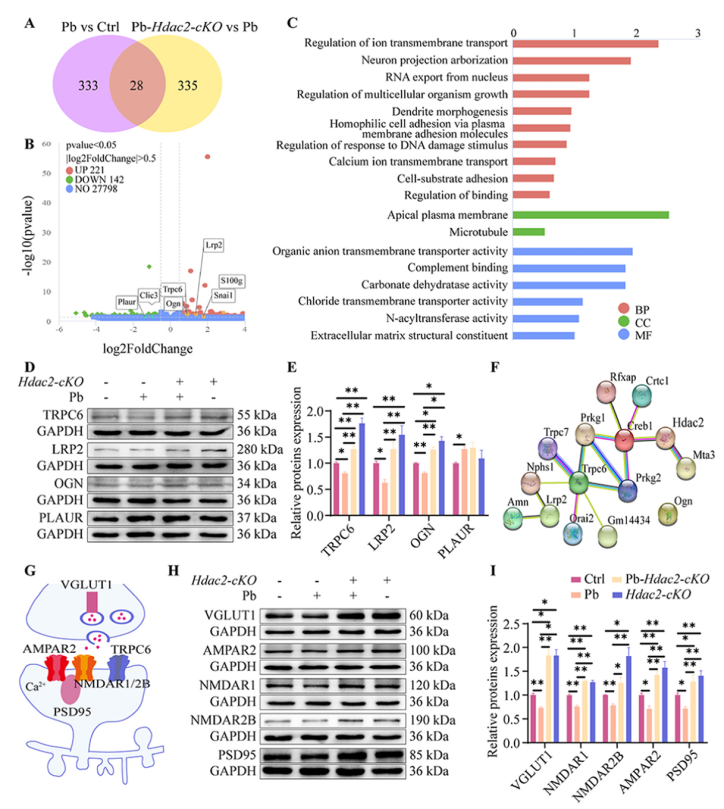
To investigate the molecular mechanisms by which HDAC2 reprogramming regulates Pb-induced damage in CA1 excitatory neurons, mRNA sequencing was performed on CA1 tissues from Pb-exposed vs. Ctrl mice, and Pb-Hdac2-cKO vs. Pb mice. A total of 28 differentially expressed genes were identified. Gene Ontology (GO) enrichment analysis revealed that these genes are involved in pathways related to neuronal structure and function, including calcium ion transmembrane transport and dendrite morphogenesis, which may mediate Pb-induced damage and the reparative effects of HDAC2 knockout.
Protein-level analyses showed that Pb exposure reduced expression of proteins such as TRPC6 and LRP2, whereas HDAC2 knockout reversed this trend, confirming the RNA sequencing results. STRING analysis predicted interactions between HDAC2 and target genes including TRPC6 and LRP2. Conditional knockout of Hdac2 was able to reverse Pb-induced reductions in CA1 excitatory neurons of VGLUT1, NMDAR1, NMDAR2B, AMPAR2, and PSD95 protein levels. Together, these results suggest that Pb impairs neuronal structure and functional activity by elevating HDAC2-mediated epigenetic reprogramming, which suppresses the protein expression of synaptic plasticity-related target genes.
Figure 7. Hdac2-cKO restores Pb-induced reduction of neuroplasticity molecules in hippocampal excitatory neurons.
Summary
Lead exposure increases HDAC2-mediated epigenetic reprogramming in hippocampal CA1 excitatory neurons, leading to abnormal expression of synaptic plasticity-related molecules, structural and functional impairments of neurons, and ultimately non-spatial memory deficits. This finding not only fills a mechanistic gap linking environmental factors and epigenetic modifications to non-spatial memory disorders, but also suggests that targeting HDAC2 to reverse neurotoxicity could represent a new strategy for preventing and treating memory deficits induced by environmental factors.