- E-mail:BD@ebraincase.com
- Tel:+8618971215294
Genetically encoded Ca2+ indicators (GECIs) are widely used to monitor neural activity. With the development of GECIs, the response kinetics, sensitivity and brightness of Ca2+ sensors have been further optimized, among which GCaMP is the most prominent. Ideally, GECIs are uniformly expressed across neuronal populations, have sufficient sensitivity and a suitable fluorescence range for in vivo applications.
Currently, a commonly used strategy is to use viral vectors to deliver GECIs to neurons. However, intracerebral injection (IC) of viral vectors often mediates expression of different strengths depending on the concentration of viral particles, resulting in local accumulation of GCaMP within the cell, thereby affecting cell function and causing Cell death; the number of infected virus particles per cell is lower when injected systemically. In addition, widely used GECIs (such as GCamp6f) are not bright enough to be compatible with systemic drug delivery for two-photon imaging.
On February 4, 2023, the team of S. Grødem and I. Nymoen from the Department of Biological Sciences at Oslo University in Norway published "An updated suite of viral vectors for in vivo calcium imaging using intracerebral and retro-orbital injections in male mice" in the journal Nature Communications. This article reveals that PHP.eB serotype-packaged jGCaMP8 and cell body-targeted EE-RR-jGCaMP8 can be used for systemic administration to achieve in vivo Ca2+ imaging. In addition, the team also constructed cytoplasm-localized RiboL1-jGCaMP8, and its intracerebral injection showed unprecedented labeling density and signal-to-noise ratio without contamination of the neuropil.
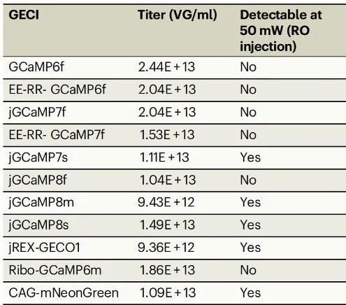
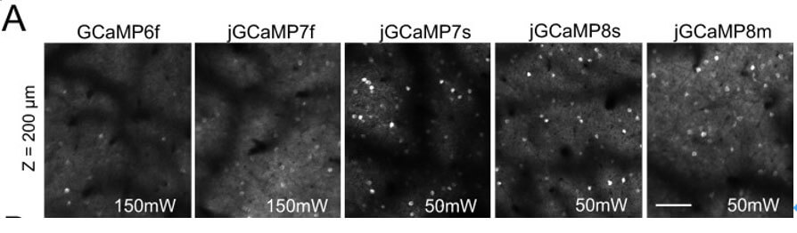
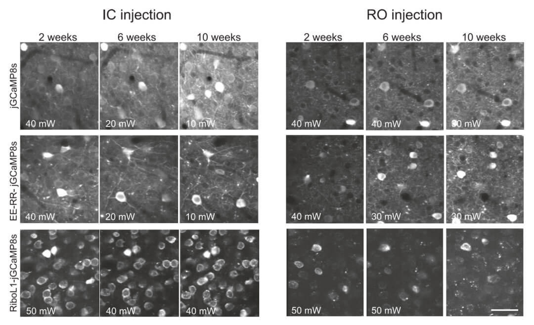
Under reasonable laser power (output power 40-50 mW), when the PHP.eB serotype and IC injection method are selected, the fluorescence signal of Ca2+ changes can be detected in different variants of GCaMP. When systemic administration is chosen, most GECIs detect Ca2+ in vivo and are not bright enough for imaging. The recently developed GECI jGCaMP7s, jGCaMP8s and jGCaMP8m are all bright enough to be used with RO injections (retroorbital injections).
Due to the fast kinetics and higher sensitivity of GCaMP, signals can also be detected from nerve fibers such as dendrites, which both interferes with the quantification of Ca2+ transients in the cell body and introduces noise that reduces the number of cells that can be recorded. Therefore, the authors first constructed EE-RR and RPL10a-tagged jGCaMP8 to alleviate these effects by limiting the cell body localization of GECI. EE-RR cell body-labeled jGCaMP8 can detect strong calcium ion signals when injected with RO or IC, but similar neuropil signals also exist with IC injection. When Ribo-jGCaMP8 is injected with RO, it is expressed in somatic cells, but the expression is slow and fluorescence aggregation occurs. The brightness of GCaMP is greatly weakened, and high laser intensity is required for in vivo imaging; while it takes 6 weeks after IC injection, Ribo-jGCaMP8 can Displays bright and dynamic signal and stable expression for up to 10 weeks.
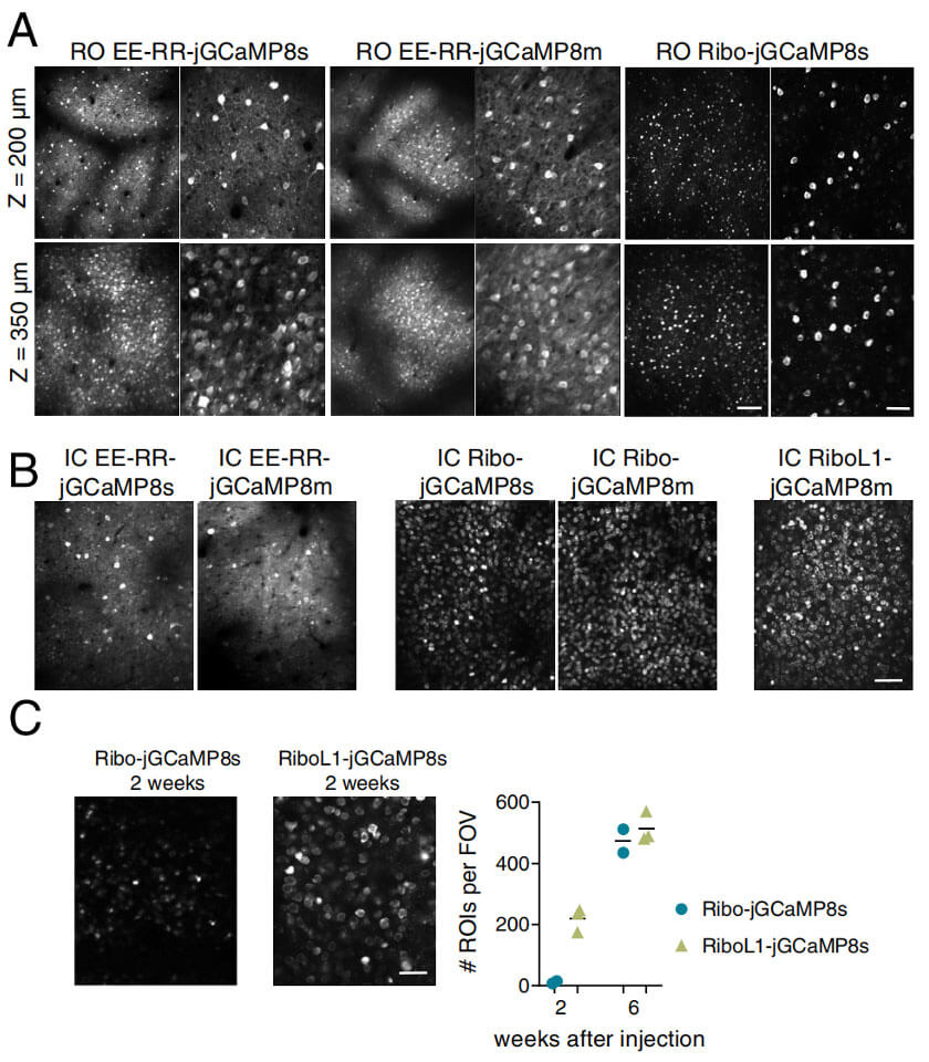
In order to improve the expression rate, the authors chose to replace the linker region of Ribo-GCaMP8 with three different linker sequences: a longer, more flexible sequence and two rigid helical linkers of RiboL1-GCaMP8. A more flexible and longer linker has the same amino acid sequence as EE-RR-GCaMP, which can greatly improve the expression rate of the protein. Strong expression in a large number of single cells could be detected two weeks after IC injection of RiboL1-GCaMP8 virus, while the fluorescent signal showed no accumulation for several weeks (Fig. 2). When RiboL1-GCaMP8 was selected for RO injection, the brightness of RiboL1-jGCaMP8 was relatively dim, similar to Ribo-jGCaMP8.
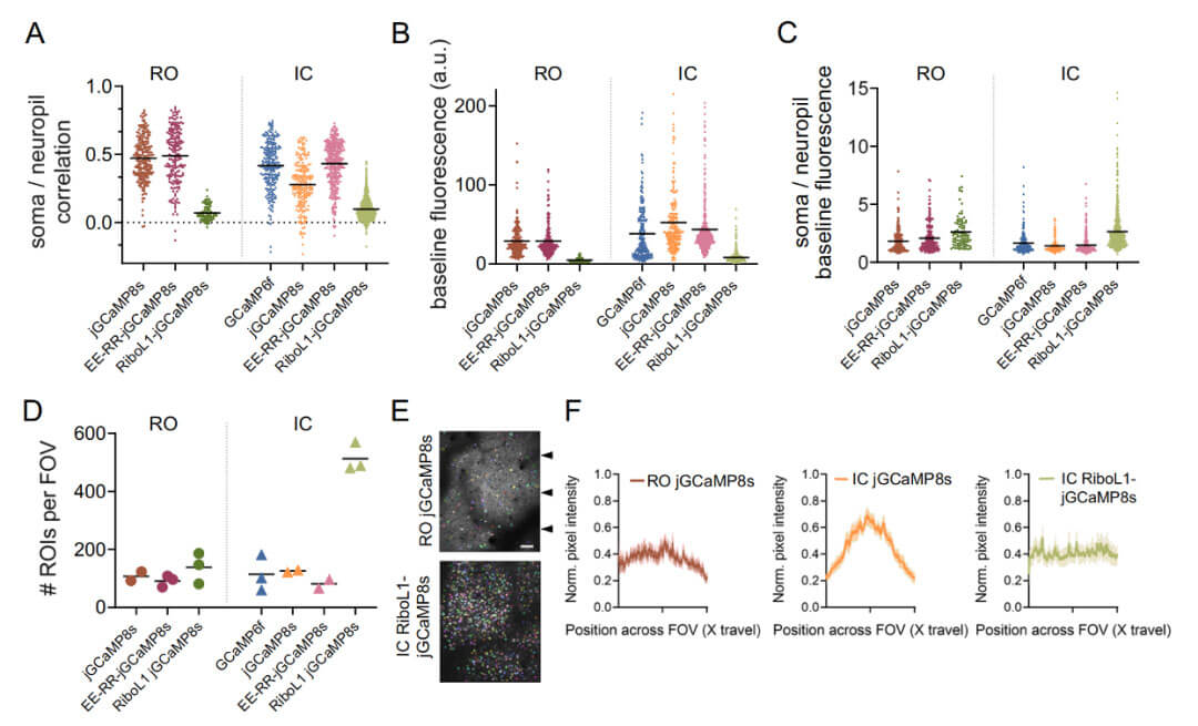
The high sensitivity and brightness of jGCaMP8 may cause neural fiber network signals, affecting the ΔF/F values used to measure neuronal activity. Further experiments found that jGCaMP8s and EE-RR-jGCaMP8s were highly correlated with neural fiber network signals, while RiboL1-jGCaMP8s had a lower correlation. In addition, during RO and IC injection, only RiboL1-jGCaMP8s had the highest ratio between somatic cells and nerve pulvinar signal brightness, the signal distribution was even, and the number of identified cells was the highest during IC injection. Compared with IC injection, jGCaMP8s and EE-RR-jGCaMP8 RO injection produced a higher ratio between somatic and pulvinar signal brightness, with no difference in cell identification number but more even signal distribution. Therefore, compared with the GECI structure, iRiboL1-jGCaMP8s can better estimate the fluctuations of Ca2+ dynamics in the cell body when choosing the IC injection method, without interference from neuropil signals.
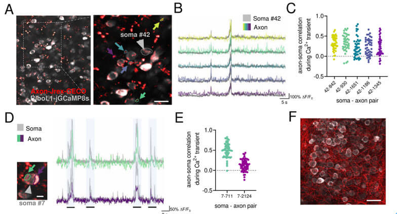
The authors further injected EE-RR-jGCaMP8 by RO and Cre-dependent hM4D receptor (DREADD) by IC in PV-Cre mice. The results showed that EE-RR-jGCaMP8 is expressed throughout the brain and mediates hM4D in PV neurons in the target brain area. Express. Therefore, RO injection is suitable for using transgenic mice to achieve whole-brain expression of GECI in specific cell types and to manipulate the activity of specific cell populations. In addition, axon-localized red GECI (hSyn-Axon-jREX-GECO1) was injected into the dorsal geniculate nucleus, and RiboL1-jGCaMP8 was injected IC into V1 (primary visual cortex). Two weeks later, the axons and cell bodies of V1 were imaged, and strong signals from two GECIs were found, enabling dual-color imaging of cell bodies and axonal activity.
The above data demonstrate that jGCaMP8 and EE-RR-jGCaMP8 are well suited for systemic delivery and produce brain-wide expression within two weeks and remain stable for months, while RiboL1-jGCaMP8 exhibits unprecedented labeling density and signal-to-noise ratio and can be used for Intracerebral viral injection.
In summary, the authors proposed a set of GECIs that can be used for systemic and local administration within the brain, showing high performance and sustainable expression over a longer period of time. The method is simple, the experiment has high flexibility and the cost is low.


WhatsApp Business Account
Address:-
Address:-


![[Research Tool Highlight] Featured in Cell! Brain Case launches new product — Enhancer AAV Vectors, a breakthrough for research Tool !](/web/allimg/250826/0R61432331JL.png)