E-mail:BD@ebraincase.com
Tel:+8618971215294
English 中文版
Virus Vector - Viral Vector Production - BrainCaseVirus Vector - Viral Vector Production - BrainCase
  • Home
  • Virus product library
    CRISPRRNAiHSV-helperRV-helperNeurophilic virusCalcium SensorsOptogenetics activationOptogenetics inhibitionChemical geneticsSparse labelingFluorescent proteinBiosensorsRecombinaseApoptosis & AutophagyDisease ModelNeurotoxicityOther
  • Products & Service

    Product Center

    Virus

    VSV-vaccine and gene therapy research
    Retrovirus-RCAS-TVA
    Lentivirus Vector-Lentivirus Production
    Rabies Virus Vector-RBV Vector
    Herpes simplex virus-Oncolytic and anterograde tracing
    PRV-retrograd multisynaptic-Peripheral
    AAV-gene therapy vectors-neuroscience

    Animal Model

    Neurological Disease Models
    Tumor animal models-anti-tumor
    Digestive System Disease Animal Model
    Cardiovascular System Disease Animal Models

    Plasmid Construction

    Library Construction
    Plasmid design and construction

    Popular Applications

    Gene Regulation

    Gene Overexpression-Brain Case
    RNA interference(RNAi)-siRNA-Brain Case
    Gene Editing - CRSIPR cloning - BrainCase

    Neural Circuit Function Research

    Optogenetics - BrainCase
    Chemical genetics-DREADDs-Brain Case
    Calcium signal recording-Gels- Brain Case
    GRAB Neurotransmitter Fluorescent Sensor- Brain Case
    Functional Magnetic Resonance Imaging Technology- Brain Case

    Research on the structure of neural circuits

    Direct Input and Output-viral vectors- Brain Case
    Anterograde Mono-synaptic Tracing -HSV- Brain Case
    Antrograde Muti-synaptic Tracing-HSV & VSV-Brain Case
    Retrograde Mono-synaptic Tracing-Rabies Virus-Brain Case
    Retrograde Muti-synaptic Tracing-PRV-Brain Case

    Featured Services

    Nervous System Disease Drug Effect

    Alzheimer's disease-AD
    Depression-mental disorders
    Parkinson's disease--PD
    Epilepsy-an ancient neurological disorder

    AAV Serotype Screening

    AAV Serotypes screening-gene therapy

    Collaboration Products

    Kiryl Piatkevich lab
    Zhifei Fu lab
    Yulong Li lab

    Efficacy of Oncolytic Virus

    Oncolytic virus-for cancer therapy-Brain Case
    Herpes Virus Vector-anti-tumor- BrainCase
    Vesicular stomatitis virus-killing tumor cells-Brain Case
  • News
    Corporate News New Product Launch Team Investor News
  • Support
    Literature interpretation Customer article Video Zone FAQs
  • About Us
    Virus product library Products & Service News Support About Us Contact
  • Contact
    Contact Us Join us
  • 中文
    English 中文版
  • Home
  • Support
  • Customer article
  • Support
  • Literature interpretation
  • Customer article
  • Video Zone
  • FAQs

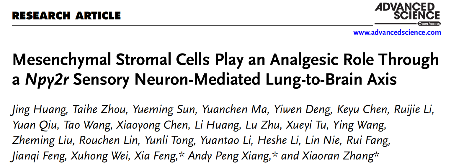
Client Publication | Adv. Sci. | Sun Yat-sen University Teams of Peng Xiang, Xiaoran Zhang, and Xia Feng Reveal Analgesic Mechanism of MSCs via a Lung–Brain Axis Activating Npy2r Sensory Neurons

Release time:2025-09-28 17:03:49
Neuropathic pain (NP), caused by injury or disease of the somatosensory nervous system, is characterized by abnormal pain perception and negative emotions. It severely impairs quality of life and often leads to anxiety and depression. Current pharmacological treatments provide insufficient relief and can have adverse effects with long-term use, while non-pharmacological interventions show variable efficacy. Developing targeted analgesics to alleviate disease remains an unmet medical need.

Mesenchymal stromal cells (MSCs), with multipotent differentiation capacity, immunomodulatory properties, and paracrine functions, can be isolated from various tissues and have shown efficacy in alleviating multiple types of pain. However, their therapeutic outcomes are influenced by many factors, and the analgesic mechanisms are not fully understood.

On August 28, 2025, a collaborative team led by Peng Xiang and Xiaoran Zhang of Sun Yat-sen University Zhongshan School of Medicine and Xia Feng of the First Affiliated Hospital of Sun Yat-sen University published a study in Advanced Science titled “Mesenchymal Stromal Cells Play an Analgesic Role Through a Npy2r Sensory Neuron-Mediated Lung-to-Brain Axis.” The study reveals that peripherally delivered MSCs activate vagal sensory neurons in the lung expressing Npy2r. These neurons project to the nucleus tractus solitarius and the ventrolateral periaqueductal gray, producing analgesic effects through a vagus nerve–mediated lung–brain pathway.
 

01. MSCs Alleviate Nerve-Injury–Induced Pain via vlPAG Neurons

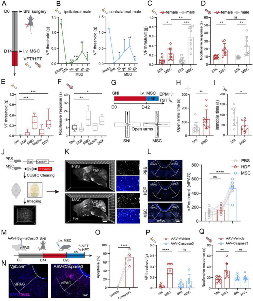
Using a spared nerve injury (SNI) mouse model, 1×10⁶ bone marrow–derived MSCs were intravenously injected two weeks after SNI. Von Frey test (VFT) results showed that, compared with the sham group, male SNI mice exhibited reduced bilateral mechanical pain thresholds. MSC administration alleviated mechanical allodynia on both sides within 1 hour and peaked at 4 hours. The analgesic effect on mechanical allodynia was stronger in males than in females, while the hot plate test (HPT) revealed similar effects on thermal pain in both sexes (Fig. 1C,D).

Compared with analgesics, human dermal fibroblasts (HDFs) produced no significant pain relief, whereas MSCs achieved significant analgesia in male SNI mice 4 hours post-injection—comparable to dexmedetomidine (DEX) and superior to aspirin (Fig. 1E,F). MSC infusion also improved anxiety-like and depression-like behaviors in SNI mice, as assessed by the elevated plus maze (EPM) and tail suspension test (TST) (Fig. 1G–I).

Using Fos^CreERT2; Loxp-tdTomato transgenic mice and whole-brain clearing, immediate early gene Fos expression was significantly activated in the ventrolateral periaqueductal gray (vlPAG), hippocampus, and other regions shortly after MSC injection (Fig. 1J,K), with vlPAG known to be involved in pain modulation. Targeted ablation of vlPAG neurons via AAV-hSyn-taCasp3 injection (Fig. 1M) induced apoptosis confirmed by TUNEL staining (Fig. 1N,O), abolishing MSC-induced analgesia (Fig. 1P,Q). Recombinant pseudorabies virus (PRV-EGFP) neural tracing further confirmed neural connections between vlPAG and the peripheral paw. Conclusion: MSCs exert analgesic effects in SNI mice through vlPAG neurons.

Figure 1. MSCs relieve nerve-injury–induced pain via vlPAG neurons.


02. MSCs Regulate vlPAG Neurons via a Lung Vagus Nerve → NTS → vlPAG Pathway

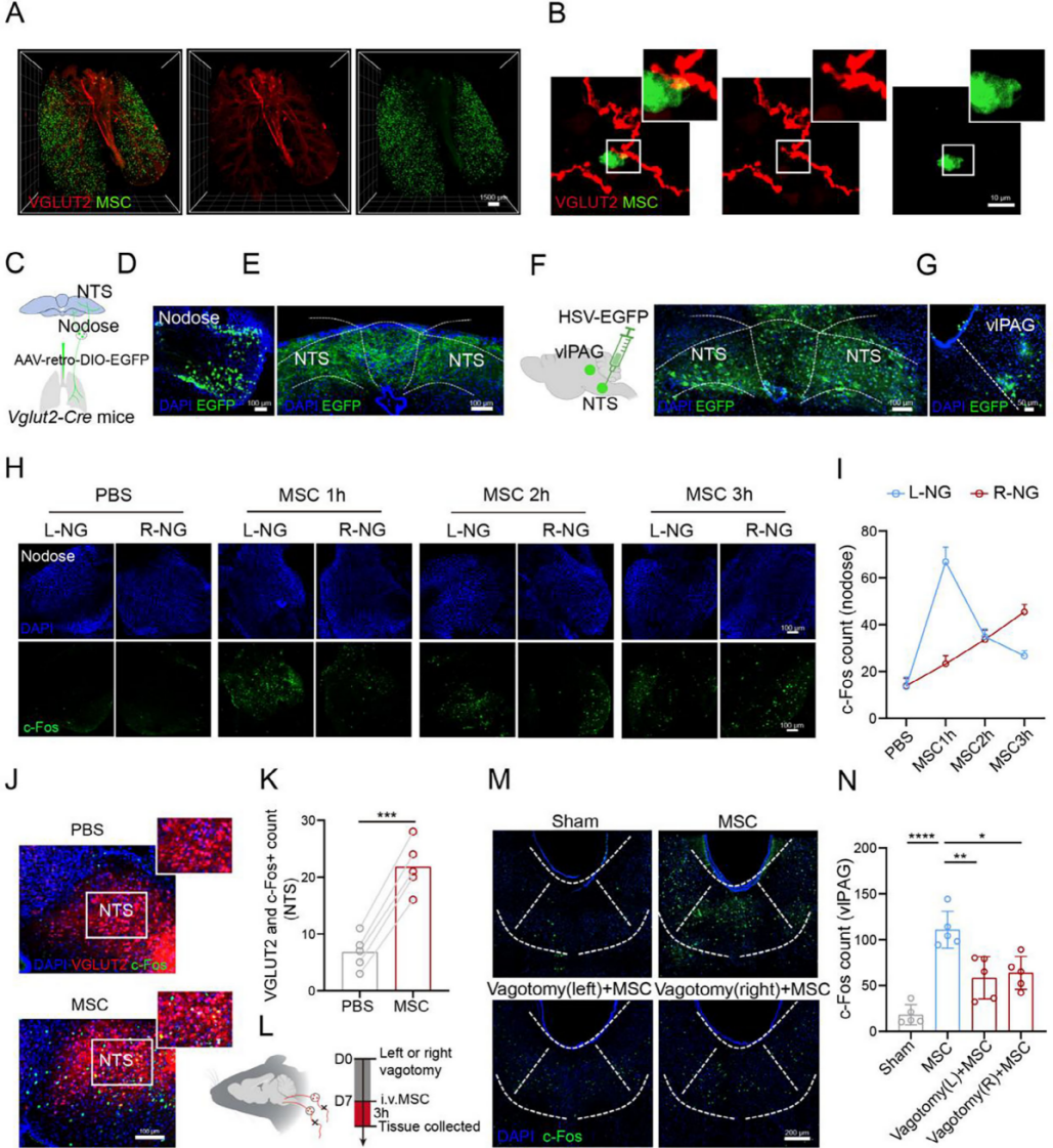
Intravenously infused MSCs predominantly localize to the lungs, which are richly innervated by vagal sensory branches. When GFP-labeled MSCs were injected into VGLUT2-Cre; Loxp-tdTomato mice, tissue clearing and imaging revealed that most VGLUT2-positive fibers were distributed along the major airways beneath the smooth muscle layer and ran parallel to it. GFP-MSCs were found in close proximity to vagal sensory nerves in the lung (Fig. 2A,B). Moreover, GFP⁺ cells in the lung expressed MSC-specific markers CD105 and CD90, indicating that the infused cells retained their MSC identity and remained viable and functionally active within the 3-day treatment window.

These findings suggest that intravenously delivered MSCs may modulate vlPAG activity through vagal afferents from the lung. To map this circuit, AAV-retro-DIO-EGFP was injected into the lungs of VGLUT2-Cre mice to label vagal neurons innervating the lung (Fig. 2C). Strong signals were observed in the nodose ganglion and the nucleus tractus solitarius (NTS) (Fig. 2D,E). Anterograde tracer HSV-EGFP was then injected into the NTS (Fig. 2F), and viral spread was detected in the vlPAG target area (Fig. 2G). After MSC injection, c-Fos signals in bilateral nodose ganglia increased in a time-dependent manner, with earlier responses on the left side (Fig. 2H,I).

In VGLUT-Cre; Loxp-GCaMP6 mice, calcium imaging showed that HDF injection triggered only sparse calcium transients in vagal sensory neurons, whereas MSC injection induced calcium responses in nearly all recorded neurons. Post-imaging analysis revealed higher c-Fos expression in VGLUT2-GCaMP6–positive sensory neurons of the MSC group compared with the HDF group. MSC injection also activated NTS neurons, with increased co-localization of c-Fos and VGLUT2, indicating activation of glutamatergic neurons in the NTS (Fig. 2J,K).

Pre-injection vagotomy (Fig. 2L) partially suppressed MSC-induced activation of both NTS and vlPAG neurons (Fig. 2M,N). Conclusion: MSCs likely transmit signals to the vlPAG through a lung vagus nerve–brain axis involving the NTS.

Figure 2. MSCs Regulate vlPAG Neurons via the Lung Vagus Nerve → NTS → vlPAG Pathway


03. Pulmonary Npy2r Sensory Neurons Exert Analgesic Effects

Afferent sensory ganglia contain diverse neuron subtypes with distinct molecular profiles. Vagal sensory neurons are the primary fibers innervating the lung and airways. Studies revealed that P2ry1 and Npy2r vagal sensory neurons are associated with different lung–brain connections: P2ry1 neurons are fast-conducting A-fibers projecting to the lateral NTS, whereas Npy2r neurons target the medial–posterior NTS region receiving lung C-fiber input.

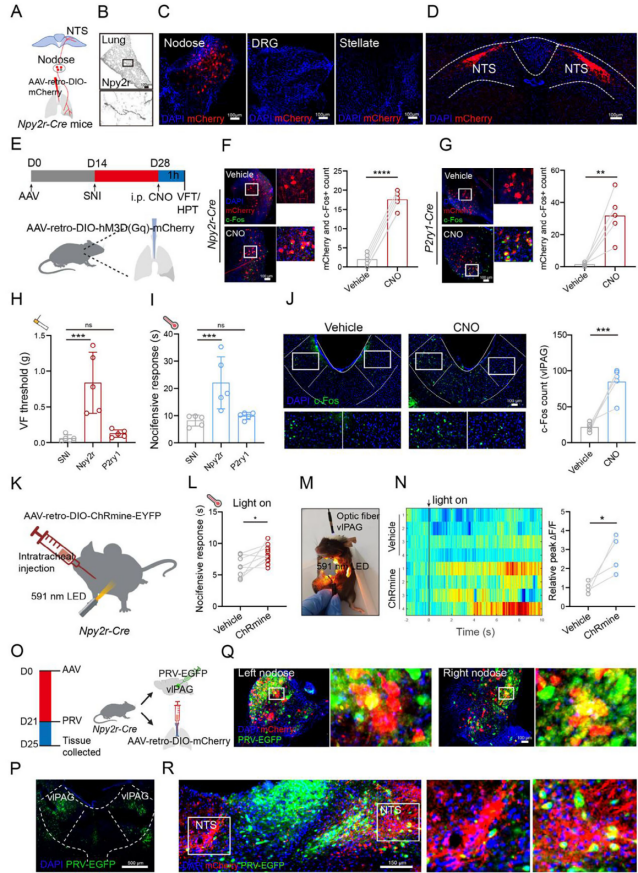
To identify lung-connected Npy2r and P2ry1 sensory neurons, AAV-retro-DIO-mCherry was injected into the lungs of Npy2r-Cre mice (Fig. 3A,B), showing infection signals only in the nodose ganglion and NTS (Fig. 3C,D). To clarify their analgesic role, chemogenetic activation was performed by injecting AAV-retro-DIO-hM3Dq-mCherry into the lungs of Npy2r-Cre mice (Fig. 3E). After CNO administration, c-Fos expression increased in mCherry⁺ neurons of the nodose ganglion (Fig. 3F). Similarly, activating P2ry1 sensory neurons via mixed AAV injection (AAV-retro-DIO-hM3Dq-mCherry and AAV-retro-P2ry1-Cre) in wild-type mice also increased c-Fos in nodose ganglion neurons after CNO injection (Fig. 3G).

Selective activation of Npy2r sensory neurons alleviated SNI-induced mechanical allodynia and thermal hyperalgesia (Fig. 3H,I) in a CNO dose-dependent manner and increased c-Fos expression in the vlPAG (Fig. 3J). In Npy2r-Cre mice injected with AAV-retro-DIO-ChRmine-EYFP (Fig. 3K), 591 nm LED chest illumination for 1 minute improved SNI-induced thermal hyperalgesia (Fig. 3L). Fiber photometry showed increased calcium signals and firing rates in vlPAG neurons upon stimulation of lung-innervating Npy2r vagal sensory neurons (Fig. 3M,N), indicating that selective activation of these neurons produces analgesia and establishes a potential lung–brain pain-modulation circuit.

To confirm whether lung-innervating Npy2r vagal sensory neurons project to the vlPAG, a dual-virus strategy was used: on day 0, AAV-retro-DIO-mCherry was injected into the lungs of Npy2r-Cre mice, followed by PRV-EGFP injection into the vlPAG on day 21 (Fig. 3P). After 96 hours of infection, overlapping red and green fluorescence was observed in the nodose ganglion and NTS (Fig. 3Q,R), suggesting projections to the vlPAG. Similar results were obtained in wild-type mice co-injected with cholera toxin B and PRV-EGFP. Conclusion: Npy2r sensory neurons drive analgesia through a lung–brain pathway.

Figure 3. Pulmonary Npy2r sensory neurons mediate analgesic effects.

04. MSCs Activate Npy2r Sensory Neurons to Exert Analgesic Effects

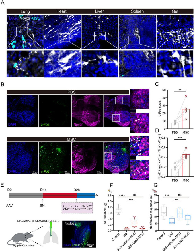
Since pulmonary Npy2r vagal sensory neurons project to the vlPAG, it was hypothesized that MSCs might induce analgesia by activating these neurons. Examination of GFP-MSC distribution alongside Npy2r sensory neurons in the lung, heart, liver, spleen, and intestine revealed co-localization only in the lung, suggesting a potential interaction (Fig. 4A). In Npy2r-Cre; Loxp-tdTomato mouse lungs, co-staining with the neuronal marker TUBB3 confirmed Npy2r expression within pulmonary nerves.

MSC infusion increased c-Fos expression in Npy2r sensory neurons of the nodose ganglion (Fig. 4B–D), indicating possible activation of these lung-innervating vagal sensory neurons. To test their functional role, AAV-retro-DIO-hM4D(Gi)-EGFP was injected into the lungs of Npy2r-Cre mice to silence pulmonary Npy2r sensory neurons (Fig. 4E). Von Frey testing showed that MSCs elevated mechanical pain thresholds in SNI mice in the control group, whereas hM4Di-mediated silencing of Npy2r sensory neurons abolished this analgesic effect (Fig. 4F). Similarly, hot plate tests revealed that suppression of Npy2r sensory neurons eliminated MSC-induced relief of thermal hyperalgesia (Fig. 4G). Conclusion: Activation of lung-innervating Npy2r sensory neurons is required for MSCs to exert analgesic effects.

Figure 4. MSCs activate Npy2r sensory neurons to mediate analgesia.

05. Peripheral MSCs Enhance Calcium Signaling in Pulmonary Npy2r Sensory Neurons

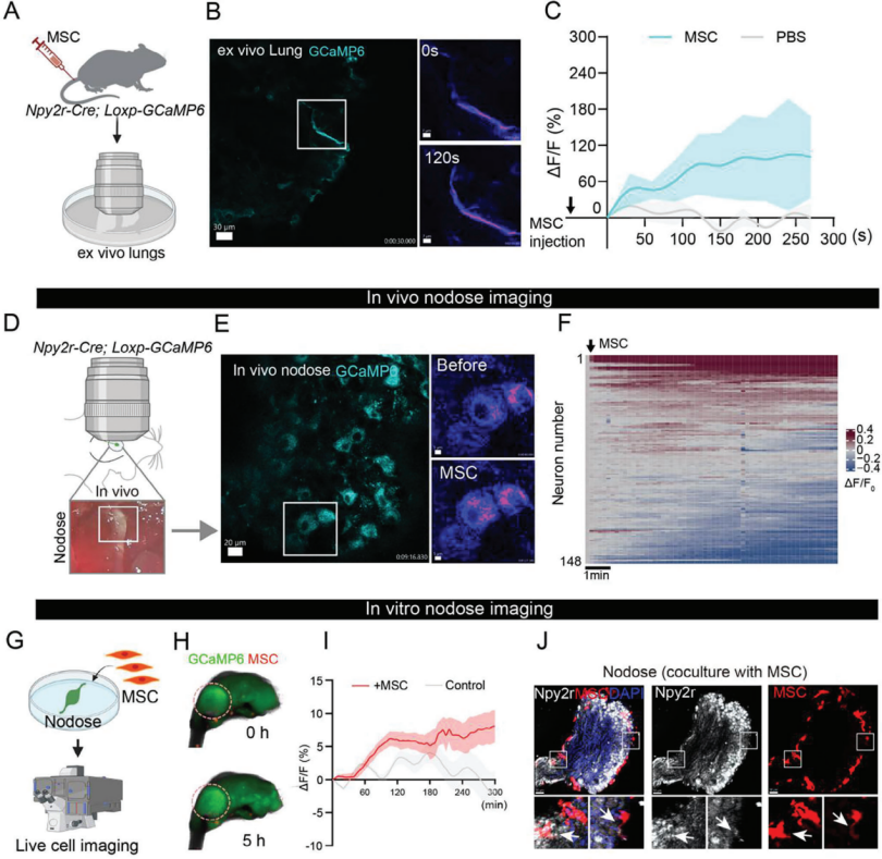
To investigate the effect of MSC infusion on pulmonary Npy2r sensory fibers, Npy2r-Cre; Loxp-GCaMP6 mice received intravenous MSCs, and the activity of Npy2r sensory fibers in freshly isolated lungs was recorded (Fig. 5A). GCaMP6 fluorescence intensity showed that MSCs stimulated Npy2r sensory fibers compared with the PBS group (Fig. 5B,C).

In vivo vagal ganglion calcium imaging was performed to examine the response of Npy2r vagal sensory neurons to MSC injection while maintaining lung connections (Fig. 5D). MSC administration induced calcium transients in most Npy2r vagal sensory neurons (Fig. 5E,F). Furthermore, when AAV-retro-DIO-GCaMP6 was injected into the lungs of Npy2r-Cre mice, calcium imaging of the nodose ganglion confirmed that the majority of lung-innervating neurons responded to MSC infusion.

To further validate this interaction, nodose ganglia were collected from Npy2r-Cre; Loxp-GCaMP6 mice and co-cultured with RFP-labeled MSCs for several hours (Fig. 5G). In vitro, MSCs elicited a mild but sustained increase in neuronal calcium activity lasting at least 5 hours (Fig. 5H,I). Immunofluorescence analysis of the co-cultured nodose ganglia showed MSCs positioned nearby (Fig. 5J). Conclusion: Intravenously delivered MSCs enhance calcium signaling in lung-innervating Npy2r vagal sensory neurons.

Figure 5. Peripheral MSCs enhance calcium signaling in pulmonary Npy2r sensory neurons.
 

06. MSCs Activate Npy2r Sensory Neurons via ATP Signaling

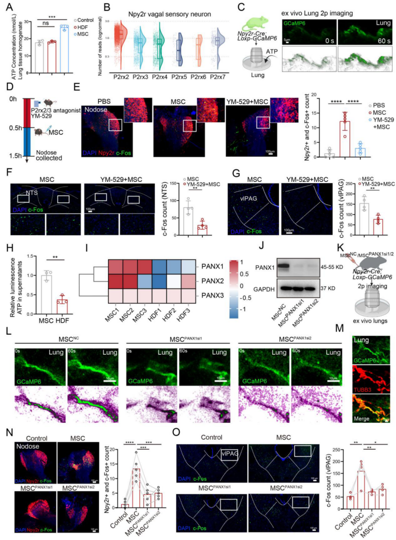
Previous studies have shown that extracellular ATP can stimulate pulmonary vagal C-fibers through P2rx2/3 receptors at vagal sensory nerve terminals. In this study, ATP levels in lung tissue homogenates were higher in MSC-injected mice than in HDF controls (Fig. 6A), suggesting that MSCs may activate pulmonary Npy2r sensory neurons through ATP signaling. Reanalysis of nodose ganglion single-cell RNA-seq data revealed that Npy2r sensory neurons express higher levels of P2rx2 and P2rx3 than other ligand-gated ionotropic purinergic receptors (Fig. 6B). In Npy2r-Cre; Loxp-GCaMP6 mice, ATP application further increased fluorescence intensity of pulmonary Npy2r sensory neurons (Fig. 6C).

Using the P2rx2/3 receptor antagonist minodronic acid (YM-529), it was confirmed that aerosol inhalation of YM-529 before MSC infusion blocked MSC-induced activation of Npy2r neurons in the nodose ganglion (Fig. 6E) and reduced MSC-induced c-Fos expression in the NTS (Fig. 6F) and vlPAG (Fig. 6G). These findings indicate that MSCs partially activate Npy2r vagal sensory neurons through ATP signaling, positively modulating the central nervous system.

ATP is known to be released via vesicles or channel proteins such as pannexin 1 (PANX1). Quinacrine staining combined with flow cytometry showed no significant difference in the number of ATP-containing vesicles between MSC and HDF supernatants, but ATP levels were higher in MSC supernatant (Fig. 6H). Sequencing analysis further revealed higher PANX1 expression in MSCs compared with HDFs (Fig. 6I). Knockdown of PANX1 in MSCs (MSC PANX1si1 and MSC PANX1si2) (Fig. 6J) markedly reduced the response of Npy2r sensory fibers in freshly isolated lungs of Npy2r-Cre; Loxp-GCaMP6 mice compared with MSC NC controls (Fig. 6L). In Npy2r-Cre; Loxp-GCaMP6 mouse lungs, TUBB3 co-staining confirmed Npy2r-GCaMP6 expression in pulmonary nerves (Fig. 6M). Immunofluorescence analysis further demonstrated that PANX1-knockdown MSCs failed to activate Npy2r vagal sensory neurons in the nodose ganglion (Fig. 6N), with a similar decrease in c-Fos expression in the vlPAG (Fig. 6O). Conclusion: MSCs activate Npy2r sensory neurons and exert analgesic effects through ATP signaling mediated in part by PANX1-dependent ATP release.

Figure 6. MSCs activate Npy2r sensory neurons via ATP signaling.
 

07. ATPγS Activation of Npy2r Sensory Neurons Reduces Pain Responses

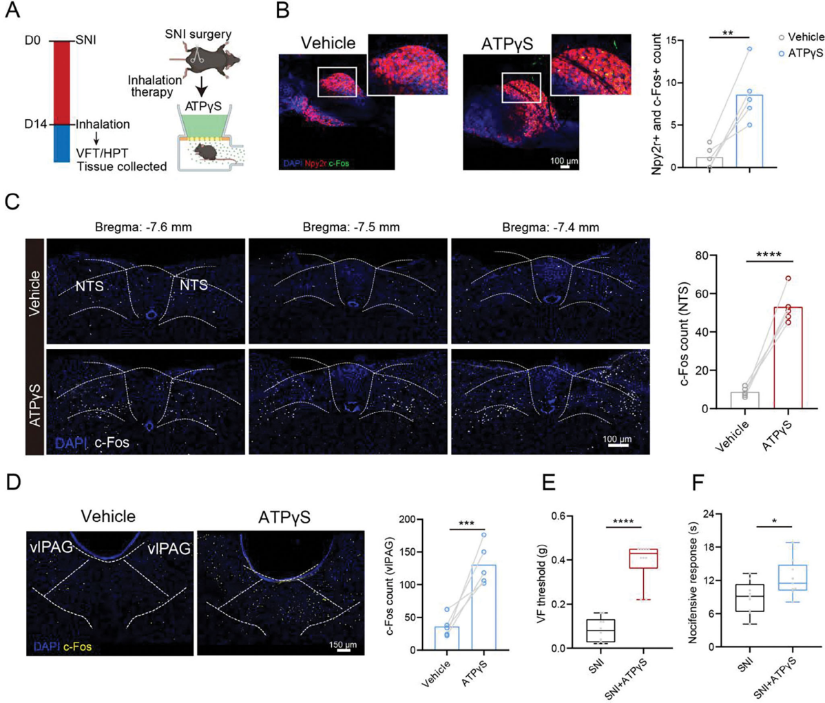
To explore a more convenient method for pathway activation, the study tested whether direct inhalation of adenosine 5′-O-(3-thiotriphosphate) trilithium salt (ATPγS, a P2rx2 agonist) could activate the lung–brain axis associated with Npy2r sensory neurons and produce analgesic effects in SNI mice.

As shown in Fig. 7A, SNI mice were placed in a sealed chamber and exposed to aerosolized PBS or ATPγS. Inhalation of ATPγS activated Npy2r vagal sensory neurons in the nodose ganglion (Fig. 7B) and induced neuronal activation in the NTS (Fig. 7C) and vlPAG (Fig. 7D).

Importantly, ATPγS inhalation significantly alleviated SNI-induced mechanical allodynia and thermal hyperalgesia (Fig. 7E,F). Dose–response testing revealed that 10 mg/kg inhalation markedly increased mechanical and thermal pain thresholds, while 15 mg/kg offered no additional benefit. Serum IL-6 levels remained unchanged after 10 mg/kg ATPγS administration, indicating no significant systemic inflammation.

In untreated mice, ATPγS inhalation also elevated thermal pain thresholds in the hot-plate test and enhanced c-Fos expression in the vlPAG, confirming its analgesic potential. Conclusion: Direct inhalation of a P2rx2 agonist produces significant analgesic effects under various conditions.

Figure 7. ATPγS activates Npy2r sensory neurons to reduce pain responses.

Summary

This study reveals that mesenchymal stromal cells (MSCs) activate Npy2r sensory neurons innervating the lung through the lung vagus nerve → nucleus of the solitary tract (NTS) → ventrolateral periaqueductal gray (vlPAG) pathway, a process dependent on ATP signaling. Moreover, direct inhalation of a P2rx2 agonist effectively alleviates pain, offering a novel therapeutic target and a convenient potential intervention for the treatment of chronic pain, with significant clinical translational value.

The viral vectors used in this study are all available from Brain Case Biotech
(bd@ebraincase.com)


Product Information
Product No. Product Name
BC-0653 rAAV-hSyn-DIO-taCasp3-T2A-TEVp
BC-0015 rAAV2-retro-DIO-EGFP
BC-0016 rAAV2-retro-DIO-mCherry
BC-0623 rAAV2-retro-DIO-hM4D(Gi)-EGFP
BC-0143 rAAV2-retro-DIO-hM3D(Gq)-mCherry
BC-0238 rAAV-retro-DIO-GCaMP6s
BC-0077 rAAV-hSyn-GCaMP6s
BC-PRV-801 PRV-CAG-3Gc
BC-HSV-HBEGFP H129-hUbC-HBEGFP

Service Type :

Select the service you'd like to purchase.

Order Information(Premade-AAVs)

Please provide us some information about the service you'd like to order.

How did you hear about BrainCase Biotech or our products?

Detailed requirements:

scroll

Order Information(Custom AAV/Lentivirus)

Please provide us some information about the service you'd like to order.

Gene ID or gene information:

Selection of the reporting gene:

Special Instructions:

scroll

Order Information(Others)

Please provide us some information about the service you'd like to order.

Virus name/Detailed requirements:

scroll

Related products

Client Publication | Adv. Sci. | Sun Yat-sen University Teams of Peng Xiang, Xiaoran Zhang, and Xia Feng Reveal Analgesic Mechanism of MSCs via a Lung–Brain Axis Activating Npy2r Sensory Neurons

Client Publication | Adv. Sci. | Sun Yat-sen University Teams of Peng Xiang, Xiaoran Zhang, and Xia Feng Reveal Analgesic Mechanism of MSCs via a Lung–Brain Axis Activating Npy2r Sensory Neurons

Mol. Psychiatry | Team Led by Miao He and Ling Gong Reveals Independent Circuits and Behavioral Regulation Mechanisms of Two Neuronal Subtypes in the Medial Mammillary Body

Mol. Psychiatry | Team Led by Miao He and Ling Gong Reveals Independent Circuits and Behavioral Regulation Mechanisms of Two Neuronal Subtypes in the Medial Mammillary Body

Client Article | Cell Metabolism | Liu Danqian’s Team at the Institute of Neuroscience, CAS, Decodes the Oxidation-Sleep Code in Substantia Nigra Neurons

Client Article | Cell Metabolism | Liu Danqian’s Team at the Institute of Neuroscience, CAS, Decodes the Oxidation-Sleep Code in Substantia Nigra Neurons

Client Publication | Neuron | Liping Wang and Xuemei Liu’s Team at SIAT Uncovers Neural Mechanisms Behind Individual Differences in Innate Fear Responses

Client Publication | Neuron | Liping Wang and Xuemei Liu’s Team at SIAT Uncovers Neural Mechanisms Behind Individual Differences in Innate Fear Responses

map
{dede:global.cfg_webname/}

Virus product library

CRISPR
RNAi
Neurophilic virus
Optogenetics activation
Biosensors

News

Corporate News
New Product Launch
Team
Investor News

Support

Literature interpretation
Customer article
Video Zone
FAQs
微信

WhatsApp Business Account

Tel: +8618971215294
E-mail: BD@ebraincase.com

Address:-

Address:-

  • Copyright © 2024 Brain Case All Rights Reserved.