Client Article | Cell | High-Resolution Whole-Body Imaging of the Mouse Nervous System by the Guoqiang Bi and Beiming Liu Teams
Release time:2025-09-19 15:23:31
Mesoscale connectomics of the central nervous systemhas seen significant advances at subcellular resolution. However, the peripheral nervous systempresents substantial challenges due to the body’s large size and structural heterogeneity. Current whole-body tissue clearing techniques combined with traditional light-sheet microscopy struggle to resolve fine nerve branches and single-axon fibers evenly throughout the mouse body. Meanwhile, block-face imaging using confocal microscopy is too slow to meet the demands of high-resolution imaging across an entire adult mouse.
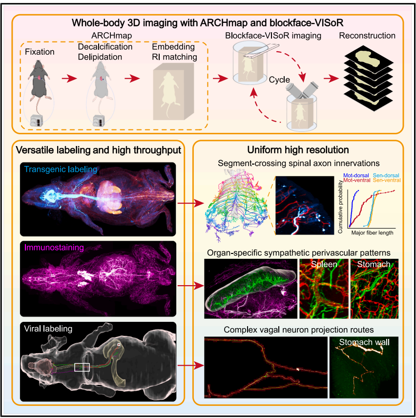
On July 10, 2025, Professor Guoqiang Bi, Associate Researcher Cheng Xu, Professor Beiming Liu, and Senior Engineer Qingyuan Zhu from the University of Science and Technology of China published a groundbreaking study in the prestigious journal Cell, titled “High-speed mapping of whole-mouse peripheral nerves at subcellular resolution.” In this work, the team introduced a novel high-speed, whole-body 3D imaging technique with subcellular resolution, termed blockface-VISoR. This approach enabled comprehensive, high-resolution 3D reconstruction of the entire peripheral nervous system in mice—resulting in an unprecedentedly detailed peripheral nerve atlas. By extending connectomics beyond the brain, the study provides a powerful new tool for investigating peripheral neural circuits and disease mechanisms.
The researchers developed the blockface-VISoR imaging system, which integrates a precision vibratome, along with a sample preparation pipeline called ARCHmap that includes whole-body tissue clearing and hydrogel embedding of the mouse. The key innovation lies in the imaging strategy: only the uppermost ~600 μm of the tissue block is imaged in 3D at a time, followed by automatic removal of a 400 μm thick slice. This process is repeated iteratively until the entire sample is imaged. Then, automated algorithms stitch the overlapping ~200 μm zones between adjacent slices into a seamless 3D reconstruction. Since each scanned depth is only a few hundred microns and tissue clearing minimizes light scattering, this method achieves consistently high resolution.
Using this optimized workflow, the team completed uniform whole-body subcellular-resolution imaging of an adult mouse within 40 hours, acquiring approximately 70 TB of raw image data per channel. To date, the team has imaged dozens of mice, amassing over 4 PB of data in total.
Thanks to the excellent fluorescence preservation of the ARCHmap protocol, the blockface-VISoR system is compatible with commonly used fluorescent proteins delivered via transgenic techniques or neurotropic viruses, as well as immunofluorescent labeling. Combining these labeling methods with the advanced imaging platform, the researchers successfully mapped the fine structure and single-fiber projection paths of various types of peripheral nerves throughout the mouse body. They revealed—for the first time—cross-segmental projections from individual spinal neurons, organ-specific perivascular distribution patterns of the sympathetic nervous system, and the overall projection architecture and intricate single-fiber paths of the vagus nerve.
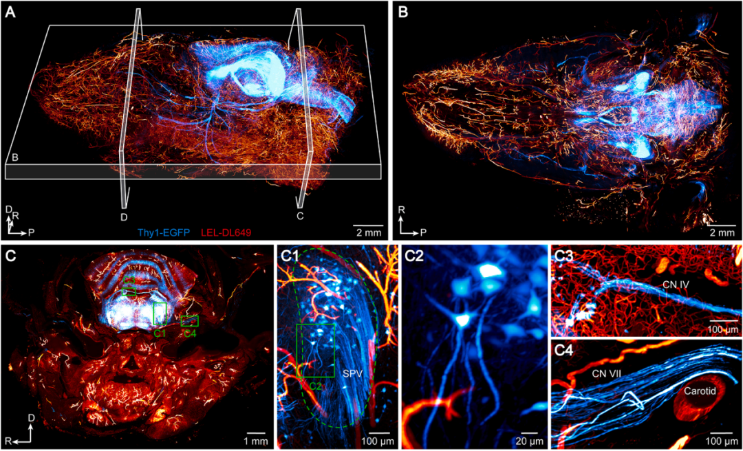
Cranial Nerve Visualization
In Thy1-EGFP mice, 3D reconstruction clearly reveals the fine structures of various cranial nerves, including the trochlear nerve (CN IV), facial nerve (CN VII), and trigeminal nerve (CN V). The motor branch of the trigeminal nerve was observed to innervate head and facial muscles through extensive claw-like neuromuscular junctions, while a single axon bundle from the sensory branch innervates the periodontal tissue of the incisors and molars.
Figure 1. Visualization of Cranial Nerves
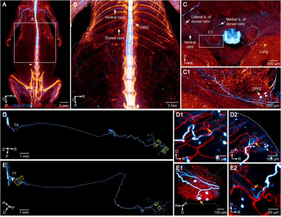
Spinal Nerve Reconstruction
Long projections (~8 cm) of thoracic motor neurons (within the spinal cord) and dorsal root ganglionsensory neurons were traced, revealing both neuromuscular junctions and sensory nerve endings. A total of 191 thoracic spinal neurons (T2–T13) expressing EGFP were reconstructed from two Thy1-EGFP mice, including 66 motor and 66 sensory neurons from the ventral rami, and 30 motor and 29 sensory neurons from the dorsal rami. These findings highlight segment-specific innervation patterns among different neuron types.
Figure 2. Visualization of Individual Thoracic Spinal Sensory and Motor Neurons
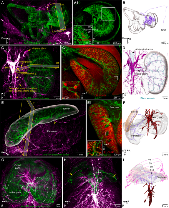
Sympathetic Nervous System Imaging
Using an anti-tyrosine hydroxylaseantibody, widespread labeling of the sympathetic nervous system was achieved, revealing the sympathetic chain, nerve plexuses, and fine branches connecting to ganglia throughout the body. A perivascular innervation pattern was observed in skeletal structures of the lumbar and sacral regions as well as leg muscles. Similar patterns were found in visceral organs such as the kidneys and spleen. In contrast, in the stomach and intestines, the sympathetic nerves formed a mesh-like structure that did not overlap with blood vessels.
Figure 3. Visualization of Sympathetic Innervation of Organs
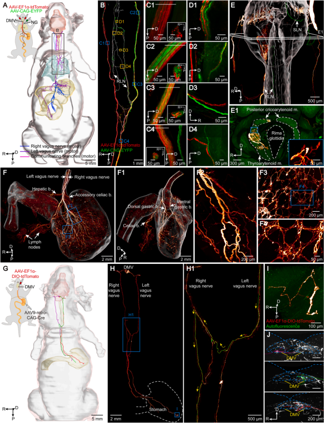
Vagus Nerve Visualization
Using viral labeling, both motor and sensory components of the vagus nerve and their projection paths to organs such as the pharynx, larynx, and esophagus were traced. Multiple interconnections between the left and right recurrent laryngeal nerves (RLNs) and the vagus nerve trunk were observed. Notably, fibers from the left RLN were found to cross over and innervate the larynx on the right side—anatomical feature not previously described. Sparse labeling revealed that individual motor and sensory neurons of the vagus nerve do not branch before entering their target organs, exhibiting a one-to-one innervation pattern, though their projection paths are complex.
Figure 4. Visualization and Single-Neuron Tracing of the Vagus Nerve via Viral Labeling
All viral vectors used in this study are available from Brain Case Biotech. For packaging or customization services, please contact bd@ebraincase.com