Client Publication | Cell | Jun Yan, Chun Xu, Zhiming Shen, and Xiaoquan Yang Collaboratively Map Whole-Brain Projections of Prefrontal Cortex Neurons in Macaques
Release time:2025-09-19 14:27:23
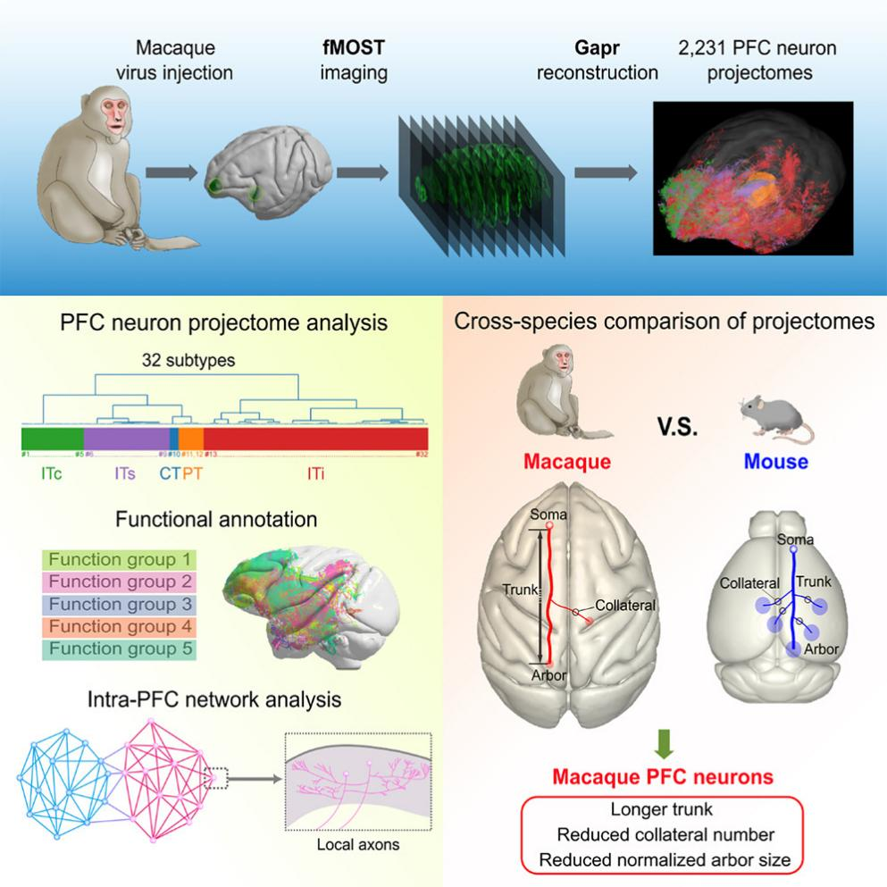
The prefrontal cortex (PFC), positioned at the highest level of the cortical hierarchy, is responsible for integrating sensory input with memory and executing cognitive control. Previous studies have mapped interareal connections in the macaque cortex using anterograde and retrograde dye tracing, as well as bulk-tracing techniques. However, these approaches lack resolution in analyzing individual axonal branches and arborization patterns. This study aims to reconstruct whole-brain projectomes of single neurons in the macaque PFC to uncover the neural circuit mechanisms underlying primate-specific cognitive functions and to compare them with those in mice, in order to explore both conserved and divergent features.
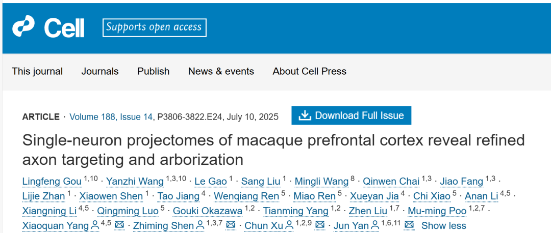
On July 10, 2025, Jun Yan, Chun Xu, and Zhiming Shen from the Center for Excellence in Brain Science and Intelligence Technology, Chinese Academy of Sciences, in collaboration with Professor Xiaoquan Yang from the Suzhou Institute of Brain Spatial Information at Huazhong University of Science and Technology, published a research article in the top-tier journal Cell, titled “Single-neuron projectomes of macaque prefrontal cortex reveal refined axon targeting and arborization.” The study presents a whole-brain projection atlas of prefrontal cortex neurons in macaques.
Neurons were sparsely labeled at 19 sites within the prefrontal cortex (PFC) of 7 adult female macaques, using MRI-guided viral injections. Whole-brain axonal projection data were acquired at a spatial resolution of 0.65 × 0.65 × 3 μm³ using the LV-fMOST system—an independently developed large-volume, submicron-resolution, continuous 3D imaging technology by the Suzhou Institute of Brain Spatial Information, Huazhong University of Science and Technology. A single-channel dataset of a typical macaque brain reached a data volume of 432 TB.
Single-neuron tracing was performed using the Gapr system, a neuron reconstruction software developed by Jun Yan’s research group. Over 50 annotators worked for 12 weeks (approximately 7,200 person-hours) to reconstruct 576 single-neuron projectomes from dataset G97-1, with a total axonal length of 94,985 mm. The reconstruction results were consistent with fMRI and bulk-tracing data, indicating that the sampling coverage was sufficient to represent the overall axonal projection patterns.
In this study, single-neuron labeling was performed via AAV viral injections. After the animals were anesthetized and secured, the skull was exposed, and the craniotomy site was determined based on MRI images. Targeted brain regions were then injected using stereotaxic injection techniques.
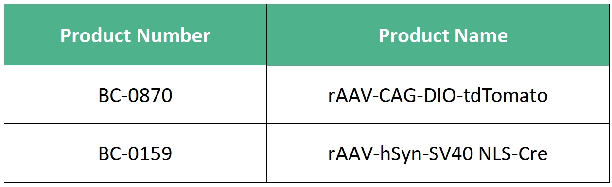
Viruses carrying fluorescent protein markers—either AAV-CAG-DIO-EGFP (titer: 4.57 × 10¹² vg/mL) or rAAV-CAG-DIO-tdTomato (titer: 1.10 × 10¹³ vg/mL)—were mixed at a 1:1 ratio with a Cre-expressing virus (rAAV-hSyn-SV40 NLS-Cre, original titer: 1.05 × 10¹³ vg/mL). The final dilution factor of the viral mixture ranged from 40,000 to 80,000 times. The final injection volume per site ranged from 0.2 to 2 μL. A microinfusion pump was used to deliver the virus precisely to the target locations.
All the viral vectors used in this study were provided by Brain Case Biotech.