E-mail:BD@ebraincase.com
Tel:+8618971215294
English 中文版
Virus Vector - Viral Vector Production - BrainCaseVirus Vector - Viral Vector Production - BrainCase
  • Home
  • Virus product library
    CRISPRRNAiHSV-helperRV-helperNeurophilic virusCalcium SensorsOptogenetics activationOptogenetics inhibitionChemical geneticsSparse labelingFluorescent proteinBiosensorsRecombinaseApoptosis & AutophagyDisease ModelNeurotoxicityOther
  • Products & Service

    Product Center

    Virus

    VSV-circuit research-vaccine and gene therapy research-BrainCase
    Retrovirus-RCAS-TVA-BrainCase
    Lentivirus Vector - Lentivirus Production - BrainCase
    Rabies Virus Vector - RBV Vector - BrainCase
    Herpes simplex virus-Oncolytic and anterograde tracing-Brain Case
    PRV-retrograd multisynaptic-Peripheral-Btain Case
    AAV-gene therapy vectors-BrainCase

    Animal Model

    Neurological Disease Models-BrainCase
    Tumor animal models-anti-tumor-BrainCase
    Digestive System Disease Animal Model-Brain Case
    Cardiovascular System Disease Animal Models-Brain Case

    Plasmid Construction

    Library Construction
    Plasmid design and construction

    Popular Applications

    Gene Regulation

    Gene Overexpression-Brain Case
    RNA interference(RNAi)-siRNA-Brain Case
    Gene Editing - CRSIPR cloning - BrainCase

    Neural Circuit Function Research

    Optogenetics - BrainCase
    Chemical genetics-DREADDs-Brain Case
    Calcium signal recording-Gels- Brain Case
    GRAB Neurotransmitter Fluorescent Probe- Brain Case
    Functional Magnetic Resonance Imaging Technology- Brain Case

    Research on the structure of neural circuits

    Direct Input and Output-viral vectors- Brain Case
    Anterograde Mono-synaptic Tracing -HSV- Brain Case
    Antrograde Muti-synaptic Tracing-HSV & VSV-Brain Case
    Retrograde Mono-synaptic Tracing-Rabies Virus-Brain Case
    Retrograde Muti-synaptic Tracing-PRV-Brain Case

    Featured Services

    Nervous System Disease Drug Effect

    Alzheimer's disease-AD-Brain Case
    Depression-mental disorders-Brain Case
    Parkinson's disease--PD-Brain Case
    Epilepsy-an ancient neurological disorder-Brain Case

    AAV Serotype Screening

    AAV Serotypes screening-gene therapy-Brain Case

    Tumorigenicity Test

    Tumorigenicity Testing-Evaluation of tumor models-Brain Case

    Efficacy of Oncolytic Virus

    Oncolytic virus-for cancer therapy-Brain Case
    Herpes Virus Vector-anti-tumor- BrainCase
    Vesicular stomatitis virus-killing tumor cells-Brain Case
  • News
    Corporate News New Product Launch Media Activity Investor News
  • Support
    Literature interpretation Customer article Video Zone FAQs
  • About Us
  • Contact
    Contact UsJoin us
  • 中文
    English 中文版
  • Home
  • Support
  • Customer article
  • Support
  • Literature interpretation
  • Customer article
  • Video Zone
  • FAQs

Customer Article|Cortical regulation of two-stage rapid eye movement sleep

Release time:2024-10-15 17:23:19

The sleep–wake cycle, including rapid eye movement (REM) sleep and non-rapid eye movement (NREM) sleep, is generally considered to be primarily regulated by distributed subcortical circuits, whereas cortical regions are involved in generating sleep-related electroencephalogram (EEG) oscillations. However, recent studies showed that cortical neurons could be actively involved in NREM sleep generation and homeostatic sleep regulation. Although cortical activation during REM sleep is considered to be associated with vivid dreaming, the role for the cortex in REM sleep regulation remains unknown. The transition of activity patterns temporally matched the transition of two sequential REM substages, identified by contrasting facial movement and distinguishable EEG theta oscillation. Closed-loop optogenetic inactivation of excitatory neurons in RSC during REM sleep shortened REM sleep duration, via inhibition of the REM substage transition. By contrast, inactivation of other cortical areas—the secondary motor cortex (M2) or the anterior cingulate cortex (ACC)—had no effect on REM sleep duration. These results uncover two distinct substages in REM sleep and highlight a role for RSC in regulating REM substage transition.

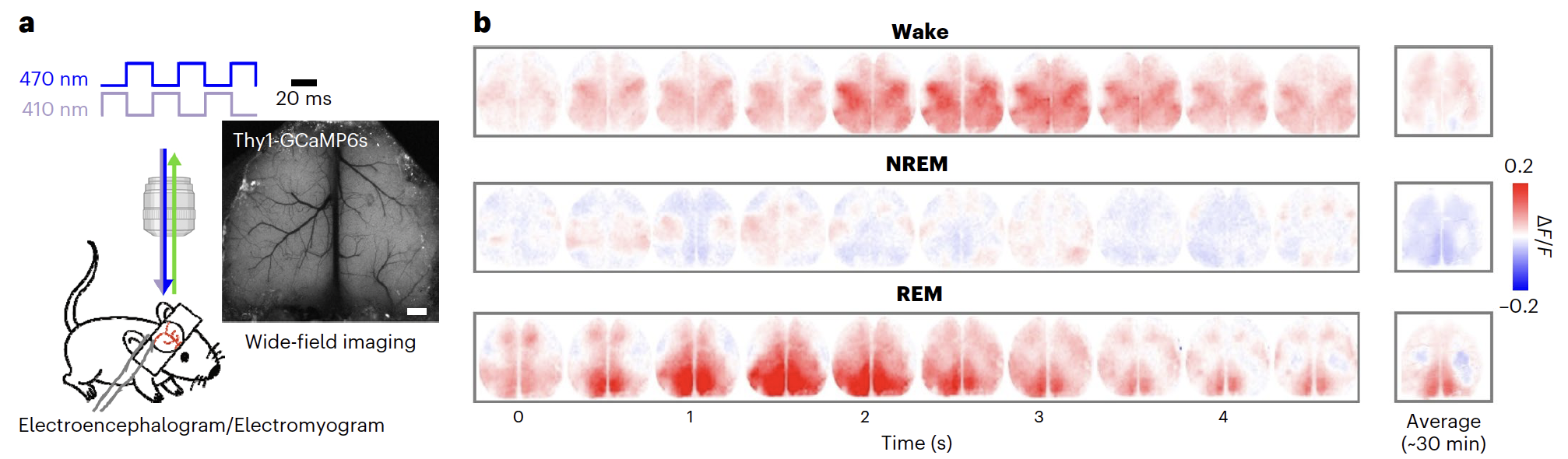
Fig 1. Researchers implanted a chronic transparent window covering the entire dorsal cortex of the Thy1-GCaMP6s mice and recorded Ca2+signals together with EEG, electromyogram (EMG) and video recording. They found that cortical activity during REM sleep was higher than that during other sleep states, with posterior and medial cortical regions more active, these results indicate that the RSC was selectively activated during REM sleep.

Analysis of mouse facial movements reveals two distinct substages of REM sleep

By inspecting mouse facial characteristics during REM sleep, we found that the eye, whisker and cheek movements also exhibited two distinguishable patterns
 

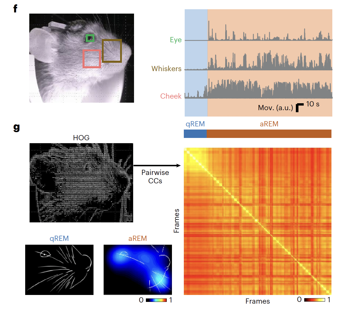
Fig2: The results revealed two main clusters during REM sleep, corresponding to a state with and a state without active facial movements, defined here as active REM (aREM) and quiescent REM (qREM) sleep, respectively.

RSC inactivation alters cortical activity dynamics during REM sleep

To examine whether the RSC plays an active role in REM sleep, researchers performed optogenetic inactivation experiments. They first characterized the effect of RSC inactivation on cortical dynamics. Adeno-associated virus expressing halorhodopsin (NpHR) fused with mCherry (AAV-CaMKIIα-NpHR3.0-mCherry) was injected bilaterally into the L2/3 of the RSC in Thy1-GCaMP6s mice, allowing simultaneous GCaMP6s imaging (with blue light) and RSC inhibition.

We found that RSC activity was markedly reduced by the optogenetic inactivation during REM sleep. By analyzing cortex-wide Ca2+ activities, under RSCREM inactivation, we observed two new motifs with Ca2+ activity initiated at FrA, followed by either sustained local FrA activation or propagated waves to posterior cortical regions. These data showed that RSC inactivation markedly reorganized global cortical activity dynamics during REM sleep.


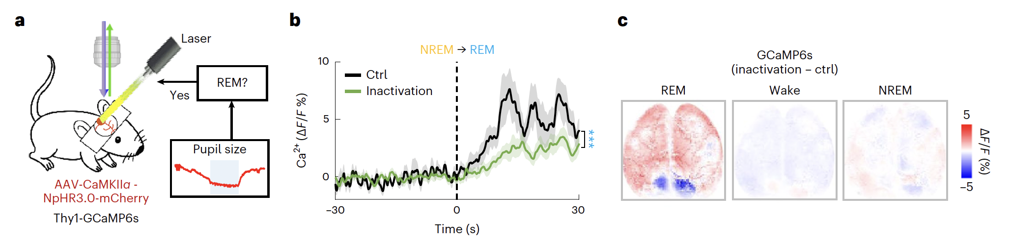
Fig 3. Adeno-associated virus expressing halorhodopsin (NpHR) fused with mCherry (AAV-CaMKIIα-NpHR3.0-mCherry) was injected bilaterally into the L2/3 of the RSC in Thy1-GCaMP6s mice, allowing simultaneous GCaMP6s imaging (with blue light) and RSC inhibition.

The RSC is required for substage transition during REM sleep

To achieve high inhibition efficiency for behavioral measurements, we next injected RSC L2/3 bilaterally with AAV expressing light-gated chlorine channel GtACR2 (AAV-CaMKIIα-GtACR2-EGFP). They found that RSCREM inactivation markedly reduced the duration of aREM sleep and increased the duration of qREM sleep, resulting a shortening of total REM duration. This effect of RSCREM inactivation was largely due to the inhibition of qREM-to-aREM transitions, rather than simply the induction of wakefulness, indicating that aREM initiation is crucial for maintaining REM sleep. RSC neurons were also important for aREM sleep maintenance, as RSCaREM inactivation largely reduced aREM duration.

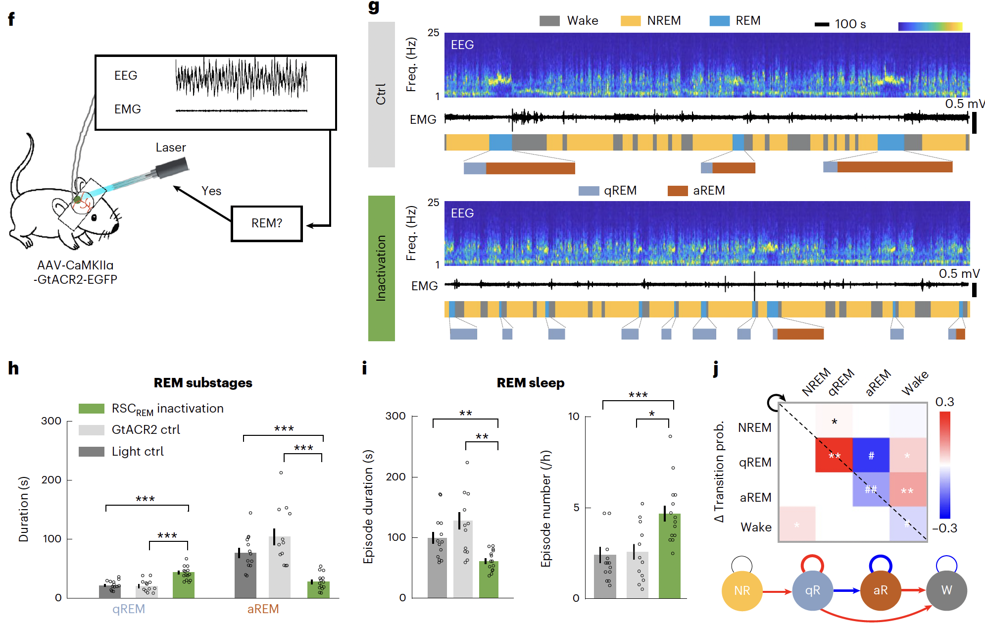
Fig.4 RSCREM inactivation markedly reduced the duration of aREM sleep and increased the duration of qREM sleep, resulting a shortening of total REM duration. This effect of RSCREM inactivation was largely due to the inhibition of qREM-to-aREM transitions, rather than simply the induction of wakefulness, indicating that aREM initiation is crucial for maintaining REM sleep

Article Source:
https://www.nature.com/articles/s41593-022-01195-2

The virus produced by Brain Case that used in this article

AAV9-hSyn-DIO-GCaMP6s
AAV9-CaMKIIα-NPHR3.0-mCherry
AAV9-hSyn-DIO-GtACR2-EGFP
AAV9-CaMKIIα-GtACR2-EGFP
AAV9-CaMKIIα-mCherry
AAV9-CaMKIIα-EYFP

AAV9-hSyn-jRGECO1a

Brain Case Biotech provided all viruses used in this study. If interested, please contact us at BD@ebraincase.com


💥Holiday

Special

Offers

Click to fill in the requirements and submit them to us!

Service Type :

Select the service you'd like to purchase.

Order Information(Premade-AAVs)

Please provide us some information about the service you'd like to order.

Detailed requirements:

scroll

Order Information(Custom AAV/Lentivirus)

Please provide us some information about the service you'd like to order.

Gene ID or gene information:

Selection of the reporting gene:

Special Instructions:

scroll

Order Information(Others)

Please provide us some information about the service you'd like to order.

Virus name/Detailed requirements:

scroll

Related products

Client Article | Cell Metabolism | Liu Danqian’s Team at the Institute of Neuroscience, CAS, Decodes the Oxidation-Sleep Code in Substantia Nigra Neurons

Client Article | Cell Metabolism | Liu Danqian’s Team at the Institute of Neuroscience, CAS, Decodes the Oxidation-Sleep Code in Substantia Nigra Neurons

Client Publication | Nature Communications | USTC Team Led by Zhi Zhang, Juan Li, and Peng Cao Unveils Neural Circuit Mechanism of Male-Specific Conditioned Pain Hypersensitivity

Client Publication | Nature Communications | USTC Team Led by Zhi Zhang, Juan Li, and Peng Cao Unveils Neural Circuit Mechanism of Male-Specific Conditioned Pain Hypersensitivity

Customer Publication | Sci Adv | Team of Zhong Chen & Yi Wang from Zhejiang Chinese Medical University Reveals Astrocyte-Mediated Mechanism in Fear Extinction

Customer Publication | Sci Adv | Team of Zhong Chen & Yi Wang from Zhejiang Chinese Medical University Reveals Astrocyte-Mediated Mechanism in Fear Extinction

Client Article | Cell Stem Cell Teams Led by Jun Qin, Qi Han, Moubin Lin, and Xiao Su Reveal How Chronic Stress Induces Intestinal Stem Cell Aging via the Vagus Nerve–Cholinergic Pathway

Client Article | Cell Stem Cell Teams Led by Jun Qin, Qi Han, Moubin Lin, and Xiao Su Reveal How Chronic Stress Induces Intestinal Stem Cell Aging via the Vagus Nerve–Cholinergic Pathway

map
{dede:global.cfg_webname/}

Virus product library

CRISPR
RNAi
Neurophilic virus
Optogenetics activation
Biosensors

News

Corporate News
New Product Launch
Media Activity
Investor News

Support

Literature interpretation
Customer article
Video Zone
FAQs
微信

WhatsApp Business Account

Tel: +8618971215294
E-mail: BD@ebraincase.com

Address:-

Address:-

  • Copyright © 2024 Brain Case All Rights Reserved.