Nature Methods|mYongHong: A New Generation of Ultra-Stable Monomeric Red Fluorescent Protein
Time:2025-09-02 11:33:24
The discovery of fluorescent proteins has revolutionized the research paradigm of modern biology. In 1962, Japanese scientist Osamu Shimomura discovered green fluorescent protein (GFP) in jellyfish. In 1994, Martin Chalfie’s team successfully expressed GFP in Caenorhabditis elegans; that same year, Roger Tsien’s team engineered GFP into a blue fluorescent protein through directed evolution. Since then, biological research has seen explosive growth driven by fluorescent proteins. At the same time, the development of fluorescent proteins evolved into a distinct scientific discipline, with fluorescent proteins of various colors being created one after another. In 2002, Jennifer Lippincott-Schwartz’s team discovered PhotoActivatable Green Fluorescent Protein (PA-GFP), paving the way for the development of photo-controllable fluorescent proteins. Subsequently, PhotoSwitchable Fluorescent Proteins (PSFPs) and Photo-Convertible Fluorescent Proteins (PCFPs) were successively developed. Alongside these advances, super-resolution imaging techniques based on photo-controllable fluorescent proteins were also developed, such as PhotoActivated Localization Microscopy (PALM) and Reversible Saturable Optical Fluorescence Transition (RESOLFT). In recent years, with the advancement of microscopy techniques, researchers have demanded even higher performance from fluorescent proteins. For example, Correlative Light and Electron Microscopy (CLEM) combines the spatial and distribution information provided by light microscopy with the ultrastructural information from electron microscopy, leveraging the strengths of both imaging modalities. However, osmium tetroxide (OsO₄), a strong oxidant used in electron microscopy sample preparation, causes rapid quenching of fluorescent protein signals. Furthermore, subsequent dehydration and high-temperature embedding steps severely diminish the remaining fluorescence in ultrathin sections. Although some OsO₄-resistant fluorescent proteins have been reported, their fluorescence retention typically does not exceed 20%, greatly limiting the development of CLEM technology. Tissue clearing techniques, which remove lipids to render tissues optically transparent, facilitate 3D tissue imaging and the construction of mesoscale brain connectomes. However, the limited thermal stability of existing fluorescent proteins means that clearing usually must be performed at room temperature or below 37°C, significantly prolonging the process, often requiring one to several weeks to achieve optimal transparency, thereby limiting imaging efficiency. Meanwhile, live-cell super-resolution imaging demands high photostability from fluorescent proteins to enable long-term live imaging. In 2020, the successful development of mEosEM shattered the long-held belief that fluorescent proteins could not withstand conventional electron microscopy sample preparation, enabling super-resolution CLEM imaging on conventionally prepared sections for the first time. In 2022, the chemically ultra-stable yellow fluorescent protein hfYFP was introduced for CLEM and expansion microscopy applications. That same year, the extraordinarily bright and ultra-stable green fluorescent protein StayGold stunned the entire field. Subsequently, three monomeric versions of StayGold were reported. Thus, fluorescent proteins officially entered a new era—the "Ultra-Stable Fluorescent Protein Era." Compared to green and yellow fluorescent proteins, red fluorescent proteins offer natural advantages, especially in terms of reduced phototoxicity, lower background autofluorescence, decreased light scattering, and better tissue penetration. However, the stability of existing red fluorescent proteins has been substantially lacking. Atsushi Miyawaki, a renowned expert in fluorescent proteins, once remarked at a conference that whoever develops an ultra-stable monomeric red fluorescent protein would be remembered in history. On April 17, 2025, a research team led by Zhifei Fu from Fujian Medical University, in collaboration with Kiryl Piatkevich's team from Westlake University, Qingbing Zheng (from Professor Ning-Shao Xia’s group) at Xiamen University, and Hu Zhao's team from the Beijing Institute for Brain Science and Brain-Inspired Intelligence, published a full-length article titled "A highly stable monomeric red fluorescent protein for advanced microscopy" in Nature Methods. Based on breakthroughs in protein crystal structure analysis, the team developed the ultra-stable monomeric red fluorescent protein mScarlet3-H, also known as mYongHong (the name used in the preprint and widely adopted in various laboratories, meaning "forever red").
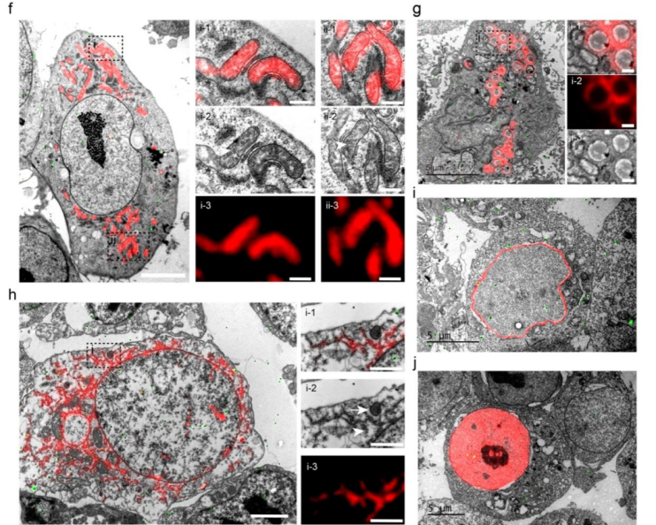
First, compared to the first-generation electron microscopy–resistant fluorescent protein mEosEM, mYongHong demonstrates a 500% improvement in osmium tetroxide resistance and a 20-fold enhancement in fluorescence signal-to-noise ratio after conventional electron microscopy sample preparation, establishing it as the current probe of choice for correlative light and electron microscopy (CLEM). Utilizing mYongHong, the research team successfully achieved high signal-to-noise ratio CLEM imaging of various subcellular organelles, including mitochondria, endoplasmic reticulum, lipid droplets, nuclear envelope, and nucleus.
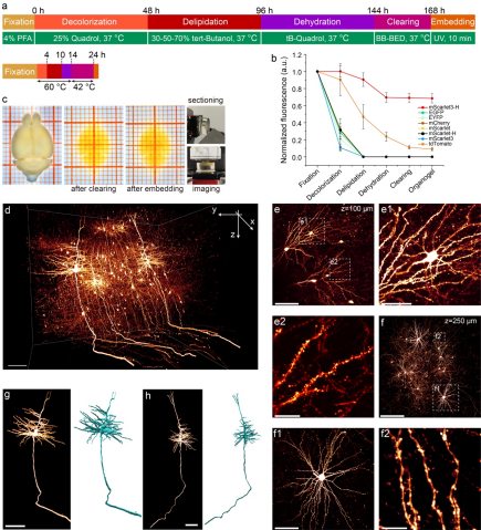
Moreover, mYongHong exhibits exceptional thermal stability, retaining 90% of its fluorescence signal after treatment at 90 °C for 1 hour. Based on this property, the research team developed a rapid whole-brain clearing method for mice, shortening the sample preparation period from the traditional one week to just 24 hours, while preserving sufficient fluorescence signals for subsequent high-quality three-dimensional imaging. This advancement significantly improved the efficiency of mesoscopic brain atlas construction by approximately sevenfold.
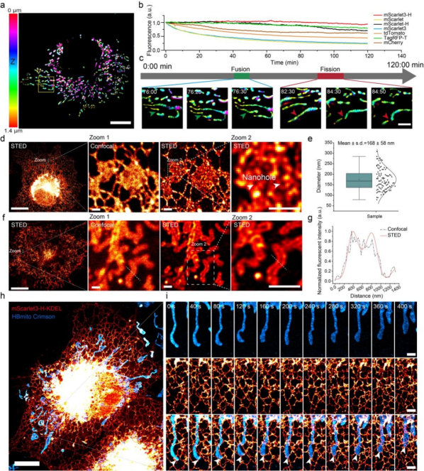
Finally, mYongHong demonstrates excellent resistance to photobleaching and is currently the most stable monomeric red fluorescent protein available. It enables long-term live-cell imaging using 3D-SIM, and notably, its superior photostability makes long-term dual-color live-cell STED super-resolution imaging feasible.
The successful development of mYongHong has overcome the common technical bottleneck of poor stability in monomeric red fluorescent proteins, expanded the spectral range of ultrastable fluorescent proteins, and laid a solid foundation for the development of future red functional fluorescent probes.
All plasmids related to mYongHong have been made freely available through the Wekwikgene Plasmid Sharing Library: https://wekwikgene.wllsb.edu.cn/plasmids?search=mYongHong. The corresponding AAV vectors can be obtained from Brain Case Biotech. Cre recombinase-dependent fluorescent reporter mice can be acquired from the domestic supplier, Jin Zhi He. Alternatively, all plasmids, viral vectors, and mice can also be requested by contacting fuzhifei@fjmu.edu.cn.
References 1. Shimomura, O., Johnson, F.H., and Saiga, Y. (1962). Extraction, Purification and Properties of Aequorin, a Bioluminescent Protein from the Luminous Hydromedusan, Aequorea. J. Cell. Comp. Physiol. 59, 223–239. https://doi.org/10.1002/jcp.1030590302. 2. Chalfie, M., Tu, Y., Euskirchen, G., Ward, W.W., and Prasher, D.C. (1994). Green Fluorescent Protein as a Marker for Gene Expression. Science 263, 802–805. https://doi.org/10.1126/science.8303295. 3. Heim, R., Prasher, D.C., and Tsien, R.Y. (1994). Wavelength mutations and posttranslational autoxidation of green fluorescent protein. Proc. Natl. Acad. Sci. 91, 12501–12504. https://doi.org/10.1073/pnas.91.26.12501. 4. Patterson, G.H., and Lippincott-Schwartz, J. (2002). A Photoactivatable GFP for Selective Photolabeling of Proteins and Cells. Science 297, 1873–1877. https://doi.org/10.1126/science.1074952. 5. Betzig, E., Patterson, G.H., Sougrat, R., Lindwasser, O.W., Olenych, S., Bonifacino, J.S., Davidson, M.W., Lippincott-Schwartz, J., and Hess, H.F. (2006). Imaging Intracellular Fluorescent Proteins at Nanometer Resolution. Science 313, 1642–1645. https://doi.org/10.1126/science.1127344. 6. Hofmann, M., Eggeling, C., Jakobs, S., and Hell, S.W. (2005). Breaking the diffraction barrier in fluorescence microscopy at low light intensities by using reversibly photoswitchable proteins. Proc. Natl. Acad. Sci. 102, 17565–17569. https://doi.org/10.1073/pnas.0506010102. 7. Fu, Z., Peng, D., Zhang, M., Xue, F., Zhang, R., He, W., Xu, T., and Xu, P. (2020). mEosEM withstands osmium staining and Epon embedding for super-resolution CLEM. Nat. Methods 17, 55–58. https://doi.org/10.1038/s41592-019-0613-6. 8. Campbell, B.C., Paez-Segala, M.G., Looger, L.L., Petsko, G.A., and Liu, C.F. (2022). Chemically stable fluorescent proteins for advanced microscopy. Nat Methods, 1–10. https://doi.org/10.1038/s41592-022-01660-7. 9. Hirano, M., Ando, R., Shimozono, S., Sugiyama, M., Takeda, N., Kurokawa, H., Deguchi, R., Endo, K., Haga, K., Takai-Todaka, R., et al. (2022). A highly photostable and bright green fluorescent protein. Nat. Biotechnol. 40, 1132–1142. https://doi.org/10.1038/s41587-022-01278-2. 10. Ivorra-Molla, E., Akhuli, D., McAndrew, M.B.L., Scott, W., Kumar, L., Palani, S., Mishima, M., Crow, A., and Balasubramanian, M.K. (2023). A monomeric StayGold fluorescent protein. Nat. Biotechnol., 1–4. https://doi.org/10.1038/s41587-023-02018-w. 11. Zhang, H., Lesnov, G.D., Subach, O.M., Zhang, W., Kuzmicheva, T.P., Vlaskina, A.V., Samygina, V.R., Chen, L., Ye, X., Nikolaeva, A.Yu., et al. (2024). Bright and stable monomeric green fluorescent protein derived from StayGold. Nat. Methods, 1–9. https://doi.org/10.1038/s41592-024-02203-y. 12. Ando, R., Shimozono, S., Ago, H., Takagi, M., Sugiyama, M., Kurokawa, H., Hirano, M., Niino, Y., Ueno, G., Ishidate, F., et al. (2023). StayGold variants for molecular fusion and membrane-targeting applications. Nat. Methods, 1–9. https://doi.org/10.1038/s41592-023-02085-6. Brain Case Biotech offers a full range of LV-mYongHong organelle fluorescent probes (Table 1) to support your high-precision subcellular research with ease.
Brain Case Biotech offers a full range of LV-mYongHong organelle fluorescent probes (see Table 1) to support your high-precision subcellular research with ease.
Subcellular Localization
Product Number
Name
Mitochondrial Inner Membrane
BM-0445
rLV-CAG-PHB2-mYongHong-WPRE
Mitochondrial Outer Membrane
BM-0447
rLV-CAG-Tom20-mYongHong-WPRE
Microtubules
BM-0438
rLV-CAG-EMTB-mYongHong-WPRE
Nuclear Chromatin
BM-0439
rLV-CAG-H2B-mYongHong-WPRE
Nuclear Lamina
BM-0440
rLV-CAG-LaminA-mYongHong-WPRE
Lysosomal Membrane
BM-0441
rLV-CAG-LAMP1-mYonghong-WPRE
Cytoskeleton
BM-0442
rLV-CAG-Lifeact-mYongHong-WPRE
Golgi Apparatus Membrane
BM-0443
rLV-CAG-mYongHong-Giantin-WPRE
Endoplasmic Reticulum Lumen
BM-0444
rLV-CAG-mYongHong-KDEL-WPRE
Lipid Droplet Membrane
BM-0446
rLV-CAG-Plin5-mYongHong-WPRE
For products suitable for whole-brain clearing imaging and various neuroscience research applications, please refer to the AAV-mYongHong product list (Table 2).
Table 2. AAV-mYongHong Product List
Product Number
Product Name
BC-3987
rAAV-CAG-mYongHong
BC-3997
rAAV-cFos-mYongHong
BC-3782
rAAV-CAG-DIO-mYongHong
BC-3412
rAAV-EF1α-DIO-mYongHong
BC-3415
rAAV-EF1α-FDIO-mYongHong
BC-SL055
NCSP-mYH-2E5 (Sparse Labeling)
At the same time, Brain Case also offers various customization services. If you have any related needs, please contact us at: bd@ebraincase.com
Service Type :
Select the service you'd like to purchase.
Order Information(Premade-AAVs)
Please provide us some information about the service you'd like to order.
Order Information(Custom AAV/Lentivirus)
Please provide us some information about the service you'd like to order.
Order Information(Others)
Please provide us some information about the service you'd like to order.


![[Research Tool Highlight] Featured in Cell! Brain Case launches new product — Enhancer AAV Vectors, a breakthrough for research Tool !](/web/allimg/250826/0R61432331JL.png)