Brain Case Research Services | PRV Neural Circuit Tracing Service Empowering Peripheral–Central Research
Time:2025-09-11 16:41:37
1.What is Pseudorabies Virus (PRV)?
After infecting neurons, PRV replicates intracellularly and expresses the target gene. The progeny virus is transported to the postsynaptic terminal and then crosses the synapse retrogradely to infect upstream neurons, initiating a new round of replication and retrograde trans-synaptic transmission. Pseudorabies virus (PRV), also known as Suid herpesvirus type I, is a linear double-stranded DNA virus with a genome of approximately 150 kb. The Bartha strain of PRV possesses only retrograde, trans-synaptic properties. Recombinant tracing viral tools derived from the Bartha strain can label neural networks strictly in a retrograde, multi-synaptic manner.
2. What Can PRV Do in Neural Circuit Research?
PRV plays an important role in studies of both peripheral–central and central–central neural connections. It is commonly used to explore central neural nuclei that are directly or indirectly associated with specific peripheral organs.
For example, to investigate which brain nuclei initiate or participate in the command to lift the thigh, PRV can be injected into the target muscle group. After a short infection window of several days, animals are perfused and brain tissue is sectioned. Fluorescent signals observed in central brain slices allow identification of the neural nuclei involved in controlling thigh lifting.
Case 1
Experimental Objective:
To investigate the neural circuits associated with thigh lifting and to evaluate differences in neural regulation between the left and right limbs.
Experimental Design:
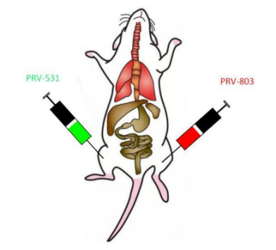
PRV viral tracers were used. Mice received intramuscular injections of 2.5 μL each of PRV-803 (red, expressing mScarlet) and PRV-531 (green, expressing GFP) into the left and right thigh muscles, respectively. The skin was incised for the injection procedure and sutured after completion. Following injection, animals were observed at least twice daily for 1–3 days to ensure postoperative recovery. From Day 3 onward, animals were monitored at high frequency, with observation intervals not exceeding 4 hours.
Figure 1: Schematic diagram of viral injection
Experimental Results:
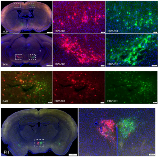
Approximately 5 days post-injection, animals developed evident neurological symptoms, after which cardiac perfusion was performed and brain tissue collected. Frozen brain sections yielded the following data. Results suggest that control of the target muscle group is directly or indirectly connected to nuclei including the motor cortex (MOp), suprachiasmatic nucleus (SCN), and periaqueductal gray (PAG). Further validation could be achieved by calcium signal recording to assess the critical role of these nuclei, or by applying optogenetic and chemogenetic manipulations to selectively regulate them, thereby identifying central nuclei more closely related to the target muscle group.
Figure 2: Multisynaptic retrograde viral tracing of muscle (Brain Case Test Data)
Case 2
Experimental Objective:
To investigate the neural circuits involved in the control of urination and identify the key brain regions that regulate voluntary micturition.
Experimental Design:
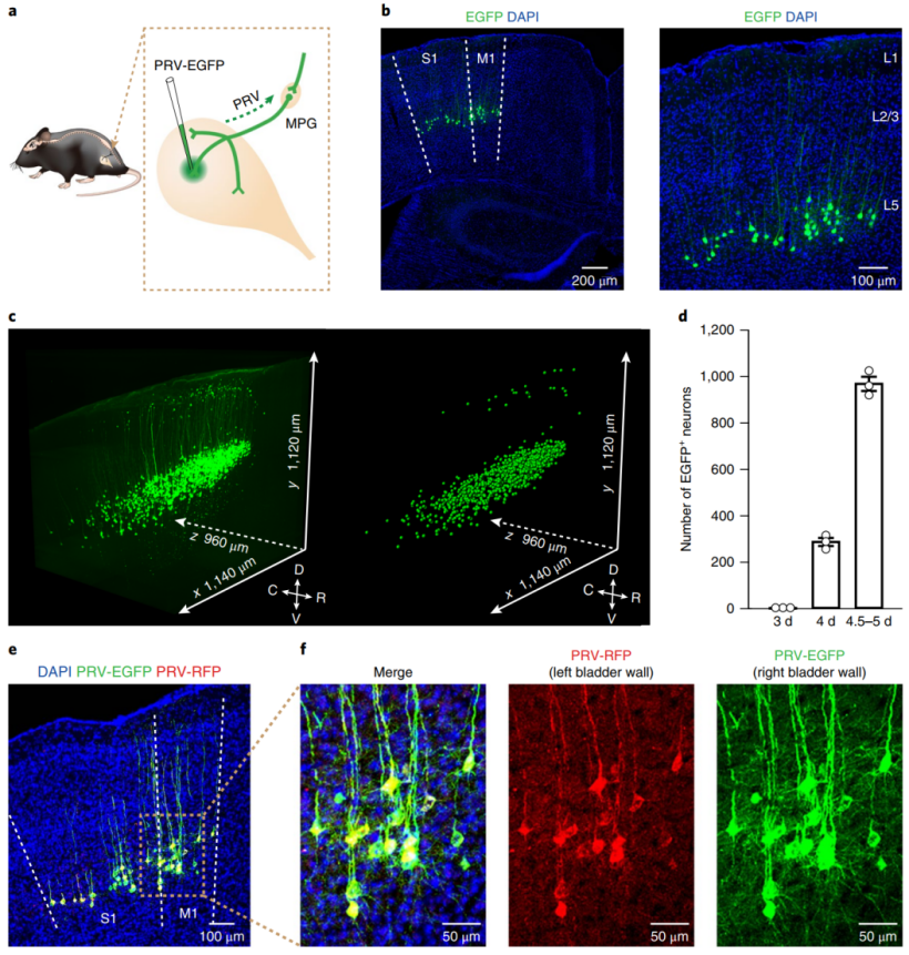
PRV viral tracers were used. Microinjections of PRV-531 (green) and PRV-724 (red, expressing mRuby3) were administered into the left and right sides of the bladder wall, respectively. Because PRV infection causes severe neurological symptoms leading to death, animals were closely monitored at high frequency to ensure normal recovery after surgery. Cardiac perfusion and brain collection were performed when animals reached a moribund state.
Experimental Results:
Approximately 5 days post-injection, animals developed distinct neurological symptoms. Researchers then carried out cardiac perfusion and brain collection as appropriate. Frozen brain sections provided the following data: results revealed that the primary motor cortex (M1), primary somatosensory cortex (S1), and the pontine micturition center (PMC) are directly or indirectly connected to bladder control.
3. PRV Neural Circuit Tracing Services Offered by Brain Case
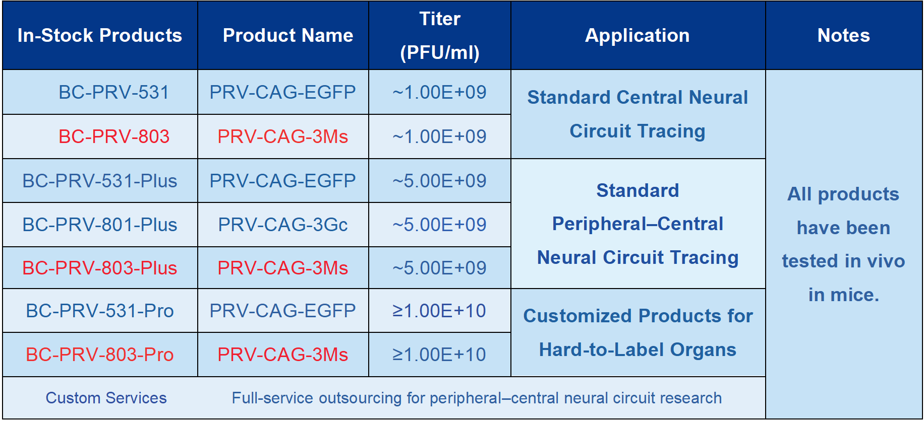
In recent years, research on peripheral–central neural circuits has gained increasing attention, with more peripheral organs becoming key study targets. Brain Case is committed to providing customer-oriented solutions to support peripheral–central neural circuit research. We offer a comprehensive one-stop PRV neural circuit tracing service, including stable legacy tracers PRV-531 (green) and PRV-724 (red), as well as highly efficient and bright new tracers PRV-801 (green, expressing mClover) and PRV-803 (red). These products have been successfully applied to studies of various peripheral tissues.
Examples of research sites we have worked on include: Digestive system: duodenum, cecum, colon, rectum, pancreas Endocrine system: thyroid Reproductive system:testes, ovaries, epididymis, prostate Urinary system:bladder wall Musculoskeletal system:muscles, bone marrow cavity Cardiovascular system:heart Immune system:spleen Circulatory system: femoral cavity Nervous system:brain and spinal cord
If you are interested in neural circuit tracing research and would like to learn more about practical applications, specific case studies, or the principles of viral tools and their use in central–peripheral interaction studies, please contact BD@ebraincase.com
Service Type :
Select the service you'd like to purchase.
Order Information(Premade-AAVs)
Please provide us some information about the service you'd like to order.
Order Information(Custom AAV/Lentivirus)
Please provide us some information about the service you'd like to order.
Order Information(Others)
Please provide us some information about the service you'd like to order.