E-mail:BD@ebraincase.com
Tel:+8618971215294
English 中文版
Virus Vector - Viral Vector Production - BrainCaseVirus Vector - Viral Vector Production - BrainCase
  • Home
  • Virus product library
    CRISPRRNAiHSV-helperRV-helperNeurophilic virusCalcium SensorsOptogenetics activationOptogenetics inhibitionChemical geneticsSparse labelingFluorescent proteinBiosensorsRecombinaseApoptosis & AutophagyDisease ModelNeurotoxicityOther
  • Products & Service

    Product Center

    Virus

    VSV-vaccine and gene therapy research
    Retrovirus-RCAS-TVA
    Lentivirus Vector-Lentivirus Production
    Rabies Virus Vector-RBV Vector
    Herpes simplex virus-Oncolytic and anterograde tracing
    PRV-retrograd multisynaptic-Peripheral
    AAV-gene therapy vectors-neuroscience

    Animal Model

    Neurological Disease Models
    Tumor animal models-anti-tumor
    Digestive System Disease Animal Model
    Cardiovascular System Disease Animal Models

    Plasmid Construction

    Library Construction
    Plasmid design and construction

    Popular Applications

    Gene Regulation

    Gene Overexpression-Brain Case
    RNA interference(RNAi)-siRNA-Brain Case
    Gene Editing - CRSIPR cloning - BrainCase

    Neural Circuit Function Research

    Optogenetics - BrainCase
    Chemical genetics-DREADDs-Brain Case
    Calcium signal recording-Gels- Brain Case
    GRAB Neurotransmitter Fluorescent Sensor- Brain Case
    Functional Magnetic Resonance Imaging Technology- Brain Case

    Research on the structure of neural circuits

    Direct Input and Output-viral vectors- Brain Case
    Anterograde Mono-synaptic Tracing -HSV- Brain Case
    Antrograde Muti-synaptic Tracing-HSV & VSV-Brain Case
    Retrograde Mono-synaptic Tracing-Rabies Virus-Brain Case
    Retrograde Muti-synaptic Tracing-PRV-Brain Case

    Featured Services

    Nervous System Disease Drug Effect

    Alzheimer's disease-AD
    Depression-mental disorders
    Parkinson's disease--PD
    Epilepsy-an ancient neurological disorder

    AAV Serotype Screening

    AAV Serotypes screening-gene therapy

    Tumorigenicity Test

    Tumorigenicity Testing-Evaluation of tumor models

    Efficacy of Oncolytic Virus

    Oncolytic virus-for cancer therapy-Brain Case
    Herpes Virus Vector-anti-tumor- BrainCase
    Vesicular stomatitis virus-killing tumor cells-Brain Case
  • News
    Corporate News New Product Launch Media Activity Investor News
  • Support
    Literature interpretation Customer article Video Zone FAQs
  • About Us
    Virus product library Products & Service News Support About Us Contact
  • Contact
    Contact Us Join us
  • 中文
    English 中文版
  • Home
  • News
  • Corporate News
  • News
  • Corporate News
  • New Product Launch
  • Media Activity
  • Investor News

Research Service | Whole-Brain Clearing Imaging & Analysis Service

Time:2025-08-06 17:04:07
Service Overview
Using efficient sample clearing and imaging techniques, we analyze the complete neural network of the mouse brain. Our process achieves high transparency of the whole-brain tissue while preserving fluorescent protein signals, enabling rapid, uniform, subcellular-resolution imaging of the entire mouse brain. This supports high-precision 3D reconstruction of neural networks. Compatible with multiple techniques such as immunolabeling, viral tracing, and transgenic labeling, this service precisely reveals the distribution features of brain neural networks as well as their interactions with other brain tissues, providing strong support for whole-brain connectomics research.
 

Service Workflow

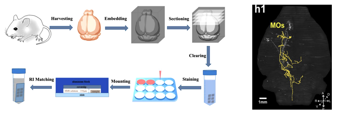
1.Sample Preparation

Includes fixation & embedding, sectioning & mounting, and tissue clearing to ensure morphological stability and optimized light transmittance.

Figure 1: Sample preparation workflow diagram
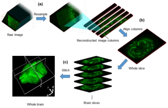

2. 3D Imaging

Whole-brain 3D imaging is performed using VISoR high-resolution microscopy, with resolutions up to 0.5 × 0.5 × 2 μm³, ensuring accurate capture of neuronal structures and connections.

Figure 2: Stitching process schematic


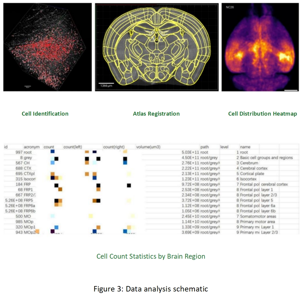
3. Data Processing & Analysis

Includes whole-brain cell detection, image stitching & reconstruction, and registration to the Allen CCFv3 atlas, enabling precise 3D reconstruction and analysis of brain structures.
 

4. Deliverables

2D Imaging: Maximum intensity projection images of individual slices for quick sample quality review and preliminary assessment.
3D Reconstruction: 4 μm / 1 μm / 0.5 μm resolution data upon request, visualizing the 3D structures of tissues, cells, and neural networks.
Final Project Report: Professional report including data charts and analysis conclusions, based on cell identification and brain-region cell count results.


Service Turnaround Time

From sample receipt to final results delivery, the overall turnaround is approximately 10–15 working days (subject to adjustment based on experiment complexity).
Sample fixation & embedding: 1–2 days
Sectioning & tissue clearing: 2 days
Mounting: 1–2 days
Refractive index matching & imaging: 2 days
3D reconstruction & data analysis: 3–10 days

Service Highlights

  1. High-Transparency Sample Preparation: Incorporates innovative clearing techniques to ensure maximum optical clarity.
  2. Antibody Labeling Support: Compatible with various antibodies, including tyrosine hydroxylase (TH) and c-Fos antibodies.
  3. High-Throughput Imaging: Enables rapid, high-throughput data acquisition and visualization — complete mouse whole-brain imaging in under 2 hours; supports processing and imaging of large or whole-body samples.
 

Case Studies

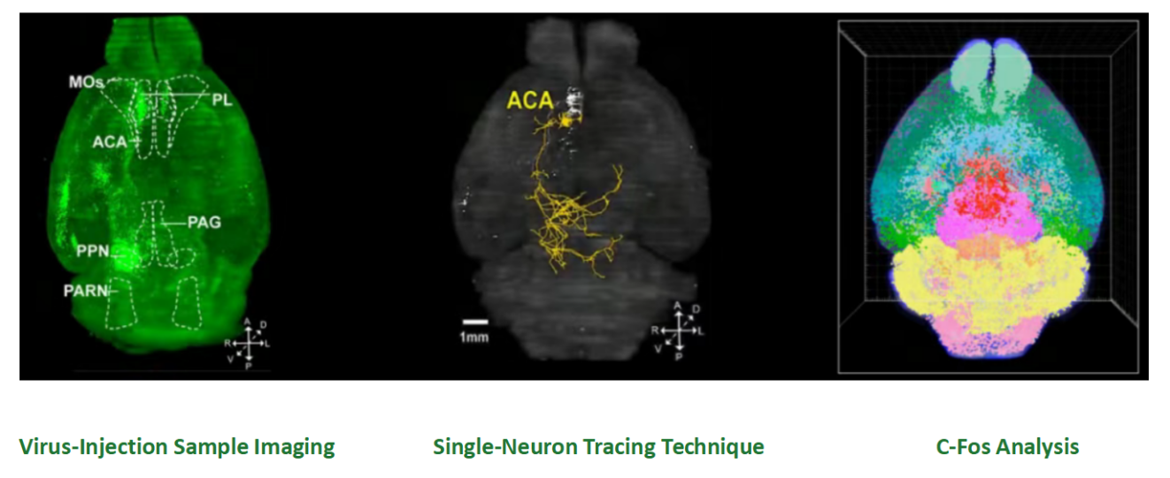
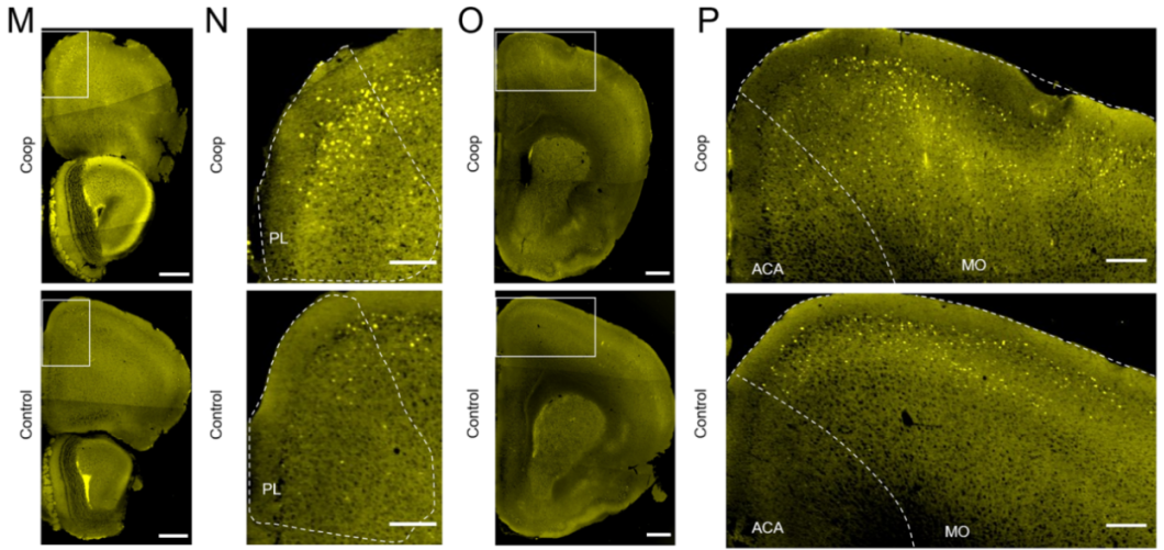
Case 1: Localization of Brain Regions Related to Cooperative Behavior in Mice

Using c-Fos immunostaining combined with VISoR whole-brain imaging, we precisely detected high c-Fos expression in the frontal pole (FRP), motor cortex (MO), and anterior cingulate cortex (ACA), indicating these regions are active during cooperative behavior. This provides key evidence for the involvement of functions such as decision-making and social interaction in cooperative tasks.

Guo-Qiang Bi,Molecular Brain,2023

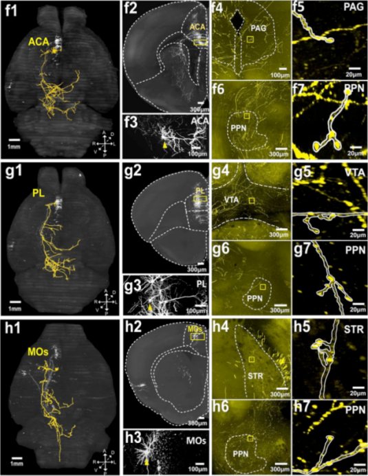
Case 2: Projection Study of Neurons Targeting the Pedunculopontine Nucleus (PPN)

By applying dual-virus labeling and VISoR imaging, we clearly traced individual neuronal axons projecting from cortical regions such as the ACA and prelimbic area (PL) to the PPN, revealing distinctive collateral branching patterns. These findings offer a new perspective for understanding the neural circuit mechanisms by which the PPN participates in motor control, sleep, and related functions.

Guo-Qiang Bi,Mol Brain,2022


For more information on related products and services, please contact  bd@ebraincase.com

Service Type :

Select the service you'd like to purchase.

Order Information(Premade-AAVs)

Please provide us some information about the service you'd like to order.

How did you hear about BrainCase Biotech or our products?

Detailed requirements:

scroll

Order Information(Custom AAV/Lentivirus)

Please provide us some information about the service you'd like to order.

Gene ID or gene information:

Selection of the reporting gene:

Special Instructions:

scroll

Order Information(Others)

Please provide us some information about the service you'd like to order.

Virus name/Detailed requirements:

scroll

Related products

Research Service | Whole-Brain Clearing Imaging & Analysis Service

Research Service | Whole-Brain Clearing Imaging & Analysis Service

Research Services | Neural Circuit Tracing + Targeted Spatial Omics + Neuronal Classification Analysis (with Case Studies)

Research Services | Neural Circuit Tracing + Targeted Spatial Omics + Neuronal Classification Analysis (with Case Studies)

🔬 SUMMER PROMOTION 2025 | Brain Case Biotech Unlock Exclusive Savings on Viral Vectors & Custom Services — Limited Time Only!

🔬 SUMMER PROMOTION 2025 | Brain Case Biotech Unlock Exclusive Savings on Viral Vectors & Custom Services — Limited Time Only!

2025 Frontier Symposium on Neuroscience and Cell & Gene Therapy Technologies

2025 Frontier Symposium on Neuroscience and Cell & Gene Therapy Technologies

map
{dede:global.cfg_webname/}

Virus product library

CRISPR
RNAi
Neurophilic virus
Optogenetics activation
Biosensors

News

Corporate News
New Product Launch
Media Activity
Investor News

Support

Literature interpretation
Customer article
Video Zone
FAQs
微信

WhatsApp Business Account

Tel: +8618971215294
E-mail: BD@ebraincase.com

Address:-

Address:-

  • Copyright © 2024 Brain Case All Rights Reserved.