E-mail:BD@ebraincase.com
Tel:+8618971215294
English 中文版
Virus Vector - Viral Vector Production - BrainCaseVirus Vector - Viral Vector Production - BrainCase
  • Home
  • Virus product library
    CRISPRRNAiHSV-helperRV-helperNeurophilic virusCalcium SensorsOptogenetics activationOptogenetics inhibitionChemical geneticsSparse labelingFluorescent proteinBiosensorsRecombinaseApoptosis & AutophagyDisease ModelNeurotoxicityOther
  • Products & Service

    Product Center

    Virus

    VSV-vaccine and gene therapy research
    Retrovirus-RCAS-TVA
    Lentivirus Vector-Lentivirus Production
    Rabies Virus Vector-RBV Vector
    Herpes simplex virus-Oncolytic and anterograde tracing
    PRV-retrograd multisynaptic-Peripheral
    AAV-gene therapy vectors-neuroscience

    Animal Model

    Neurological Disease Models
    Tumor animal models-anti-tumor
    Digestive System Disease Animal Model
    Cardiovascular System Disease Animal Models

    Plasmid Construction

    Library Construction
    Plasmid design and construction

    Popular Applications

    Gene Regulation

    Gene Overexpression-Brain Case
    RNA interference(RNAi)-siRNA-Brain Case
    Gene Editing - CRSIPR cloning - BrainCase

    Neural Circuit Function Research

    Optogenetics - BrainCase
    Chemical genetics-DREADDs-Brain Case
    Calcium signal recording-Gels- Brain Case
    GRAB Neurotransmitter Fluorescent Sensor- Brain Case
    Functional Magnetic Resonance Imaging Technology- Brain Case

    Research on the structure of neural circuits

    Direct Input and Output-viral vectors- Brain Case
    Anterograde Mono-synaptic Tracing -HSV- Brain Case
    Antrograde Muti-synaptic Tracing-HSV & VSV-Brain Case
    Retrograde Mono-synaptic Tracing-Rabies Virus-Brain Case
    Retrograde Muti-synaptic Tracing-PRV-Brain Case

    Featured Services

    Nervous System Disease Drug Effect

    Alzheimer's disease-AD
    Depression-mental disorders
    Parkinson's disease--PD
    Epilepsy-an ancient neurological disorder

    AAV Serotype Screening

    AAV Serotypes screening-gene therapy

    Tumorigenicity Test

    Tumorigenicity Testing-Evaluation of tumor models

    Efficacy of Oncolytic Virus

    Oncolytic virus-for cancer therapy-Brain Case
    Herpes Virus Vector-anti-tumor- BrainCase
    Vesicular stomatitis virus-killing tumor cells-Brain Case
  • News
    Corporate News New Product Launch Media Activity Investor News
  • Support
    Literature interpretation Customer article Video Zone FAQs
  • About Us
    Virus product library Products & Service News Support About Us Contact
  • Contact
    Contact Us Join us
  • 中文
    English 中文版
  • Home
  • News
  • Corporate News
  • News
  • Corporate News
  • New Product Launch
  • Media Activity
  • Investor News

Research Services | Neural Circuit Tracing + Targeted Spatial Omics + Neuronal Classification Analysis (with Case Studies)

Time:2025-07-22 11:35:20

Service Overview

As neuroscience research advances into a multidimensional era of “cell type–circuit–behavior” analysis, traditional neuron classification methods based on antibody labeling face several limitations—such as insufficient antibody specificity, high cost, and low throughput (e.g., difficulty labeling multiple proteins on the same brain slice). These challenges significantly hinder the in-depth investigation of complex neural circuits and disease mechanisms.

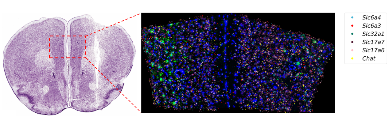
To overcome these barriers, we have launched an innovative research service: “Neural Circuit Tracing + Targeted Spatial Omics + Neuronal Classification Analysis.” After performing neural circuit tracing, we apply spatial transcriptomics to the brain tissue containing the labeled neurons. This approach enables the integration of anatomical connectivity data from tracing with spatially resolved gene expression profiles.

Such integration allows researchers to directly associate labeled neurons with their molecular characteristics—such as expression levels of specific genes or neuronal subtypes—thereby uncovering direct links between neuron types, circuit connectivity, and functional states.


Technical Principles and Advantages

This service is built upon three core technologies:

01 Neural Circuit Tracing Technology

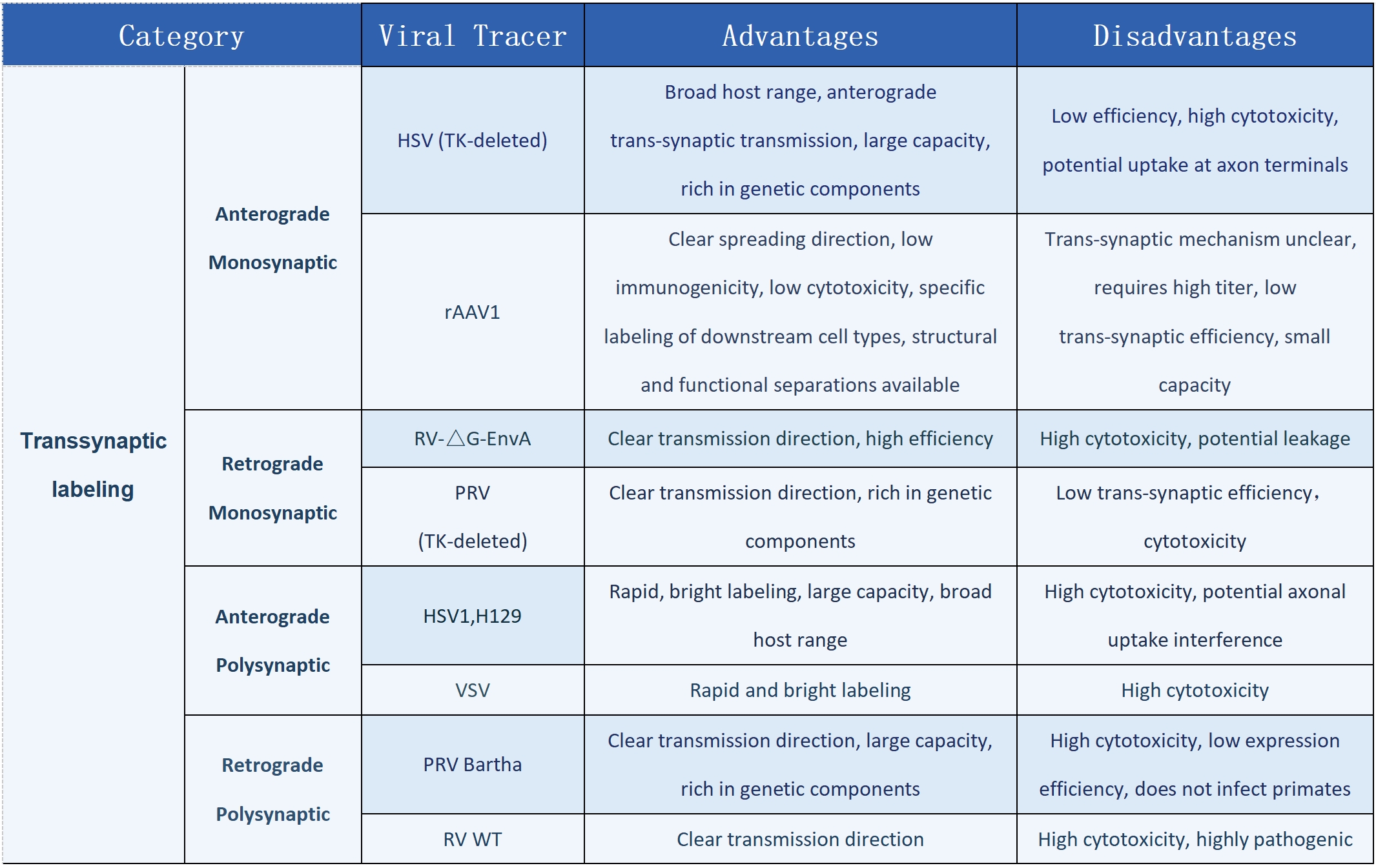
Utilizing viral tools such as AAV, RV, and PRV, this technique enables anterograde/retrograde and trans-synaptic labeling, allowing precise mapping of neuronal projection pathways.


2.Targeted Spatial Transcriptomics Technology (MiP-seq):

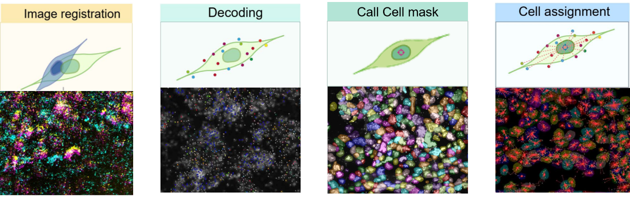
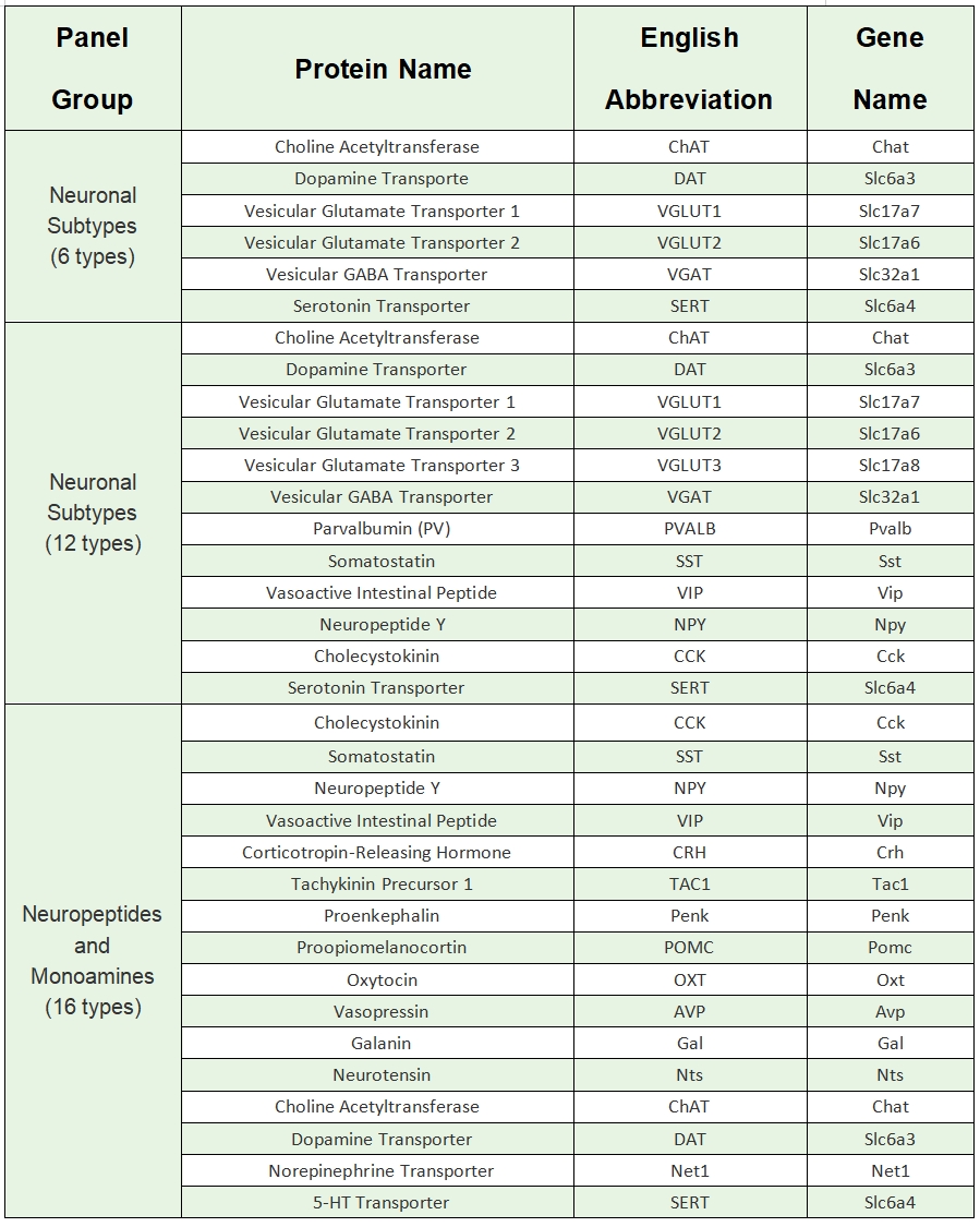
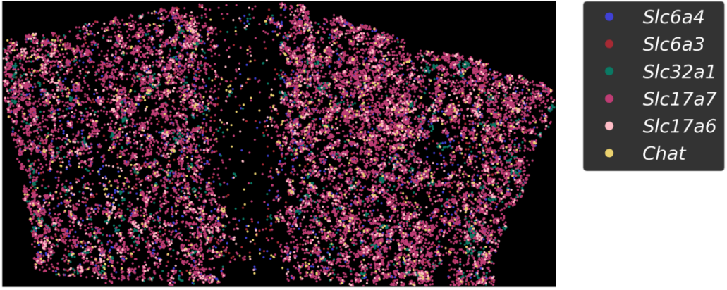
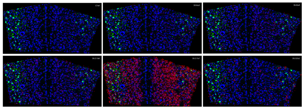
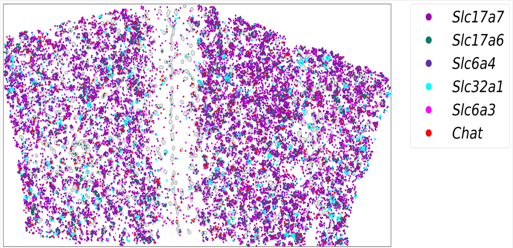
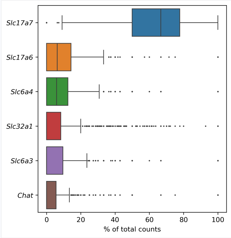
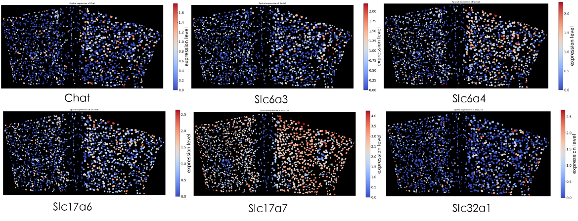
A novel targeted spatial omics technology featuring high sensitivity, high specificity, and high resolution. It enables qualitative and quantitative detection at single-base and subcellular levels, allowing the construction of single-cell spatial maps. MiP-seq is widely applied in fields such as neuroscience, development, and oncology.

3.Multidimensional Data Analysis and Visualization:

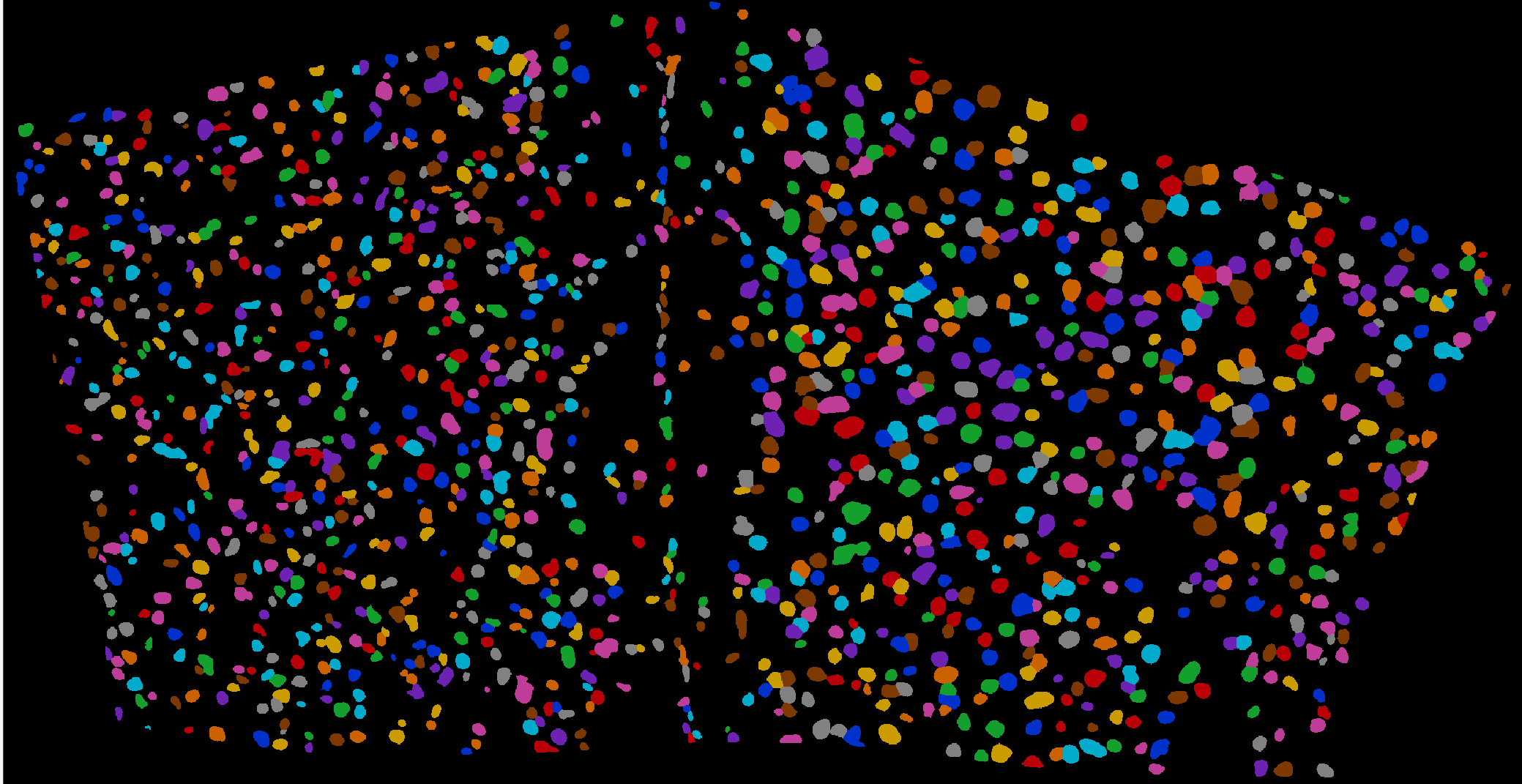
Integrating image processing, cell segmentation, gene expression quantification, and spatial localization to elucidate the relationships between neuron types and circuit functions.


Service Process

1. Experimental Design: Determine the labeling strategy based on research objectives (retrograde/anterograde viral tracing, monosynaptic/polysynaptic tracing), select the appropriate virus type, and design or choose the target marker probe panel.
2. Viral Labeling and Tissue Processing: Process samples following the workflow of “virus injection → tissue collection → fixation → sucrose dehydration and sedimentation → OCT embedding → cryosectioning.”
3. Targeted Spatial Transcriptomics: Localization and quantitative detection of mRNA at the targeted spatial transcriptomic level.
4. Multidimensional Data Integration and Analysis: Analyze the neuron types, quantities, and spatial distribution labeled by the tracing virus.
 

Service Advantages

1.High-Specificity Probes and Comprehensive Panel: Precise Identification of Neuronal Subtypes

We screen key neuronal cell types to select target sequence regions with high hybridization efficiency and strong specificity, and design dedicated hybridization probes. The integrated targeted panel includes more than 30 neuronal markers, enabling precise identification of various neuronal subtypes. This allows accurate differentiation of different neuron types within complex neuronal populations, laying a solid foundation for in-depth research on neuronal characteristics and functions.


2.Comprehensive Deliverables: One-Stop Presentation of Scientific Findings

Includes raw images (e.g., virus-labeled fluorescence images, gene in situ hybridization images), analytical charts (e.g., spatial distribution maps of neuronal subtypes, gene expression box plots/heatmaps), statistical results, and conclusions. All are compiled into a final report that visually and clearly presents the research outcomes.

3.Broad Application Prospects: Spanning Multiple Research Fields

Applicable to various areas within neuroscience. In developmental neuroscience, it enables tracking of neuronal changes during development; in neurodegenerative disease research, it helps identify disease-associated neuronal alterations and potential therapeutic targets; in the study of psychiatric disorders, it supports the exploration of abnormal brain neural circuits.


Case Results Display

To investigate the neuronal types in the prelimbic cortex (PrL) of mice that project to the ventral tegmental area (VTA) of the midbrain, RV retrograde trans-synaptic tracing virus was injected into the VTA. After tissue sectioning, in situ hybridization was performed. The detection panel included choline acetyltransferase (ChAT), dopamine transporter (DAT), vesicular glutamate transporter 1 (VGLUT1), vesicular glutamate transporter 2 (VGLUT2), vesicular GABA transporter (VGAT), and serotonin transporter (SERT).

1.Image Calibration

The acquired images were adjusted and optimized to correct or eliminate image batch deviations caused by various factors (such as equipment differences, lighting conditions, experimental operations, etc.), making the images more accurate, consistent, and reliable.

Figure 1: Merged fluorescence image (blue indicates cell nuclei, green indicates virus-labeled cells)


2.Signal Spot Decoding

Quantification and localization of fluorescence signals. Quantification refers to measuring the intensity, area, or other relevant parameters of the fluorescence signals to reflect their expression levels in biological samples. Localization refers to determining the precise position of the fluorescence signals in the image to analyze their spatial distribution and interactions.

Figure 2: Distribution map of all genes (dots in different colors represent signal spots of different genes)


Figure 3: Distribution map of a single gene (expression profile of a single gene)


3.Cell Segmentation and Signal Spot Assignment

Image processing and analysis to identify and define cell boundaries, thereby enabling the localization and segmentation of individual cells.

Figure 4: Cell segmentation map (showing identified cells)


Figure 5: Gene-cell distribution map (blue/gray indicates cell nuclei; dots in different colors represent signal spots of different genes)


4.Data Statistics

Statistical analysis of the expression of six genes in virus-traced labeled cells.

Figure 6: Gene expression box plot


Figure 7: Gene in situ expression heatmap
 
Based on the experimental results, among the neurons in the prelimbic cortex projecting to the ventral tegmental area of the midbrain, Slc17a7-positive cells represent the major subtype, accounting for approximately 63% of the total.

All the tracing viral vectors mentioned in the article are available from the Brain Case Team. In addition, we offer comprehensive experimental services related to these tracers, including in vivo viral delivery, tissue sectioning, and imaging. If you’re interested, please feel free to contact us at bd@ebraincase.com


Service Type :

Select the service you'd like to purchase.

Order Information(Premade-AAVs)

Please provide us some information about the service you'd like to order.

How did you hear about BrainCase Biotech or our products?

Detailed requirements:

scroll

Order Information(Custom AAV/Lentivirus)

Please provide us some information about the service you'd like to order.

Gene ID or gene information:

Selection of the reporting gene:

Special Instructions:

scroll

Order Information(Others)

Please provide us some information about the service you'd like to order.

Virus name/Detailed requirements:

scroll

Related products

Research Services | Neural Circuit Tracing + Targeted Spatial Omics + Neuronal Classification Analysis (with Case Studies)

Research Services | Neural Circuit Tracing + Targeted Spatial Omics + Neuronal Classification Analysis (with Case Studies)

🔬 SUMMER PROMOTION 2025 | Brain Case Biotech Unlock Exclusive Savings on Viral Vectors & Custom Services — Limited Time Only!

🔬 SUMMER PROMOTION 2025 | Brain Case Biotech Unlock Exclusive Savings on Viral Vectors & Custom Services — Limited Time Only!

2025 Frontier Symposium on Neuroscience and Cell & Gene Therapy Technologies

2025 Frontier Symposium on Neuroscience and Cell & Gene Therapy Technologies

Memorial Day Virus Vector Specials Are Here - Shop Now!

Memorial Day Virus Vector Specials Are Here - Shop Now!

map
{dede:global.cfg_webname/}

Virus product library

CRISPR
RNAi
Neurophilic virus
Optogenetics activation
Biosensors

News

Corporate News
New Product Launch
Media Activity
Investor News

Support

Literature interpretation
Customer article
Video Zone
FAQs
微信

WhatsApp Business Account

Tel: +8618971215294
E-mail: BD@ebraincase.com

Address:-

Address:-

  • Copyright © 2024 Brain Case All Rights Reserved.