- E-mail:BD@ebraincase.com
- Tel:+8618971215294
Q: What are Cre and loxP?
A: In 1981, Nat Sternberg et al. reported a protein cloned from the P1 bacteriophage of Escherichia coli—a 38 kDa recombinase encoded by 343 amino acids called Cre (Cyclization Recombination Enzyme). It specifically recognizes loxP sites and mediates sequence deletion or recombination between loxP sites. The loxP (locus of X-overP1) site is 34 bp long, consisting of two 13 bp inverted repeat sequences and an 8 bp spacer region. The inverted repeat sequences are the specific recognition sites for the Cre recombinase, while the spacer region determines the orientation of loxP.
Q: What analgesics or anti-inflammatory treatments are used after animal surgery?
A: We typically use lincomycin-lidocaine gel, applied topically to the skull immediately after the procedure. For more invasive surgeries such as optical fiber implantation, we recommend intraperitoneal injection of cefazolin for antibacterial and anti-inflammatory purposes (1 gram of cefazolin dissolved in 40 mL of saline; each mouse receives 0.1–0.2 mL per day for three consecutive days).
Q: What is the typical timeline for AAV expression? For example, expression in the soma at 2 weeks, in distal projections at 3 weeks, peaking around 3–4 weeks — how long does the stable expression phase last, and what is the attenuation pattern like?
A: Fluorescence in the soma typically begins to appear about one week after injection (surrounding fibers may also show signal). By the second week, proximal axons start to exhibit detectable fluorescence, and by the third week, fluorescence can be observed in distal axons. Stable expression is usually reached around one month post-injection.
In our experience, robust expression is evident by week 2. For mice, peak expression generally occurs around 6 weeks; for rats, around 8 weeks. After that, stable expression can be maintained for approximately 2 months.
Q: During whole-brain projection imaging, some brain regions show high levels of spontaneous green background. How can this be addressed?
A: Before mounting, briefly immerse the sections in 0.05 mol/L sodium carbonate solution for around 10 seconds, then allow them to air dry before coverslipping. This sodium carbonate treatment helps reduce background fluorescence, especially for GFP and YFP. The effect is less pronounced for RFP.
Q: How to inject virus into brain regions near the medulla?
A: Use an angled injection approach and calculate the trajectory using trigonometric functions. For targeting more posterior nuclei, using the lambda point (posterior fontanelle) as a reference generally yields more accurate positioning.
Q: When injecting a brain region located below the ventricle in non-human primates, the needle passes through the ventricle and causes virus leakage into the cerebrospinal fluid, leading to off-target AAV expression. How can this be avoided?
A: Use MRI to precisely localize the target region and fix the coordinates. Then insert the needle at an angle to avoid passing through the ventricle.
Q: What’s the recommended strategy for bilateral hippocampal AAV injection in mice?
A: Perform bilateral injections at two sites per hemisphere, totaling four injection sites. Inject 500–800 nL per site, with up to 800 nL for the ventral hippocampus.
Suggested coordinates:
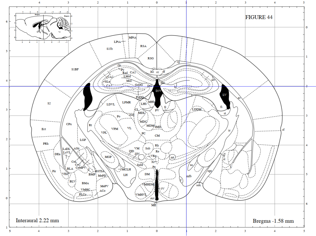
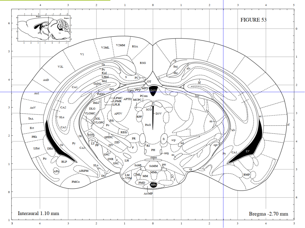
• Dorsal hippocampus: AP -1.70, ML ±1.1, DV -2.0 → inject 500 nL
• Ventral hippocampus: AP -2.90, ML ±3.0, DV -2.7 → inject 800 nL


Q: Has anyone used 10% DMSO for intraperitoneal injection in mice? Will the mice survive a 200 μL dose?
A: We’ve tried daily injections for over 10 days with no adverse effects observed.
Q: If I use intravenous injection of PHP.eB for whole-brain expression and plan to repeat the injection every four months, will immune responses or antibodies affect the second injection?
A: We recommend switching to a different BBB-crossing AAV serotype, such as AAV-B10, for the second injection. This can help reduce immune responses triggered by repeated dosing.
Q: Can PHP.eB be injected into the lateral ventricle? What is the recommended volume? Is the infection efficient?
A: You can start by injecting 1 μL of virus to test the efficiency. When injecting BBB-crossing AAV serotypes into the lateral ventricle, the distribution of fluorescent signals tends to be uneven, mainly localizing around the ventricle. In contrast, intravenous injection with a total dose of ≥1E11 vg typically results in more uniform whole-brain expression.
Q: For AAV2/9, after the virus infects the soma, can there also be expression at the synaptic terminals?
A: Yes, it’s possible. It mainly depends on the expression efficiency after infection. Once the protein is synthesized in the cytoplasm, it can be transported along the axon to the terminal. The more protein that’s produced, the more will reach the terminals.
Q: Which serotype is suitable for liver infection?
A: AAV8 is recommended, combined with a liver-specific promoter such as TBG or LP1B. Tail vein injection results in even liver.
Q: Is retrograde AAV injection via peripheral sites effective?
A: It’s a bit challenging. Some users have successfully traced back to the spinal cord or brain using AAV2-retro or AAV9-retro via peripheral injection. Multisite injections are suggested—3–5 sites with 1 μL per site.
We will be regularly updating with additional Q&As. Please follow us if you’d like to stay informed.